. 2018 Dec 21:1:236.
doi: 10.1038/s42003-018-0226-0. eCollection 2018.

Identification of genes required for eye development by high-throughput screening of mouse knockouts

Bret A Moore  1 Brian C Leonard  2 Lionel Sebbag  1 Sydney G Edwards  1 Ann Cooper  1 Denise M Imai  3 Ewan Straiton  4 Luis Santos  4 Christopher Reilly  3 Stephen M Griffey  3 Lynette Bower  5 David Clary  5 Jeremy Mason  6 Michel J Roux  7   8   9   10   11 Hamid Meziane  7   8   9   10   11 Yann Herault  7   8   9   10   11 International Mouse Phenotyping ConsortiumColin McKerlie  12   13 Ann M Flenniken  12   14 Lauryl M J Nutter  12   13 Zorana Berberovic  12   14 Celeste Owen  12   14 Susan Newbigging  12   14 Hibret Adissu  12   14 Mohammed Eskandarian  12   14 Chih-Wei Hsu  15 Sowmya Kalaga  15 Uchechukwu Udensi  15 Chinwe Asomugha  15 Ritu Bohat  16 Juan J Gallegos  16 John R Seavitt  16 Jason D Heaney  16 Arthur L Beaudet  16 Mary E Dickinson  15   16 Monica J Justice  12   13   16 Vivek Philip  17 Vivek Kumar  17 Karen L Svenson  17 Robert E Braun  17 Sara Wells  4 Heather Cater  4 Michelle Stewart  4 Sharon Clementson-Mobbs  4 Russell Joynson  4 Xiang Gao  18 Tomohiro Suzuki  19 Shigeharu Wakana  19 Damian Smedley  20 J K Seong  21 Glauco Tocchini-Valentini  22 Mark Moore  23 Colin Fletcher  24 Natasha Karp  25 Ramiro Ramirez-Solis  25 Jacqueline K White  17   25 Martin Hrabe de Angelis  26 Wolfgang Wurst  26 Sara M Thomasy  2   27 Paul Flicek  6 Helen Parkinson  6 Steve D M Brown  4 Terrence F Meehan  6 Patsy M Nishina  17 Stephen A Murray  17 Mark P Krebs  17 Ann-Marie Mallon  4 K C Kent Lloyd  5 Christopher J Murphy  28   29 Ala Moshiri  30
Collaborators, Affiliations

Identification of genes required for eye development by high-throughput screening of mouse knockouts

Bret A Moore et al. Commun Biol. .

Erratum in

  • Erratum: Author Correction: Identification of genes required for eye development by high-throughput screening of mouse knockouts.
    Moore BA, Leonard BC, Sebbag L, Edwards SG, Cooper A, Imai DM, Straiton E, Santos L, Reilly C, Griffey SM, Bower L, Clary D, Mason J, Roux MJ, Meziane H, Herault Y; International Mouse Phenotyping Consortium; McKerlie C, Flenniken AM, Nutter LMJ, Berberovic Z, Owen C, Newbigging S, Adissu H, Eskandarian M, Hsu CW, Kalaga S, Udensi U, Asomugha C, Bohat R, Gallegos JJ, Seavitt JR, Heaney JD, Beaudet AL, Dickinson ME, Justice MJ, Philip V, Kumar V, Svenson KL, Braun RE, Wells S, Cater H, Stewart M, Clementson-Mobbs S, Joynson R, Gao X, Suzuki T, Wakana S, Smedley D, Seong JK, Tocchini-Valentini G, Moore M, Fletcher C, Karp N, Ramirez-Solis R, White JK, de Angelis MH, Wurst W, Thomasy SM, Flicek P, Parkinson H, Brown SDM, Meehan TF, Nishina PM, Murray SA, Krebs MP, Mallon AM, Kent Lloyd KC, Murphy CJ, Moshiri A. Moore BA, et al. Commun Biol. 2019 Mar 7;2:97. doi: 10.1038/s42003-019-0349-y. eCollection 2019. Commun Biol. 2019. PMID: 30854487 Free PMC article.

Abstract

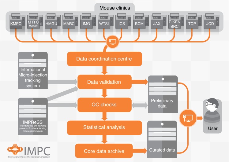
Despite advances in next generation sequencing technologies, determining the genetic basis of ocular disease remains a major challenge due to the limited access and prohibitive cost of human forward genetics. Thus, less than 4,000 genes currently have available phenotype information for any organ system. Here we report the ophthalmic findings from the International Mouse Phenotyping Consortium, a large-scale functional genetic screen with the goal of generating and phenotyping a null mutant for every mouse gene. Of 4364 genes evaluated, 347 were identified to influence ocular phenotypes, 75% of which are entirely novel in ocular pathology. This discovery greatly increases the current number of genes known to contribute to ophthalmic disease, and it is likely that many of the genes will subsequently prove to be important in human ocular development and disease.


Conflict of interest statement

The authors declare no competing interests.

Figures

Fig. 1
Fig. 1
Schematic overview of IMPC data flow from acquisition to web portal availability for public users. Data are collected from 12 phenotyping centers, validated, and processed to produce curated data accessible on the project portal. Legacy data from EuroPhenome and Sanger MGP were directly transferred to the Central Data Archive at EMBL-EBI for direct integration on the portal. https://academic.oup.com/nar/article-lookup/doi/10.1093/nar/gkt977. KMPC (Korea Mouse Phenotyping Center), MRC (Medical Research Council) Harwell Institute, HMGU (Helmholtz Zentrum Muenchen), MARC (Model Animal Research Center), IMG (Institute of Molecular Genetics), WTSI (Wellcome Trust Sanger Institute), ICS (Institut Clinique de la Souris— PHENOMIN-ICS), BCM (Baylor College of Medicine), JAX (The Jackson Laboratory), RBRC (RIKEN Bio-Resource Center), TCP (The Center for Phenogenomics), UCD (University of California Davis), IMPReSS (International Mouse Phenotyping Resource of Standardized Screens https://www.mousephenotype.org/impress)
Fig. 2
Fig. 2
Corneal abnormalities in Fam20a, Col6a2, and Nadsyn1 knockout mice. Biomicroscopy (a) of Fam20a knockout mice revealed polygonal opacities with indistinct edges and interweaving clear spaces in the corneal stroma (arrow), which were also apparent (arrow) on retro-illumination (b), scale bars = 500 µm. Histology (c) shows superficial stromal mineralization (arrow, scale bar = 50 µm). Corneal vascularization (arrow) and chronic superficial keratitis (arrowheads) were observed in Nadsyn1 knockout mice (d, scale bar = 500 µm), and red blood cells (arrow) are shown in the lumen of neovascular vessels on histology (e, scale bar = 20 µm). Mice lacking Col6a2 had subtle corneal stromal opacities seen on slit-lamp examination, which electron microscopy revealed to be a basket weaving appearance (arrowheads) of the corneal stroma (f) that was not seen in wild type (WT) controls (g), scale bars = 5 µm
Fig. 3
Fig. 3
Lenticular abnormalities in Ndrg1, Adamts18, and Cdkn2a knockout mice. a Retro-illumination highlights the well-defined concentric annular anterior and posterior cortical optical discontinuities in Ndgr1 knockout mice (arrow, scale bar = 500 µm). b Mice deficient in Adamts18 knockout mice had clinically evident vitreous crystalline deposits, which represented extruded lens material (arrow, scale bar = 100 µm). c Cdkn2a knockout mice had ocular lesions consistent with persistent tunica vasculosa lentis, with most severe cases having posterior lenticonus where the lens was adhered to the retina at the optic disc. The posterior lens capsule was segmentally disrupted and there was posterior subcapsular cataract. Additionally, the retinal segment was focally dysplastic. Scale bar = 500 µm
Fig. 4
Fig. 4
Retinal thinning in Arap1 and Rnf10 knockout mice. Arap1−/− mice had normal appearing retinal tissue at 2 weeks postnatal age (a) in comparison to wild type (WT) littermate control animals (b). The outer nuclear layer (asterisks in a, c) progressively degenerated (c) by 8 weeks postnatal in Arap1−/− mice, when compared with littermate control eyes (d). Scale bars = 50 µm. Optical coherence tomography of Rnf10−/− mice (e) shows statistically significant retinal thinning by 16 weeks postnatal age when compared to control retinal images (f). Scale bar = 100 µm. Box plots of male and female Rnf10−/− mice document retinal thinning (g) in comparison to normal age-matched controls, particularly affecting the inner plexiform layer (IPL) (h), and the inner nuclear layer. Error bars represent the standard deviation (SD) for the knockout measurements. Data points outside of the mean + /- SD range are represented by black circles. The two dotted red lines delimit the reference range, defined as two SD away from the mean for controls, represented by the solid red line
Fig. 5
Fig. 5
Ocular phenotypes of homozygous C57BL/6NJ-Mpdzem1J/J (Mpdz−/−) and control C57BL/6NJ (B6NJ) mice. Bright-field fundus images at 14 weeks of age (a). Detail of a showing enhanced detection of RPE cells (b) in mutant mice. Scale bar in a = 250 µm. ERG traces (c) from the left and right eyes of B6NJ (blue) and Mpdz−/− (red) mice (n = 4 both strains) at 16 weeks of age. The mean of all traces is shown in black. Scale bars, 100µV vertical, 50 ms horizontal. Summary of ERG response amplitudes (d) for data in b. Bars show mean ± SD; p-values from t-tests are indicated

References

    1. Eye Diseases Prevalence Research Group. Causes and prevalence of visual impairment among adults in the United States. Arch. Ophthal. 2004;122:477–485. doi: 10.1001/archopht.122.4.477. - DOI - PubMed
    1. Ko F, et al. Prevalence of nonrefractive visual impairment in US adults and associated risk factors, 1999–2002 and 2005–2008. JAMA. 2012;308:2361–2368. doi: 10.1001/jama.2012.85685. - DOI - PMC - PubMed
    1. Bourne R, Price H, Stevens G. Global burden of visual impairment and blindness. Arch. Ophthalmol. 2012;130:645–647. doi: 10.1001/archophthalmol.2012.1032. - DOI - PubMed
    1. Stone EM, et al. Clinically focused molecular investigation of 1000 consecutive families with inherited retinal disease. Ophthalmol. 2017;124:1314–1331. doi: 10.1016/j.ophtha.2017.04.008. - DOI - PMC - PubMed
    1. Warwick, A. & Lotery, A. Genetics and genetic testing for age-related macular degeneration. Eye (Lond). 32, 849–857 (2017). - PMC - PubMed