Review
. 2019 Mar;29(2):278-300.
doi: 10.1111/bpa.12695. Epub 2019 Jan 22.

Recent advances in the histo-molecular pathology of human prion disease

Affiliations
Review

Recent advances in the histo-molecular pathology of human prion disease

Simone Baiardi et al. Brain Pathol. 2019 Mar.

Abstract

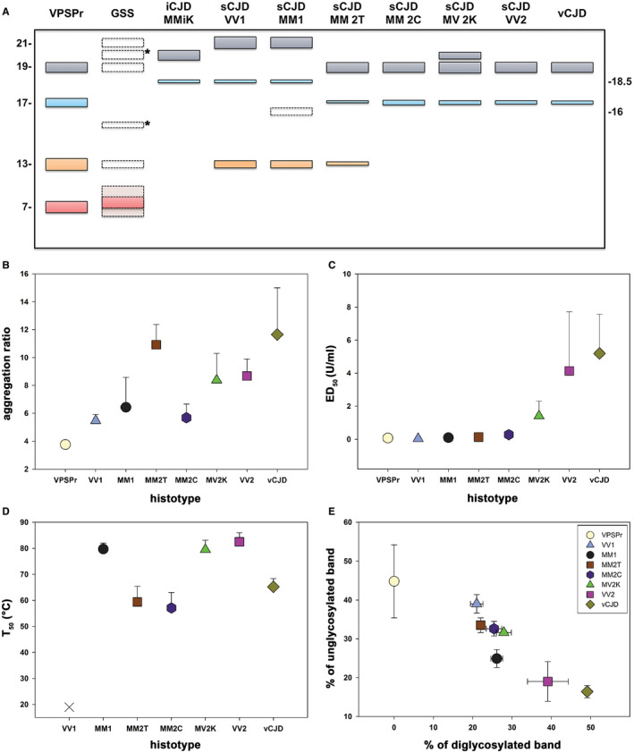
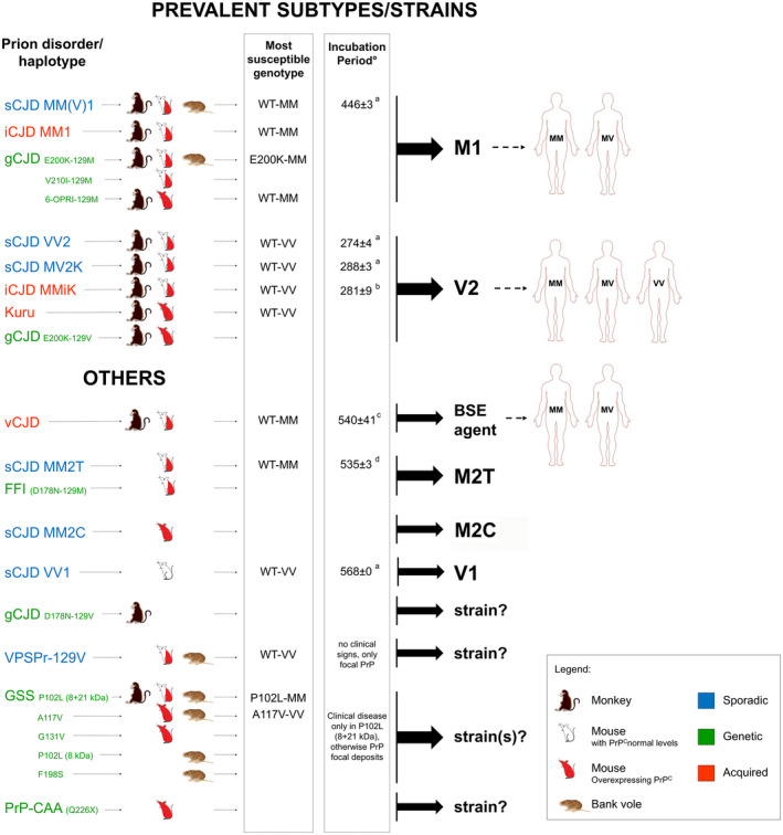
Prion diseases are progressive neurodegenerative disorders affecting humans and other mammalian species. The term prion, originally put forward to propose the concept that a protein could be infectious, refers to PrPSc , a misfolded isoform of the cellular prion protein (PrPC ) that represents the pathogenetic hallmark of these disorders. The discovery that other proteins characterized by misfolding and seeded aggregation can spread from cell to cell, similarly to PrPSc , has increased interest in prion diseases. Among neurodegenerative disorders, however, prion diseases distinguish themselves for the broader phenotypic spectrum, the fastest disease progression and the existence of infectious forms that can be transmitted through the exposure to diseased tissues via ingestion, injection or transplantation. The main clinicopathological phenotypes of human prion disease include Creutzfeldt-Jakob disease, by far the most common, fatal insomnia, variably protease-sensitive prionopathy, and Gerstmann-Sträussler-Scheinker disease. However, clinicopathological manifestations extend even beyond those predicted by this classification. Because of their transmissibility, the phenotypic diversity of prion diseases can also be propagated into syngenic hosts as prion strains with distinct characteristics, such as incubation period, pattern of PrPSc distribution and regional severity of histopathological changes in the brain. Increasing evidence indicates that different PrPSc conformers, forming distinct ordered aggregates, encipher the phenotypic variants related to prion strains. In this review, we summarize the most recent advances concerning the histo-molecular pathology of human prion disease focusing on the phenotypic spectrum of the disease including co-pathologies, the characterization of prion strains by experimental transmission and their correlation with the physicochemical properties of PrPSc aggregates.

Keywords: Creutzfeldt-Jakob disease; Gerstmann-Sträussler-Scheinker disease; amyloidosis; fatal insomnia; human prions; neurodegenerative dementia; prion strains.


Figures

Figure 1
Figure 1
Classification of human prion diseases according to the dominant PrPSc form, etiology and phenotypic features. *Atypical refers to disease lacking distinctive histopathological features. List of abbreviations: CJD, Creutzfeldt–Jakob disease; vCJD, variant CJD; iCJD, iatrogenic CJD; FI, fatal insomnia; FFI, fatal familial insomnia; sFI, sporadic FI; VPSPr, variably protease‐sensitive prionopathy; GSS, Gerstmann–Sträussler–Scheinker disease; PrP‐CAA, PrP‐cerebral amyloid angiopathy; PrP‐SA, PrP‐systemic amyloidosis; OPRIs, octapeptide repeat insertions.
Figure 2
Figure 2
Lesion profiles and histopathological hallmarks of sporadic Creutzfeldt–Jakob subtypes. Analyzed brain regions have been highlighted in yellow, orange and red to indicate, respectively, “minimal to mild” (score ≤1), “mild to moderate” (score >1–2) and “moderate to severe” (score >2) neuropathological changes. Lesion profiles were obtained by averaging the pathological score of spongiosis (not detectable = 0, mild = 1, moderate = 2, severe = 3, status spongiosus = 4), astrogliosis and neuronal loss (not detectable = 0, mild = 1, moderate = 2, severe = 3). Lesion profile data are from “typical” cases (eg, with a disease duration representative of the subtype). Distinctive histopathological features of sCJD subtypes. A. Spongiform change characterized by small, fine, microvacuoles in the neuropil (H&E, ×200, temporal cortex), and (B) synaptic pattern of PrPSc deposition in the molecular and granular layers of cerebellum in sCJDMM(V)1 (IHC, ×400). PrP staining is punctate and diffuse in the molecular layer, while it stands out the cerebellar glomeruli in the granular cell layers. C. Perineuronal PrP staining in deep layers of frontal cortex (IHC, x600) and (D) plaque‐like PrPSc deposits at transition between the molecular and the granular cell layers of cerebellum in sCJDVV2 (IHC, ×400). E. Spongiform change characterized by non‐confluent vacuoles of “intermediate” size, relatively larger than those observed in sCJDMM(V)1, but overall smaller than those of sCJDMM2C, in the occipital cortex (H&E, ×200), and (F) minimal/mild pathological changes in the cerebellum of sCJDVV1 (H&E, ×100). G, H. Unicentric kuru amyloid plaques at transition between the molecular and the granular cell layers of cerebellum in sCJDMV2K (G, H&E, ×600; H, IHC, ×400). I. Spongiform change characterized by large, confluent vacuoles (H&E, ×200) and (J) coarse, dense PrPSc deposits with perivacuolar distribution in the temporal cortex of sCJDMM2C (IHC, 400×). K, L. Severe neuronal loss and reactive gliosis in the absence of significant spongiform change in medial‐dorsal thalamic nucleus (K) and inferior olive (L) of sCJDMM2T (H&E, ×200). Legend: H&E, hematoxylin and eosin stain; IHC, PrP immunochemistry with primary mAb 3F4.
Figure 3
Figure 3
Histological features and western blot profile of iCJD MMiK. A. Unicentric kuru amyloid plaques in the molecular and granular cell layers, and in the white matter of cerebellum of iCJD MMiK (immunochemistry with primary Ab 3F4, ×200). B. Electrophoretic mobility of PK‐resistant PrPSc fragments in subtype iCJD MMiK compared with sCJDMM1 and sCJD MV2K. The molecular weight of the lower band (unglycosylated fragment) is ~20 kDa (PrPSc type “i”) in the iCJD MMiK case (courtesy from Prof. T. Kitamoto). Immunoblot was probed with the primary mAb 3F4.
Figure 4
Figure 4
Updated diagnostic flowchart for diagnosis and classification of sporadic and acquired human prion disease histotypes. Adapted from Parchi et al 125. Abbreviations: ctx = cerebral cortex; V = variant.
Figure 5
Figure 5
Spectrum of PrPSc physicochemical properties among phenotypic variants of human prion disease. A. Schematic representation of the spectrum of truncated PrPSc (unglycosylated) fragments observed in human prion disease. The classic N‐terminally truncated PrPSc fragments of ~21 (type 1), 20 (type “i”) and 19 (type 2) kDa, comprising the so‐called PrP27‐30, are shown in grey, while their anchorless counterparts of ~17 and 18.5 kDa are represented in light blue. C‐terminal associated fragments (CTF) of lower molecular weight (~13–15 kDa) are highlighted in orange, whereas the N‐ and C‐terminal ragged fragment of ~7–11 kDa linked to GSS and VPSPr are in light red. The bands with dotted lines symbolize PrPSc fragments that are only seen occasionally (eg, associated with specific GSS‐causing mutations) or in specific experimental conditions (eg, 16 kDa fragment seen in CJDMM1 in partially denaturing conditions). *Indicates PrPSc fragments (eg, 20 and 15 kDa in GSS) reported only once and lacking adequate comparison with control samples, leaving doubt about their precise molecular mass. B. Comparison of PrPSc “aggregation ratio” between CJD subtypes [data from Saverioni et al 152]. The ratio represents a semiquantitative index of the overall state of PrPSc aggregation, with values being proportional to the mean size of protein aggregates. C. Comparison of PrPSc PK‐resistance between CJD subtypes [data from Saverioni et. al. 152]. ED50 expresses the PK concentration needed to digest 50% of PrPSc. D. Comparison of PrPSc “thermostability” between CJD subtypes [data from Cescatti et al 32]. T50 expresses the temperature needed to solubilize 50% of PrPSc. The sCJDVV1 subtype is represented with a Χ since the calculated T50 is below 25°C. E. PrPSc glycoform ratio after PK digestion [data from Saverioni et al 152]. Data in BE are based on densitometric analyses of immunoblots probed with mAb 3F4 or SAF60.
Figure 6
Figure 6
Successful transmissions of human prion disease variants: Most susceptible PRNP genotypes and resulting prion strain diversity . Uncolored mice refer to animals expressing physiological levels of PrPC, red mice to animals overexpressing PrPC. Half colored mice indicate successful transmissions in both types of mice. Susceptible genotypes are those associated with the shortest incubation time. °Only studies in mice expressing physiological levels of PrPC are listed. aBishop et al 2010 17; bKobayashi et al 2007 73; cTakeuchi et al 2013 160; dModa et al 2012 104.

References

    1. Abu‐Rumeileh S, Redaelli V, Baiardi S, Mackenzie G, Windl O, Ritchie DL et al (2018) Sporadic fatal insomnia in Europe: phenotypic features and diagnostic challenges. Ann Neurol 84:347–360. - PubMed
    1. Aguzzi A, Heikenwalder M, Polymenidou M (2007) Insights into prion strains and neurotoxicity. Nat Rev Mol Cell Biol 8:552–561. - PubMed
    1. Aguzzi A, Zhu C (2017) Microglia in prion diseases. J Clin Invest 127:3230–3239. - PMC - PubMed
    1. Almer G, Hainfellner JA, Brücke T, Jellinger K, Kleinert R, Bayer G et al (1999) Fatal familial insomnia: a new Austrian family. Brain 122(Pt 1):5–16. - PubMed
    1. Asante EA, Linehan JM, Desbruslais M, Joiner S, Gowland I, Wood AL et al (2002) BSE prions propagate as either variant CJD‐like or sporadic CJD‐like prion strains in transgenic mice expressing human prion protein. EMBO J 21:6358–6366. - PMC - PubMed

MeSH terms