

A combined diffusion-weighted and electroencephalography study on age-related differences in connectivity in the motor network during bimanual performance
- PMID: 30588749
- PMCID: PMC6865498
- DOI: 10.1002/hbm.24491
A combined diffusion-weighted and electroencephalography study on age-related differences in connectivity in the motor network during bimanual performance
Abstract
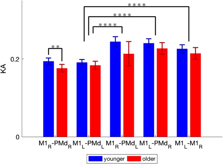
We studied the relationship between age-related differences in inter- and intra-hemispheric structural and functional connectivity in the bilateral motor network. Our focus was on the correlation between connectivity and declined motor performance in older adults. Structural and functional connectivity were estimated using diffusion weighted imaging and resting-state electro-encephalography, respectively. A total of 48 young and older healthy participants were measured. In addition, motor performances were assessed using bimanual coordination tasks. To pre-select regions-of-interest (ROIs), a neural model was adopted that accounts for intra-hemispheric functional connectivity between dorsal premotor area (PMd) and primary motor cortex (M1) and inter-hemispheric connections between left and right M1 (M1L and M1R ). Functional connectivity was determined via the weighted phase-lag index (wPLI) in the source-reconstructed beta activity during rest. We quantified structural connectivity using kurtosis anisotropy (KA) values of tracts derived from diffusion tensor-based fiber tractography between the aforementioned areas. In the group of older adults, wPLI values between M1L -M1R were negatively associated with the quality of bimanual motor performance. The additional association between wPLI values of PMdL --M1L and PMdR -M1L supports that functional connectivity with the left hemisphere mediated (bimanual) motor control in older adults. The correlational analysis between the selected structural and functional connections revealed a strong association between wPLI values in the left intra-hemispheric PMdL -M1L pathway and KA values in M1L -M1R and PMdR -M1L pathways in the group of older adults. This suggests that weaker structural connections in older adults correlate with stronger functional connectivity and, hence, poorer motor performance.
Keywords: DWI; EEG; aging; bimanual coordination; functional connectivity; motor control; structural connectivity.
© 2018 Wiley Periodicals, Inc.
Figures


Similar articles
-
Age-Related Changes in Frontal Network Structural and Functional Connectivity in Relation to Bimanual Movement Control.J Neurosci. 2016 Feb 10;36(6):1808-22. doi: 10.1523/JNEUROSCI.3355-15.2016. J Neurosci. 2016. PMID: 26865607 Free PMC article.
-
Ageing changes effective connectivity of motor networks during bimanual finger coordination.Neuroimage. 2016 Dec;143:325-342. doi: 10.1016/j.neuroimage.2016.09.014. Epub 2016 Sep 9. Neuroimage. 2016. PMID: 27616642
-
White matter microstructural organisation of interhemispheric pathways predicts different stages of bimanual coordination learning in young and older adults.Eur J Neurosci. 2018 Mar;47(5):446-459. doi: 10.1111/ejn.13841. Epub 2018 Feb 13. Eur J Neurosci. 2018. PMID: 29363832
-
The structural and functional connectivity of the posterior cingulate cortex: comparison between deterministic and probabilistic tractography for the investigation of structure-function relationships.Neuroimage. 2014 Nov 15;102 Pt 1:118-27. doi: 10.1016/j.neuroimage.2013.12.022. Epub 2013 Dec 21. Neuroimage. 2014. PMID: 24365673 Review.
-
Bimanual coordination and interhemispheric interaction.Acta Psychol (Amst). 2002 Jun;110(2-3):161-86. doi: 10.1016/s0001-6918(02)00032-x. Acta Psychol (Amst). 2002. PMID: 12102104 Review.
Cited by
-
Postmovement Beta Rebound in Real and Imagined Movement.Motor Control. 2024 Aug 22;29(1):53-68. doi: 10.1123/mc.2023-0033. Print 2025 Jan 1. Motor Control. 2024. PMID: 39179240
-
Brain Structural and Functional Connectivity: A Review of Combined Works of Diffusion Magnetic Resonance Imaging and Electro-Encephalography.Front Hum Neurosci. 2021 Oct 7;15:721206. doi: 10.3389/fnhum.2021.721206. eCollection 2021. Front Hum Neurosci. 2021. PMID: 34690718 Free PMC article. Review.
-
Bimanual motor impairments in older adults: an updated systematic review and meta-analysis.EXCLI J. 2022 Aug 16;21:1068-1083. doi: 10.17179/excli2022-5236. eCollection 2022. EXCLI J. 2022. PMID: 36381648 Free PMC article. Review.
-
White matter microstructural integrity as a key to effective propagation of gamma entrainment in humans.Geroscience. 2025 Feb;47(1):1019-1037. doi: 10.1007/s11357-024-01281-2. Epub 2024 Jul 15. Geroscience. 2025. PMID: 39004653 Free PMC article.
-
Aging and Complexity Effects on Hemisphere-Dependent Movement-Related Beta Desynchronization during Bimanual Motor Planning and Execution.Brain Sci. 2022 Oct 26;12(11):1444. doi: 10.3390/brainsci12111444. Brain Sci. 2022. PMID: 36358370 Free PMC article.
References
-
- Ballester‐Plane, J. , Schmidt, R. , Laporta‐Hoyos, O. , Junque, C. , Vazquez, E. , Delgado, I. , … Pueyo, R. (2017). Whole‐brain structural connectivity in dyskinetic cerebral palsy and its association with motor and cognitive function. Human Brain Mapping, 38, 4594–4612. 10.1002/hbm.23686 - DOI - PMC - PubMed
Publication types
MeSH terms
Grants and funding
LinkOut - more resources
Full Text Sources
Medical
