

Perilesional and homotopic area activation during proverb comprehension after stroke
- PMID: 30588768
- PMCID: PMC6346665
- DOI: 10.1002/brb3.1202
Perilesional and homotopic area activation during proverb comprehension after stroke
Abstract
Introduction: The mechanism of functional recovery in right hemisphere (RH) stroke patients when attempting to comprehend a proverb has not been identified. We previously reported that there is bilateral hemisphere involvement during proverb comprehension in the normal population. However, the underlying mechanisms of proverb comprehension following a right middle cerebral artery (MCA) infarction have not yet been fully elucidated.
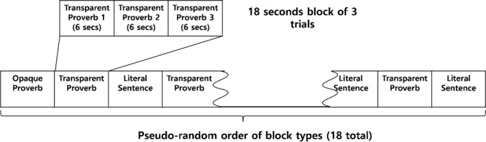
Methods: We here compared the brain regions activated by literal sentences and by opaque or transparent proverbs in right MCA infarction patients using functional magnetic resonance imaging (fMRI). Experimental stimuli included 18 opaque proverbs, 18 transparent proverbs, and 18 literal sentences that were presented pseudorandomly in 1 of 3 predesigned sequences.
Results: Fifteen normal adults and 17 right MCA infarction patients participated in this study. The areas of the brain in the stroke patients involved in understanding a proverb compared with a literal sentence included the right middle frontal gyrus (MFG) and frontal pole, right anterior cingulate gyrus/paracingulate gyrus and left inferior frontal gyrus (IFG), middle temporal gyrus (MTG), precuneus, and supramarginal gyrus (SMG). When the proverbs were presented to these stroke patients in the comprehension tests, the left supramarginal, postcentral gyrus, and right paracingulate gyrus were activated for the opaque proverbs compared to the transparent proverbs.
Conclusions: These findings suggest that the functional recovery of language in stroke patients can be explained by perilesional activation, which is thought to arise from the regulation of the excitatory and inhibitory neurotransmitter system, and by homotopic area activation which has been characterized by decreased transcallosal inhibition and astrocyte involvement.
Keywords: functional MRI; language; neural inhibition; proverb; right middle cerebral artery infarction.
© 2018 The Authors. Brain and Behavior published by Wiley Periodicals, Inc.
Figures

References
-
- Bestmann, S. , Swayne, O. , Blankenburg, F. , Ruff, C. C. , Teo, J. , Weiskopf, N. , … Ward, N. S. (2010). The role of contralesional dorsal premotor cortex after stroke as studied with concurrent TMS‐fMRI. Journal of Neuroscience, 30(36), 11926–11937. 10.1523/jneurosci.5642-09.2010 - DOI - PMC - PubMed
Publication types
MeSH terms
LinkOut - more resources
Full Text Sources
Medical
