Review
. 2019 Apr;196(1):12-27.
doi: 10.1111/cei.13256. Epub 2019 Jan 21.

Dendritic cells, T cells and their interaction in rheumatoid arthritis

Affiliations
Review

Dendritic cells, T cells and their interaction in rheumatoid arthritis

P Wehr et al. Clin Exp Immunol. 2019 Apr.

Abstract

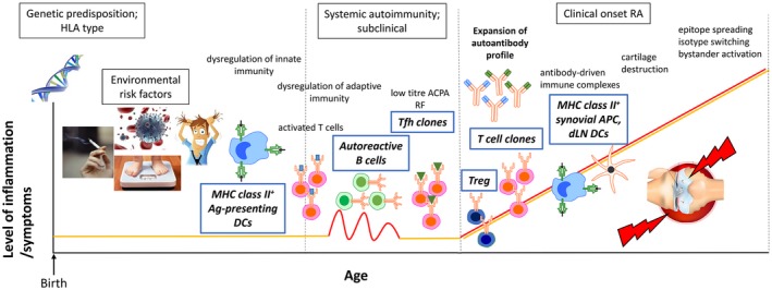
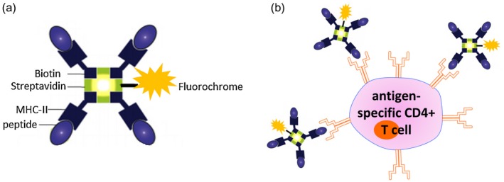
Dendritic cells (DCs) are the key professional antigen-presenting cells which bridge innate and adaptive immune responses, inducing the priming and differentiation of naive to effector CD4+ T cells, the cross-priming of CD8+ T cells and the promotion of B cell antibody responses. DCs also play a critical role in the maintenance of immune homeostasis and tolerance. DC-T cell interactions underpin the generation of an autoimmune response in rheumatoid arthritis (RA). Here we describe the function of DCs and review evidence for DC and T cell involvement in RA pathogenesis, in particular through the presentation of self-peptide by DCs that triggers differentiation and activation of autoreactive T cells. Finally, we discuss the emerging field of targeting the DC-T cell interaction for antigen-specific immunotherapy of RA.

Keywords: antigen presentation; autoantigen-specific CD4+ T cells; autoimmunity; dendritic cells; immunotherapy; rheumatoid arthritis.


Conflict of interest statement

R. T. has filed provisional patents surrounding technology for targeting DCs for antigen‐specific tolerance, and is a director of the spin‐off company, Dendright, which is commercializing immunotherapy to target DCs to suppress rheumatoid arthritis in collaboration with Janssen Biotech Inc. R. T. has also received speaker fees and/or consulting fees from Janssen and Abbvie.

Figures

Figure 1
Figure 1
A working model for the development of rheumatoid arthritis (RA). Both genetic and multiple non‐genetic risk factors predispose to the development of RA. In this context, disease initiation probably involves dysregulation of innate and adaptive immunity. Mature major histocompatibility complex (MHC) class II+ dendritic cells (DCs) probably induced by the environmal inflammatory milieu, prime autoantigen‐specific CD4+ T cells, including follicular helper T cells (Tfh) cells. Subsequent germinal centre formation, affinity maturation of B cells and expansion of the autoantibody profile may lead to antibody‐driven immune complex formation and subsequent cartilage destruction coinciding with overt RA expression. Although partially regulated, the autoimmune response persists due to ongoing stimulation of autoreactive T cell clones by a variety of synovial MHC class II+ antigen‐presenting cells (APC) and draining lymph node (dLN) DCs. Downstream processes, including neoepitope formation, isotype switching and bystander activation, may further propagate autoimmunity.
Figure 2
Figure 2
The classical and alternative nuclear factor kappa B (NF‐κB) pathways. The classical NF‐κB pathway (left): activation is induced by many extracellular signals including cytokines such as tumour necrosis factor (TNF). Activation leads to phosphorylation of inhibitory I‐κBα (I‐κB kinase) by the IKK complex, resulting in its degradation. The RelA/p50 complex is freed from the inhibitory interaction with IκBα and is an active dimer, which translocates to the nucleus to activate the transcription of target genes. c‐Rel and RelB also heterodimerize with p50. The alternative NFκB pathway (right): activation of this pathway occurs after stimulation by a more restricted set of ligands, including CD40, B cell activating factor (BAFF) and receptor activator of NF‐κB (RANK). Subsequently, NF‐κB‐inducing kinase (NIK) activation phosphorylates IKKα. pIKKα is crucial for phosphorylation of p100, leading to proteasome‐dependent processing of p100 to p52, ultimately liberating active RelB‐p52 dimers, which transactivate NF‐κB responsive genes. This figure was created using the Motfolio PPT Drawing Toolkits (www.motfolio.com), and adapted from 147, 148, 149.
Figure 3
Figure 3
Synovial CD1c+ dendritic cell (DC)‐mediated chemoattraction, T cell activation and polarization. Intra‐articular DCs secrete multiple chemokines, including chemokine (C‐C motif) ligand 3 (CCL3), CCL17, C‐X‐C motif chemokine ligand19 (CXCL19) and CXCL10, to induce T cell migration to the rheumatoid arthritis (RA) synovium. RA synovial CD1c+ DCs also secrete chemokines (including CCL3 and CXCL8) that attract proinflammatory immune cells, including macrophages, neutrophils and monocytes. RA synovial CD1c+ DCs have enhanced expression of human leucocyte antigen D‐related (HLA‐DR) and co‐stimulatory molecules CD40, CD80 and CD86 required for T cell activation. Synovial CD1c+ DCs from RA patients produce cytokines required for T cell polarization towards T helper type 17 (Th17) [interleukin (IL)‐1β, IL‐6, and IL‐23] and Th1 (IL‐12) differentiation. Synovial fluid from seropositive RA patients show high levels of serum soluble (s) programmed cell death 1 (PD‐1), which may interfere with the PD‐1/programmed death ligand 1 (PD‐L1) inhibitory signalling loop. This figure was generated using the Motfolio PPT Drawing Toolkits (www.motfolio.com), and knee image was obtained from https://www.pearsonschoolsandfecolleges.co.uk/FEAndVocational/SportsStudies/ALevel/OCRALevelPE2008/Samples/SamplepagesfromOCRASPEStudentBook/chapter1_sample.pdf, page 7, figure 1.4.
Figure 4
Figure 4
Schematic demonstrating the generation of the T cell receptor, the resultant complementarity‐determining region 3 (CDR3) of both α and β chains and T cell receptor–peptide–major histocompatibility complex (MHC) interaction. (a) Functional T cell receptors (TCRs) comprise an α and a β chain formed by somatic recombination of variable (V), diversity (D; β chain only) and junctional (J) gene segments. Recombined gene segments are then spliced together with the constant region (C) to form the functional TCR‐αβ. (b) CDR3 is formed at the V, (D), J junction, where imprecise V, (D), J joining and random addition of non‐template encoded nucleotides (N) makes this region the most diverse component of the TCR. (c) The heterodimeric TCR on a CD4+ T cell engages peptide displayed in the major histocompatibility complex class II (MHC‐II) on dendritic cells (DC). The CDR3 region is the main TCR domain in contact with the antigenic peptide. Figure (c) was created with the help of Motfolio PPT Drawing Toolkits (www.motfolio.com), and figures adapted from 74, 150.
Figure 5
Figure 5
Class II tetramers as tools for antigen‐specific CD4+ T cell detection. (a) Schematic depicting the structure of a major histocompatibility complex (MHC) class II tetramer. Four identical biotinylated MHC class II molecules loaded with a candidate (auto)antigenic peptide are linked to a central streptavidin molecule. The central streptavidin is conjugated to a fluorochrome of interest. (b) Binding of fluorescently labelled class II tetramer molecules to the antigen‐specific CD4+ T cell via the T cell receptor (TCR) enables detection of antigen‐specific T cells by flow cytometry. Figure (b) was created with the help of Motfolio PPT Drawing Toolkits (www.motfolio.com), and figures adapted from 151 and https://www.intechopen.com/books/allergen/strategies-to-study-t-cells-and-t-cell-targets-in-allergic-disease, chapter 7, page 128, figure 1.

References

    1. Merad M, Sathe P, Helft J, Miller J, Mortha A. The dendritic cell lineage: ontogeny and function of dendritic cells and their subsets in the steady state and the inflamed setting. Annu Rev Immunol 2013; 31:563–604. - PMC - PubMed
    1. Ganguly D, Haak S, Sisirak V, Reizis B. The role of dendritic cells in autoimmunity. Nat Rev Immunol 2013; 13:566–77. - PMC - PubMed
    1. Lutzky V, Hannawi S, Thomas R. Cells of the synovium in rheumatoid arthritis. Dendritic cells. Arthritis Res Ther 2007; 9:219. - PMC - PubMed
    1. Yu MB, Langridge WHR. The function of myeloid dendritic cells in rheumatoid arthritis. Rheumatol Int 2017; 37:1043–51. - PubMed
    1. Ziegler‐Heitbrock L, Ancuta P, Crowe S et al Nomenclature of monocytes and dendritic cells in blood. Blood 2010; 116:e74–80. - PubMed

Publication types