. 2019 Mar;26(3):96-103.
doi: 10.1002/jhbp.604. Epub 2019 Feb 10.

Bio-absorbable sealants for reinforcing the pancreatic stump after distal pancreatectomy are critical

Affiliations

Bio-absorbable sealants for reinforcing the pancreatic stump after distal pancreatectomy are critical

Christian M Kühlbrey et al. J Hepatobiliary Pancreat Sci. 2019 Mar.

Abstract

Background: Bio-absorbable sealants are widely used to reduce the rate and severity of postoperative pancreatic fistulas after distal pancreatectomy. However, numerous clinical trials have failed to demonstrate their clinical benefit. We therefore investigated stability and bio-compatibility of absorbable sealants in vitro and in vivo.

Methods: In vitro, polymerized compounds were incubated in pancreatic juice before their stability was tested. In vivo, two compounds were used to seal the pancreatic stump after distal pancreatectomy in nine pigs. Burst pressure of the pancreatic stump, surgical outcome, histology of the pancreatic stump, systemic inflammation, and drain fluid was examined.

Results: Products based on fibrin or collagen were unstable in the presence of active pancreatic enzymes and completely dissolved within 2 h. Sealants using chemical cross-linking of proteins showed improved stability for 7 days. In vivo, application of polyethylenglycol-based sealant leads to complete closure of the pancreatic duct after 5 days, while a glutaraldehyde-based sealant prevented physiological closure of the pancreatic main duct.

Conclusions: Many compounds used clinically to reinforce the pancreatic stump after distal pancreatectomy are inadequate due to instability in the presence of pancreatic enzymes. While selected bio-absorbable sealants inhibited the natural healing of the pancreatic stump, polyethylenglycol-based sealants should be tested in further clinical trials.

Keywords: Adhesives; Animal model; Pancreas; Pancreatic fistula; Polyethylene glycols.


Conflict of interest statement

None declared.

Figures
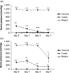
Figure 1
Figure 1
(a) Burst pressure after incubation of polymerized liquid bio‐absorbable adhesive in human plasma. (b) Burst pressure after incubation of polymerized liquid bio‐absorbable adhesive in pancreatic juice. Bioglue and Coseal both lost part of their stability after 7 days in pancreatic juice. In contrast to TissueCol, which quickly lost its integrity, the other two compounds showed sufficient stability to reasonably reinforce the pancreatic stump for up to 7 days after distal pancreatectomy. (***P < 0.001, **P < 0.01, *P < 0.05)
Figure 2
Figure 2
Change in drain amylase concentration. Drain amylase concentration after distal pancreatectomy decreased between POD 1 and POD 3. There was no difference among the groups. Interestingly, the drain amylase concentration in the control group, in which the pancreatic remnant was left unsealed, was not significantly elevated
Figure 3
Figure 3
Burst pressure of the pancreatic duct 5 days after distal pancreatectomy revealed spontaneous occlusion in untreated animals. Bioglue‐treated animals showed significantly lower burst pressure compared to control and Coseal‐treated animals which was induced by an inhibition of the natural occlusion of the pancreatic duct observed in both other groups
Figure 4
Figure 4
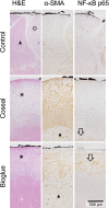
Histological presentation of the pancreatic dissection plane (black ink – surface of pancreatic remnant). H&E: the alterations of pancreatic parenchyma in untreated animals were fewer than in sealant treated animals. Here, we observed a cell‐rich layer in‐between acinar cells and the sealant (asterisk). α‐SMA: α‐SMA staining indicated increased activation of pancreatic stellate cells in the intermediate layer that was thinner in Bioglue‐treated animals. NF‐κB: the inflammatory response was most pronounced in animals with a Bioglue‐treated pancreatic stump. NF‐κB was expressed in ductal structures possibly identifying areas of acinar‐to‐ductal metaplasia
Figure 5
Figure 5
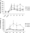
(a) Change in C‐reactive protein (CRP) concentrations. CRP was elevated after distal pancreatectomy in pigs. The elevation was noted throughout the entire experiment. The method of reinforcing the pancreatic stump had in impact on the CRP concentration. Animals treated with Bioglue showed increased CRP concentration until POD 5. (b) IL‐6 concentrations after stimulation with lipopolysaccharide (LPS). Cell sensitivity was even more pronounced when leukocytes were activated with LPS and IL‐6 release was quantified. In these experiments, Bioglue‐treated animals showed increased IL‐6 release upon LPS stimulation (*P < 0.05)
Figure 6
Figure 6
In vitro fibroblast cell growth. Fibroblast growth of human foreskin fibroblasts incubated with Coseal and Bioglue without direct contact to the sealant. Soluble components of polymerized Bioglue not only induced significant reduction in cell growth but also reduced the number of viable fibroblasts in vitro, explaining the impaired healing of the pancreatic stump after Bioglue application (**P < 0.01)
Figure 7
Figure 7
(a) Relative expression of apoptosis associated genes – Bioglue. (b) Relative expression of apoptosis associated genes – Coseal. The reduction of fibroblasts in the presence of soluble factors of bioglue is most likely due to apoptosis. Expression of anti‐apoptotic genes XIAP, BIRC2, BIRC5 was reduced pro‐apoptotic genes TNRFST10B, BCL2L11, and GADD45A were expressed at higher levels. In Coseal‐treated fibroblasts, only NOD1 showed a more than 2‐fold change in expression, possibly due to its role as an intracellular pattern recognition receptor

References

    1. Fahy BN, Frey CF, Ho HS, Beckett L, Bold RJ. Morbidity, mortality, and technical factors of distal pancreatectomy. Am J Surg. 2002;183:237–41. - PubMed
    1. Hassenpflug M, Hartwig W, Strobel O, Hinz U, Hackert T, Fritz S, et al. Decrease in clinically relevant pancreatic fistula by coverage of the pancreatic remnant after distal pancreatectomy. Surgery. 2012;152:S164–71. - PubMed
    1. Knaebel HP, Diener MK, Wente MN, Büchler MW, Seiler CM. Systematic review and meta‐analysis of technique for closure of the pancreatic remnant after distal pancreatectomy. Br J Surg. 2005;92:539–46. - PubMed
    1. Diener MK, Seiler CM, Rossion I, Kleeff J, Glanemann M, Butturini G, et al. Efficacy of stapler versus hand‐sewn closure after distal pancreatectomy (DISPACT): a randomised, controlled multicentre trial. Lancet. 2011;377:1514–22. - PubMed
    1. Sheehan MK, Beck K, Creech S, Pickleman J, Aranha GV. Distal pancreatectomy: does the method of closure influence fistula formation? Am Surg. 2002;68:264–7. - PubMed

Publication types

MeSH terms