Review
. 2019 Jul:71:26-56.
doi: 10.1016/j.preteyeres.2018.12.004. Epub 2018 Dec 25.

Insights into photoreceptor ciliogenesis revealed by animal models

Affiliations
Review

Insights into photoreceptor ciliogenesis revealed by animal models

Wolfgang Baehr et al. Prog Retin Eye Res. 2019 Jul.

Abstract

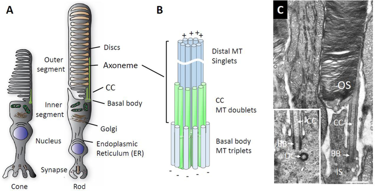
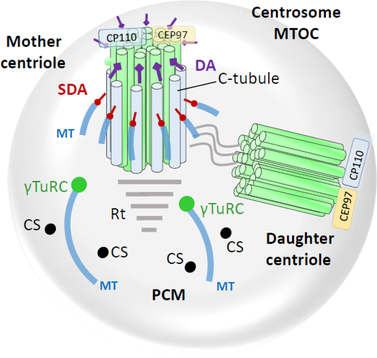
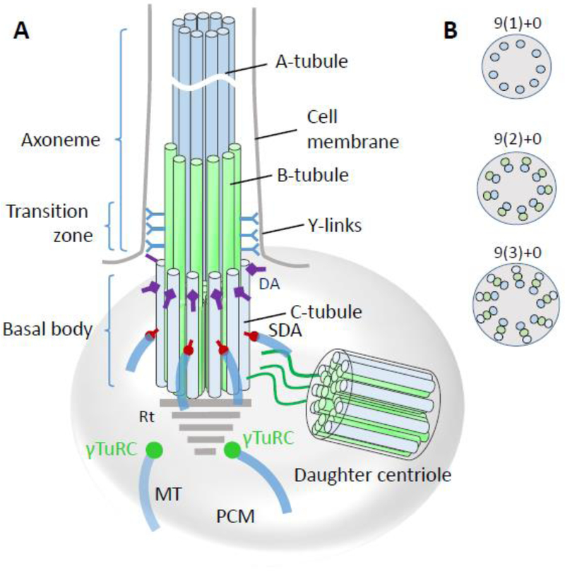
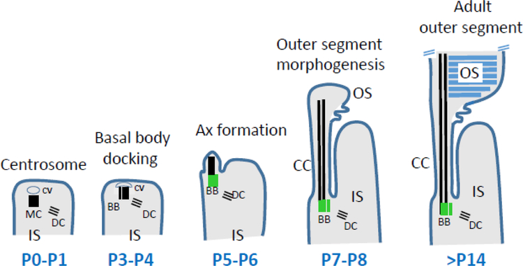
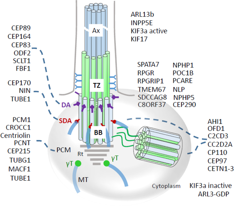
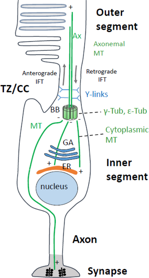
Photoreceptors are polarized neurons, with very specific subcellular compartmentalization and unique requirements for protein expression and trafficking. Each photoreceptor contains an outer segment, the site of photon capture that initiates vision, an inner segment that houses the biosynthetic machinery and a synaptic terminal for signal transmission to downstream neurons. Outer segments and inner segments are connected by a connecting cilium (CC), the equivalent of a transition zone (TZ) of primary cilia. The connecting cilium is part of the basal body/axoneme backbone that stabilizes the outer segment. This report will update the reader on late developments in photoreceptor ciliogenesis and transition zone formation, specifically in mouse photoreceptors, focusing on early events in photoreceptor ciliogenesis. The connecting cilium, an elongated and narrow structure through which all outer segment proteins and membrane components must traffic, functions as a gate that controls access to the outer segment. Here we will review genes and their protein products essential for basal body maturation and for CC/TZ genesis, sorted by phenotype. Emphasis is given to naturally occurring mouse mutants and gene knockouts that interfere with CC/TZ formation and ciliogenesis.

Keywords: Centrosome; Distal and subdistal appendages; Knockout mouse models; Microtubules and microtubule organization center; Mother and daughter centrioles; Pericentriolar matrix; Photoreceptors; Transition zone.


Figures

Figure 1.
Figure 1.
Basal body/axoneme cytoskeleton. A, Schematic of cone and rod photoreceptors depicting the outer segment where phototransduction occurs, the inner segment containing the endoplasmic reticulum and Golgi apparatus, the nuclear region and the synaptic terminal. B, enlarged detail of the axoneme cytoskeleton consisting of basal body (= mother centriole) proximally, transition zone and axoneme distally. C, electron micrograph of mouse rod, partial view, revealing the basal body, microtubule-stabilized CC and outer segment (OS) stacked with membrane discs. Note, the daughter centriole is out-of-plane and not visible; inset shows a different BB/DC pair. Scale, 0.5 µm.
Figure 2.
Figure 2.
The centrosome (microtubule organization center, MTOC) consisting of mother centriole, daughter centriole and pericentriolar matrix (PCM). Mother and daughter centriolar microtubules consist of nine triplet barrels organized in a cartwheel array. Each triplet has A-, B- and C-tubules; C-tubules are specific for mother and daughter centrioles. Cytoplasmic microtubules connect to subdistal appendages and to PCM points (green circles) anchored by the γ-tubulin ring complex. Centriolar satellites (CS) are distributed throughout the PCM. Distal ends of both mother and daughter centrioles are protected by centriolar coiled-coil protein CP110, and centrosomal protein CEP97. DA, distal appendages; SDA, subdistal appendages; MT, microtubules; γT, γ-tubulin ring complex; Rt, striated rootlet; CS, centriolar satellites.
Figure 3.
Figure 3.
Basal body/axoneme backbone. A, basal body schematic representation with transition zone and axoneme. A-tubules (blue) and B-tubules (green) emanate from the basal body to form the transition zone which is characterized by y-links connecting the tubules to the ciliary membrane. While the proximal axoneme consists of microtubule doublets, the distal axoneme has singlets. DA, distal appendages; SDA, subdistal appendages; MT, microtubules; PCM, pericentriolar matrix; γT, γ-tubulin ring complex. B, Crossections of the axoneme, transition zone and basal body, distal-to-proximal respectively, indicating the arrangement of microtubule arrays. “+0” indicates absence of a central MT.
Figure 4.
Figure 4.
Stages of rod photoreceptor ciliogenesis. At P0-P1, the centrosome consisting of mother and daughter centrioles (MC, DC) acquires a ciliary vesicle (CV) at its distal end. Soon, from P3-P4, the mother centriole docks to the cortex of the inner segment. At P5-P6, the axoneme (Ax) emanates from the MC and MC becomes a basal body (green). A- and B-tubules (black) extend at P7-P8, and the transition zone TZ is established. The cell membrane evaginates and discs are formed. The outer segment is considered mature at three weeks of age (P21). Adapted from May-Simera et al., 2018.
Figure 5.
Figure 5.
Localizations of centrosomal and centriolar proteins to the basal body, transition zone, and axoneme. CEP89, CEP164, CEP83, ODF2, SCLT1 and FBF1 are distal appendage (DA) proteins, while CEP170, NIN, and TUBE1 are subdistal appendage (SDA) proteins. PCM1, CROCC1 (rootletin), centriolin, PCNT (pericentrin), CEP215, TUBG1, TUBE1 and MACF1 are associated with the pericentriolar matrix (PCM) or ‘cloud.’ ARL13b and INPP5E are ciliary proteins. SPATA7, RPGR, RPGRIP1, NLP, centrins, CEP290, IQCB1/NPHP5, NPHP4, POC1B, TMEM67, SDCCAG8 (NPHP10) and C8ORF37 are located at the transition zone (TZ) distinguished by the presence of Y-linkers. AHI1, OFD1, C2CD3, CC2D2A, CP110, CEP97 localize to BB and DC. Inactive ARL3-GDP and KIF3a are cytoplasmic. Note that some proteins distribute to multiple subdomains (e.g., CETN2 associates with MC, DC and TZ) or occur in active and inactive forms altering their subdomain affiliation (ARL3-GTP and ARL3-GDP, or active/inactive KIF3a). Rt, rootlet; BB, basal body; Ax, axoneme.
Figure 6.
Figure 6.
Centrin 1 crystal structure and localization. A, Cetn1 (PDB 5D43) structure showing α-helical ribbons (red) and β-strand loops (green), forming the four EF-hands. Circular N (blue) and C (green) denote N- and C-terminals, respectively. Ca2+ ion positions (black dots) are indicated. B, CETN1 domain structure showing four high-affinity Ca2+ binding sites (EF-hands, blue) and DEAD-box subfamily ATP-dependent helicase signature (D, red). C, immunohistochemistry with anti-CETN1 (green) antibody to show labeling of centrioles and connecting cilia of wild-type photoreceptors (left), and absence in photoreceptors of germline knockout retina (right). Germline deletion of CETN1 does not affect photoreceptor function. D, immunohistochemistry with anti-CETN1 antibody in wild-type (left) and germline knockout (right) seminiferous tubules; Cetn1−/− mice are infertile. Scale bars: 10 μm; inset, 5 μm.
Figure 7.
Figure 7.
Transgenic expression of EGFP-CETN2 identifies photoreceptor centrioles and connecting cilia. A, rod outer segments labeled with anti-rhodopsin (red), centrioles expressing EGFP-CETN2 (green) and nuclei binding DAPI (blue). B, confocal microscopy of several connecting cilia. C, a single photoreceptor connecting cilium streak resembles the tail of a shooting star. BB, basal body; DC, daughter centriole.
Figure 8.
Figure 8.
Domain structures of mouse pericentriolar and subdistal appendage proteins. A, PCNT (pericentrin), B, CROCC1 (rootletin), C, PCM1 (pericentriolar material), D, CEP215 (CDK5RAP2), E, CEP170; F, CNTRL (centriolin), G, NIN (Ninein), H, Ninein-like (NINL) protein and I, ODF2. CD, coiled-coil domains. Myo-tail, myo tail region; CTD, centrosomal targeting domain; SHD, spectrin homology domain; NLS, nuclear localization signal; bZIP, basic leucine zipper domain; MTA, microtubule-associated region; FHA, forkhead-associated domain; Q, glutamine-rich region; E, glutamic acid-rich region; L, leucine-rich region; P, proline-rich region; EF, high-affinity Ca2+ binding site (EF-hand); P-loop, ATP/GTP-binding motif. The motifs were identified by scan “my Hits” at http://myhits.isb-sib.ch/cgi-bin/PFSCAN. Coiled-coil domains (CD) were retrieved using https://embnet.vital-it.ch/software/COILS_form.html.
Figure 9.
Figure 9.
Domain structures of mouse distal appendage (DA) proteins. A, CEP164, B, CEP83, C, SCLT1, D, FBF1, E, ODF2, F, C2CD3 and G, CEP89. WW, protein interaction domain flanked by tryptophan (W); CD, coiled-coil domains; NLS, nuclear localization signal; regions that are Q, glutamine-rich; E, glutamic acid-rich; K, lysine-rich; P, proline-rich; S, serine-rich are marked with colored lines; bZip, basic leucine zipper domain; myo-tail, myosin tail region;. Motifs were identified by motif scan “my Hits” at http://myhits.isb-sib.ch/cgi-bin/PFSCAN. Coiled-coil domains were retrieved using https://embnet.vital-it.ch/software/COILS_form.html.
Figure 10.
Figure 10.
Schematic of rod microtubule organization. Microtubules (green lines) radiate from the basal body (BB, or MTOC) to the photoreceptor periphery, i.e., the outer segment or alternatively, synaptic terminal. Microtubule (−) ends are located at the BB, (+) ends at the periphery. Plus-end directed kinesin-2 and minus-end directed cytoplasmic dynein transport “cargo” (vesicles loaded with membrane protein) to the appropriate destinations. Kinesin-2, an anterograde molecular motor, transports cargo through the TZ/CC to maintain the axoneme in the face of daily turnover of outer segment (OS) components. Cytoplasmic dynein-2 is the motor for retrograde intraflagellar transport (IFT). Y-links and ciliary proteins at the TZ/CC (CEP290) base have been proposed to form a gate controlling access to the OS, but a functional gate has not been shown to exist in photoreceptors. TZ/CC, transition zone/connecting cilium; Ax, axoneme; BB, basal body; GA, Golgi apparatus; ER, endoplasmic reticulum; MT, microtubules.
Figure 11.
Figure 11.
Distal centriole and proximal TZ protein domain structures. A, CP110; B, CEP97; C, CEP290; D, MACF1; E, CC2D2A. Ca, calmodulin- and centrin-interacting sites (Tsang, 2006); CD, coiled-coil domains; NLS, nuclear localization signal. L, leucine-rich domain; IQ, IQ calmodulin binding motif. EZRA, EZRA (bacterial scaffolding protein) domain; myo, myosin tail domain; colored bars above and below denote interaction sites with indicated proteins; Δ, area of in-frame deletion of exons 36–39 in rd16, a CEP290 mutant. S, serine-rich domain. C2, Ca2+-binding domain, a β-sandwich composed of 8 β-strands that co-ordinates two or three Ca2+ ions; Q, glutamine-rich domain. (microtubule-actin crosslinking factor 1). PR, plectin domain; SR, spectrin domain; EF, high-affinity Ca2+-binding site; CH, calponin-homology (actin-binding) domain; MT, microtubule-interacting region.
Figure 12.
Figure 12.
Retina-specific deletion of Kif3a in photoreceptors (retKif3a−/−). A, KIF3a domain schematic. Six3-Cre expression excises exon 2, thereby truncating the protein after exon 1. B, C, connecting cilium formation in retKif3a+/− (left) and retKif3a−/− (right) photoreceptors expressing the centriole marker, EGFP-CENT2, at P6 (B) and P10 (C). In C, CCs appear as 1µm-long streaks resembling shooting stars; despite presence of centrioles in the knockouts, CCs are absent. D-F, retKif3a+/− (D) and retKif3a−/− (E, F) photoreceptor ultrastructure. P10 retKif3a+/− photoreceptor with stacks of membrane discs forming at the connecting cilium distal portion (D). E,F, mother centrioles of P10 retKif3a−/− photoreceptors dock to the cortex normally but are devoid of CCs and outer segments. Mother centriole subdistal (arrow) and distal (arrowhead) appendages are indicated.
Figure 13.
Figure 13.
ARL3 distribution and function in photoreceptors. A, ARL3 activation by its GEF (ARL13b) and inactivation by its GAP (RP2) schematized. B, virally-expressed ARL3-EGFP localizes to the CC/BB area (identified by a yellow circle), inner segment (IS) and outer nuclear layer (ONL) of WT photoreceptors. Rhodopsin (red) is identified with polyclonal antibody directed against the mouse rhodopsin N-terminus (VPP-rho). Scale bar, 10µm. Enlargement of dotted area, right; scale, 5µm. C-F, ONL immunohistochemistry at P10 (C,E) and P15 (D,F). EGFP-CETN2 fluorescence (C,D), or anti-rhodopsin labeling (E,F) are illustrated. C,D (left panels) show controls with normal transition zones. C, D (right) are retina-specific knockouts with stunted or absent transition zones. Rhodopsin localizes normally to ROS (E,F left panels); rhodopsin mislocalizes to the outer nuclear layer (E,F, right). DAPI (blue) identifies extent of outer nuclear layer. Scale bar, 10µm. G, H, Ultrastructure of retArl3−/− retina. Transition zone ultrastructure of P10 (G) and P15 (H) WT and mutant retinas. Controls (G,H left panels) demonstrate intact CC, BB and DC. CCs are absent in each knockout (right). Scale bar, 500 nm.
Figure 14.
Figure 14.
Deletion of Arl13b in retina. A, ARL13b domains: hnn, hennin truncation mutation in mouse; PRR, proline-rich domain; CD, coiled-coil domain; G-domain, a canonical switch motif (‘on’ with GTP bound, ‘off’ with GDP bound). ). B, Co-crystal structure of C. reinhardtii ARL13b (residues 17–220, left) and ARL3 (residues 16–180, right) G-domains. Green circle, Mg2+ bound to GTP. C-terminal helix and switch II of ARL13B mediate GEF activity. C, Arl13b (red) immunolocalization in control (left panel) and knockout (right) photoreceptors; note, CCs of the heterozygous control are absent in the knockout. D, Egfp-Cetn2+;retArl13b+/− (a, c, e) and Egfp-Cetn2+;retArl13b−/− (b, d, f) P15 mouse retina cryosections were labeled with antibodies directed against rhodopsin (a, b), S-opsin (c, d) and ML-opsin (e, f) (red). retArl13b+/− photoreceptors develop transition zones (a, white arrowheads), while formation of the CC and OS in retArl13b−/− photoreceptors (b, d, f) is impaired. Scale bar, 5 μm. E, Ultrastructure of P10 (a, b) and P15 (c, d) WT and mutant photoreceptors. Controls (a, c) develop a CC and OS, while knockouts do not (b, d). Scale bar, 500 nm. F, tamoxifen-induced depletion of ARL13b, assayed 3 weeks post-injection (3WPI). Immunohistochemistry of 3WPI tamArl13b+/+ (left) and tamArl13b +/+ (right) cryosections with anti-IFT88 antibody. Proximal OSs (arrows) and basal bodies (arrowheads) are indicated. Enlargements, IFT88 (red) accumulates at the basal body, CC and proximal OS of control photoreceptors, while IFT88 appears predominantly restricted to the proximal OS of Arl13b-depleted cells. Scale bar, 10 μm; enlargements, 3 μm.
Figure 15.
Figure 15.
IQCB1/NPHP5 domain structure and photoreceptor localization. A, IQCB1/NPHP5 functional domains. BBS, BBSome interaction site; CD, coiled-coil domains; IQ, IQ calmodulin-binding motifs; Cep, CEP290-binding site. Crd2 dog, position of frameshift mutation in exon 12 associated with SLS. β-GEO identifies the approximate position of truncation in the Iqcb1 germline knockout. B, IQCB1/NPHP5 immunolocalization in P15 control (left) and Iqcb1/Nphp5 /− cryosections. Centrioles/CCs are identified by transgenic expression of EGFP-CETN2. Note, germline Iqcb1/Nphp5 knockout mice have photoreceptors that are unable to form OS. C, retina cryosections probed with anti-CEP290 antibody. CEP290 is unstable and degraded in the absence of IQCB1/NPHP5. D, P10 control (left) and Iqcb1/Nphp5 −/− (right) ultrastructure showing photoreceptor transition zones.
Figure 16.
Figure 16.
INPP5E and phosphoinositides. A, INPP5E domain structure. An inositide polyphosphate phosphatase, INPP5E bears a coiled-coil domain at its C-terminal region and a CAAX motif signaling farnesylation. B, INPP5E enzymatically removes a 5’-phosphate at the inositol ring of PI(3,4,5)P3; side chains R1 and R2 are acyl esters attached to glycerol. PI(3,4,5)P3 is a key secondary messenger in photoreceptors and other cells. C, virally-expressed EGFP-INPP5E (green) distributes to the Golgi apparatus of the inner segment and colocalizes partially with the Golgi marker, giantin (red). INPP5E is also found associated with the perinuclear endoplasmic reticulum (ER). D, Anti-rhodopsin labels the rod outer segments where EGFP-INPP5E is undetectable.
Figure 17.
Figure 17.
Domain structures of A, NPHP1, B, AHI1 (jouberin); C, NPHP4; D, lebercilin (LCA5); E, RPGR (RP3); F, RPGRIP1; G, SPATA7; H, TMEM67 (meckelin); I, FAM161A; J, SDCCAG8; K, POC1b. CD, coiled-coil domain; SH3, Src homology 3 domain; WD40, WD40 repeat (40 amino acids flanked by W and D); RPGR-BD, RPGR binding domain; P’ proline-rich domain; NLS, nuclear localization signal; P-loop, phosphate binding loop for interaction with ATP/GTP; RCC1, Regulator of Chromosome Condensation 1; E, glutamic acid-rich region; RID, RPGR-interacting domain; C2, β-sandwich structure that coordinates Ca2+ ions; TM, predicted transmembrane domain; S, serine-rich domain; N, asparagine-rich domain; Q- glutamine-rich domain. J, co-crystal structure of RPGR RCC1-like domain (RLD) and the RPGR-interacting domain (RID) of RPGRIP1. RPGR-RLD forms a seven-bladed propeller, each consisting of four anti-parallel β-strands (Waetzlich et al. 2013). RPGRIP1-RID consists of an eight-stranded antiparallel β-sandwich. Several mutations associated with XLRP are located at the RLD interface with RID (Remans et al., 2014).

References

    1. Acland GM, Blanton SH, Hershfield B, and Aguiree GD (1994). XLPRA: a canine retinal degeneration inherited as an X-linked trait. Am. J. Med. Genet 52, 27–33. - PubMed
    1. Adly N, Alhashem A, Ammari A, and Alkuraya FS (2014). Ciliary genes TBC1D32/C6orf170 and SCLT1 are mutated in patients with OFD type IX. Hum. Mutat 35, 36–40. - PubMed
    1. Airik R, Slaats GG, Guo Z, Weiss AC, Khan N, Ghosh A, Hurd TW, Bekker-Jensen S, Schroder JM, Elledge SJ, Andersen JS, Kispert A, Castelli M, Boletta A, Giles RH, and Hildebrandt F (2014). Renal-retinal ciliopathy gene Sdccag8 regulates DNA damage response signaling. J. Am. Soc. Nephrol 25, 2573–2583. - PMC - PubMed
    1. Aldaz H, Rice LM, Stearns T, and Agard DA (2005). Insights into microtubule nucleation from the crystal structure of human gamma-tubulin. Nature 435, 523–527. - PubMed
    1. Anand M and Khanna H (2012). Ciliary transition zone (TZ) proteins RPGR and CEP290: role in photoreceptor cilia and degenerative diseases. Expert. Opin. Ther. Targets 16, 541–551. - PMC - PubMed

Publication types

Substances