

Biopsy During Minimally Invasive Intracerebral Hemorrhage Clot Evacuation
- PMID: 30590212
- PMCID: PMC8407056
- DOI: 10.1016/j.wneu.2018.12.058
Biopsy During Minimally Invasive Intracerebral Hemorrhage Clot Evacuation
Abstract
Background: The safety and efficacy of brain parenchyma biopsy during minimally invasive (MIS) intracerebral hemorrhage (ICH) clot evacuation has not been previously reported. The objective of this study was to establish the safety and diagnostic efficacy of brain biopsy during MIS ICH clot evacuation and to validate the modified Boston criteria as a predictor of cerebral amyloid angiopathy (CAA) in this cohort.
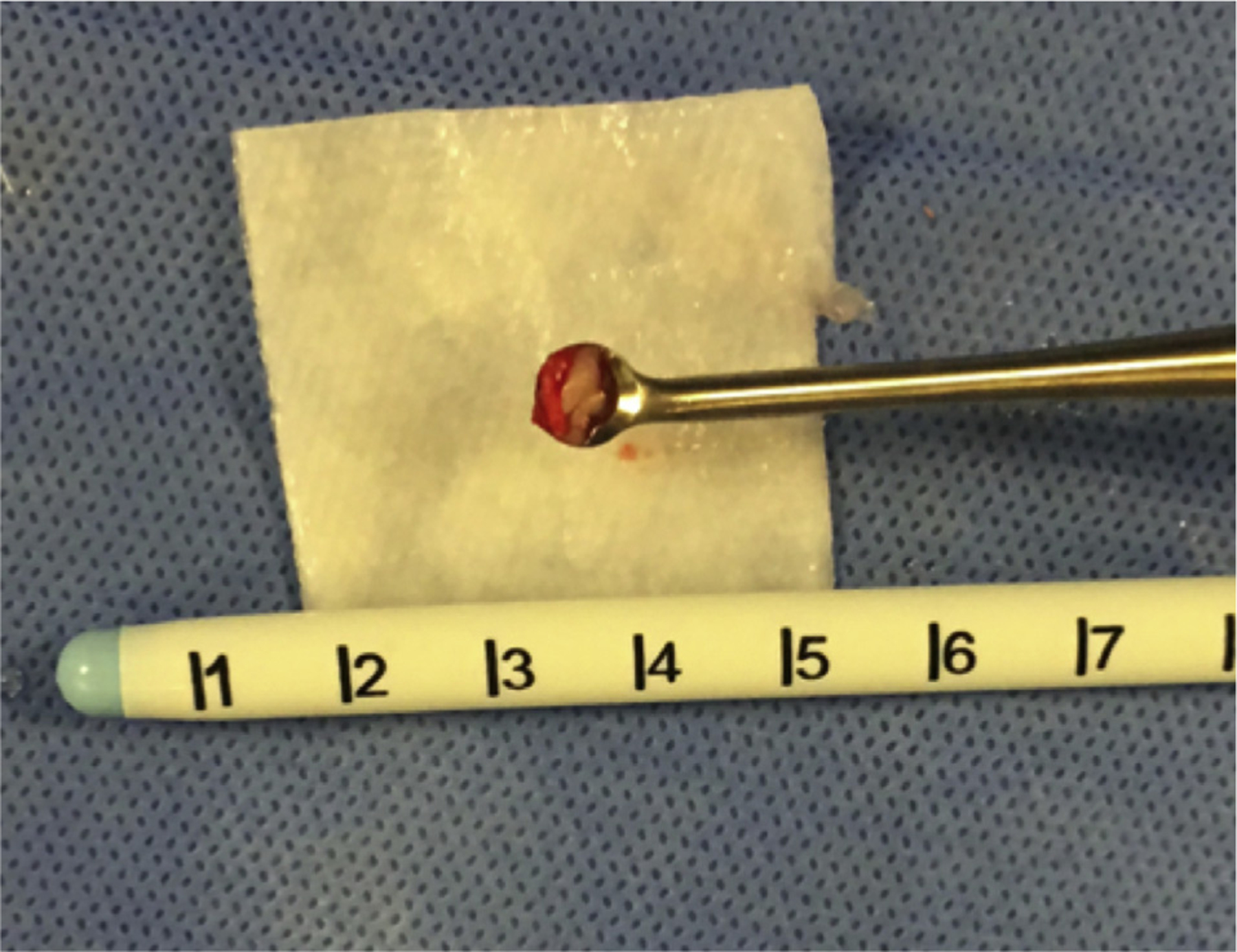
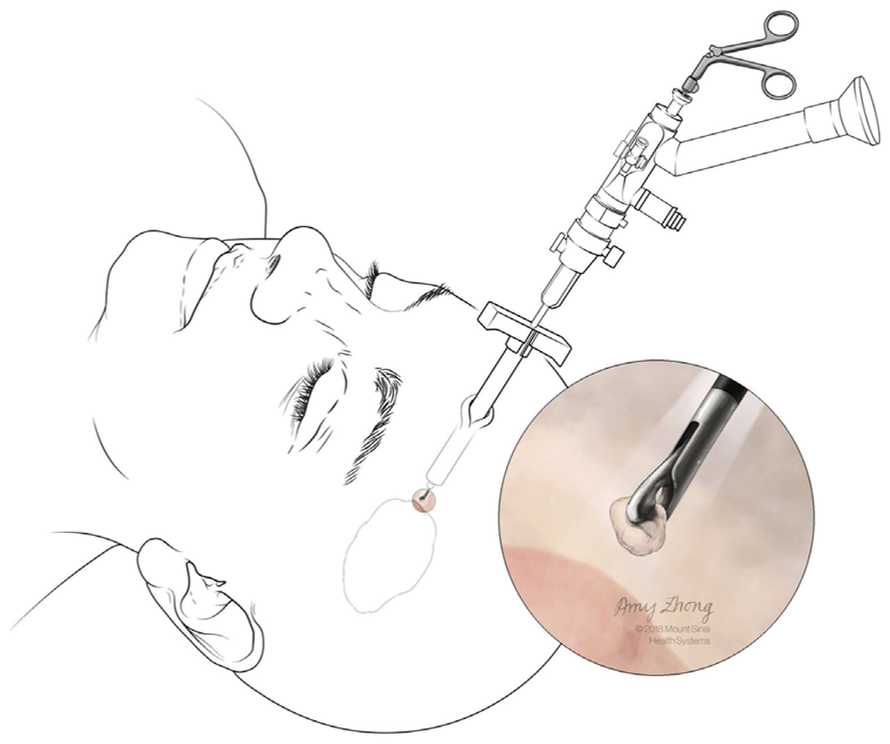
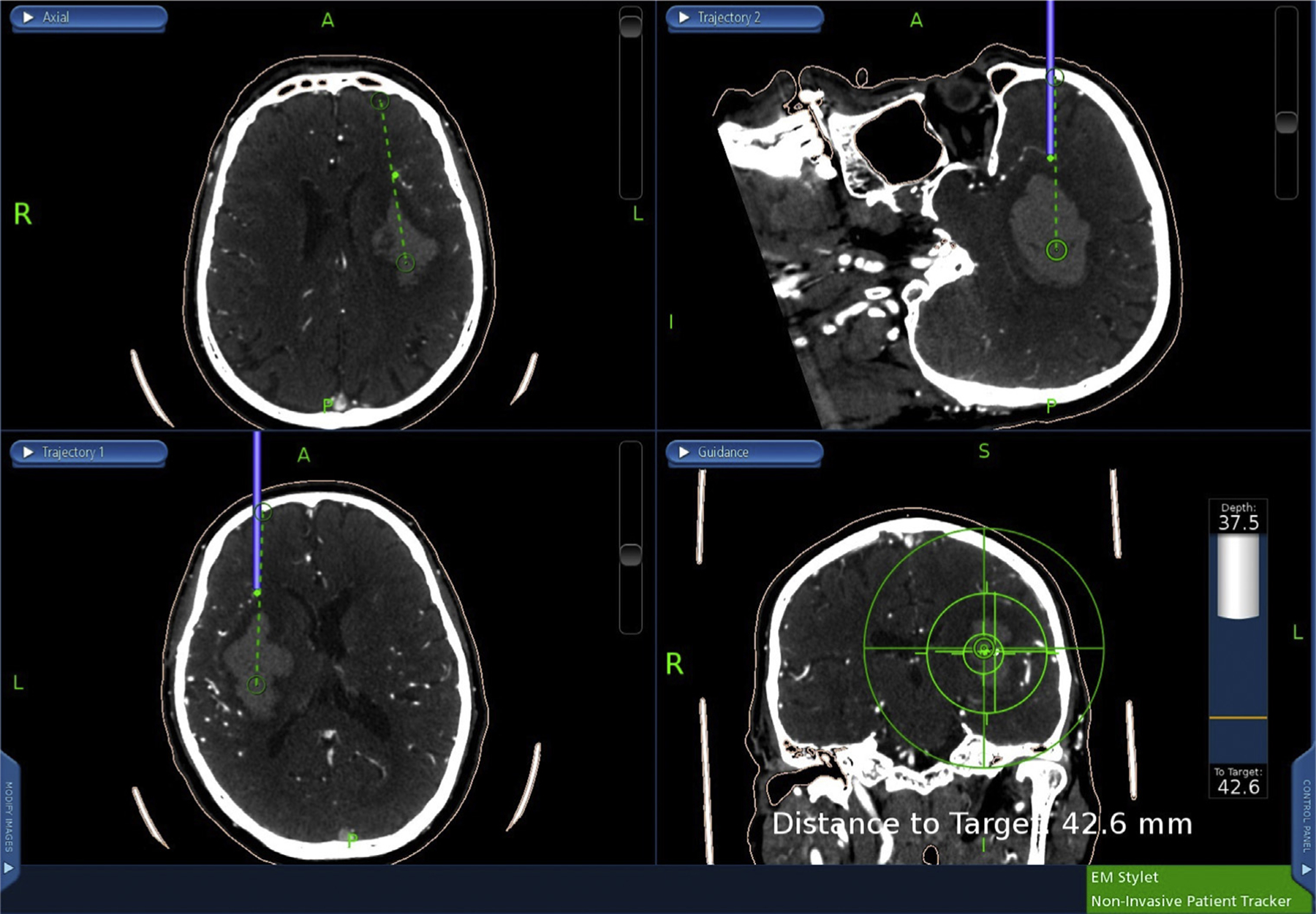
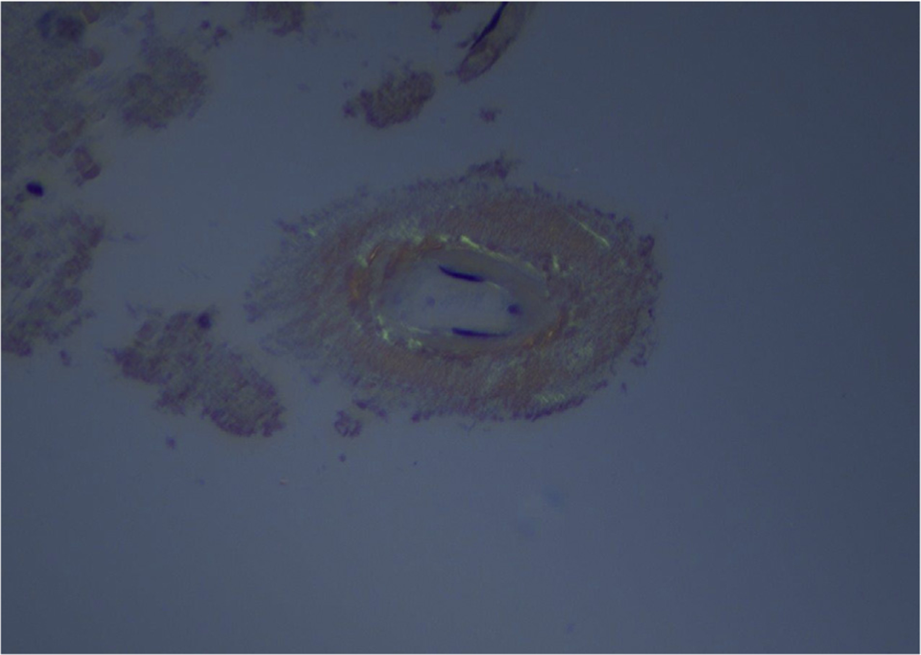
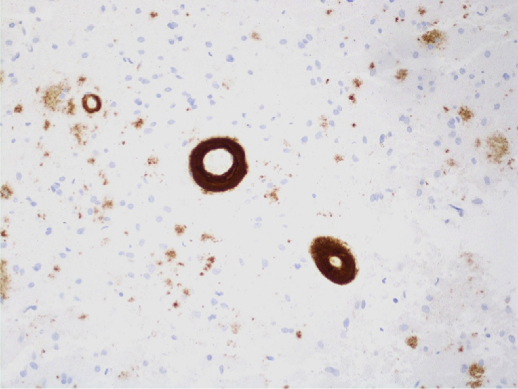
Methods: From October 2016 to March 2018, superficial and perihematomal biopsies were collected for 40 patients undergoing MIS ICH clot evacuation and analyzed by the pathology department to assess for various ICH etiologies. Additionally, the admission magnetic resonance imaging or computed tomography scan of each patient was analyzed and evaluated for the likelihood of a CAA etiology based on the modified Boston criteria. Student t test was used to analyze intergroup differences in continuous variables, and a 2-tailed Fisher exact test was used to determine intergroup differences of categorical variables, with significance set at P < 0.05.
Results: Two of the 40 patients (5%) experienced postoperative rebleed. Four of the 40 patients (10%) had evidence of CAA on biopsy. Patients with CAA on biopsy were older (P = 0.005) and had a higher prevalence of parietal lobe (P = 0.02) and occipital lobe (P = 0.001) hemorrhage. The modified Boston criteria had a sensitivity of 100% (95% confidence interval [CI], 39.6%-100%) and a specificity of 72.2% (95% CI, 54.6%-84.2%) for predicting CAA on biopsy.
Conclusions: Brain biopsy in MIS ICH clot evacuation is safe and allows for the diagnosis of various ICH etiologies.
Keywords: Biopsies; Cerebral amyloid angiopathy; Intracerebral hemorrhage; Minimally invasive surgery.
Copyright © 2018 Elsevier Inc. All rights reserved.
Conflict of interest statement
Conflict of interest statement: This research was supported in part by a grant from Arminio and Lucyna Fraga and by a grant from Mr. and Mrs. Durkovic.
Figures

References
-
- Qureshi AI, Tuhrim S, Broderick JP, Batjer HH, Hondo H, Hanley DF. Spontaneous intracerebral hemorrhage. N Engl J Med. 2001;344:1450–1460. - PubMed
-
- van Asch CJJ, Luitse MJA, Rinkel GJE, van der Tweel I, Algra A, Klijn CJM. Incidence, case fatality, and functional outcome of intracerebral haemorrhage over time, according to age, sex, and ethnic origin: a systematic review and meta-analysis. Lancet Neurol. 2010;9:167–176. - PubMed
-
- Attems J, Jellinger K, Thal DR, Van Nostrand W. Review: sporadic cerebral amyloid angiopathy. Neuropathol Appl Neurobiol. 2011;37:75–93. - PubMed
-
- González-Pérez A, Gaist D, Wallander M-A, McFeat G, García-Rodríguez LA. Mortality after hemorrhagic stroke: data from general practice (The Health Improvement Network). Neurology. 2013;81:559–565. - PubMed
-
- Sacco S, Marini C, Toni D, Olivieri L, Carolei A. Incidence and 10-year survival of intracerebral hemorrhage in a population-based registry. Stroke. 2009;40:394–399. - PubMed
Grants and funding
LinkOut - more resources
Full Text Sources
