. 2019 Oct 4;74(11):1709-1715.
doi: 10.1093/gerona/gly290.

Adipose Tissue Senescence and Inflammation in Aging is Reversed by the Young Milieu

Affiliations

Adipose Tissue Senescence and Inflammation in Aging is Reversed by the Young Milieu

Amiya Kumar Ghosh et al. J Gerontol A Biol Sci Med Sci. .

Abstract

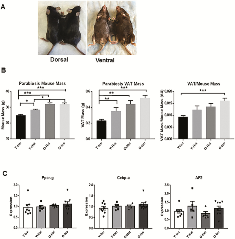
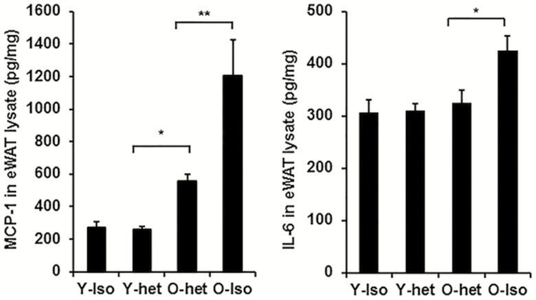
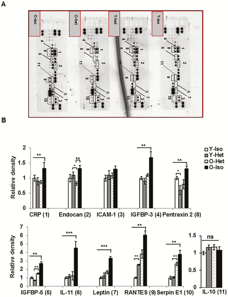
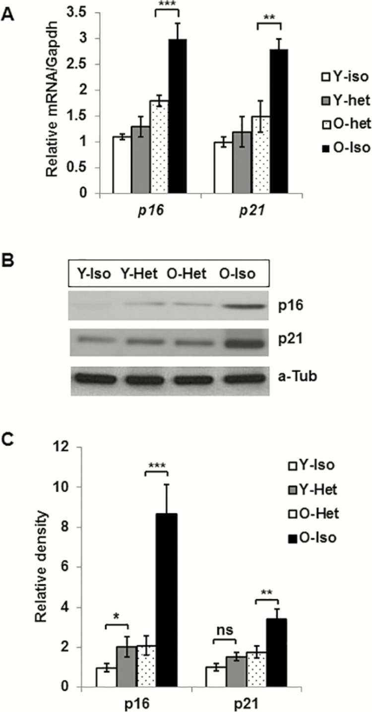
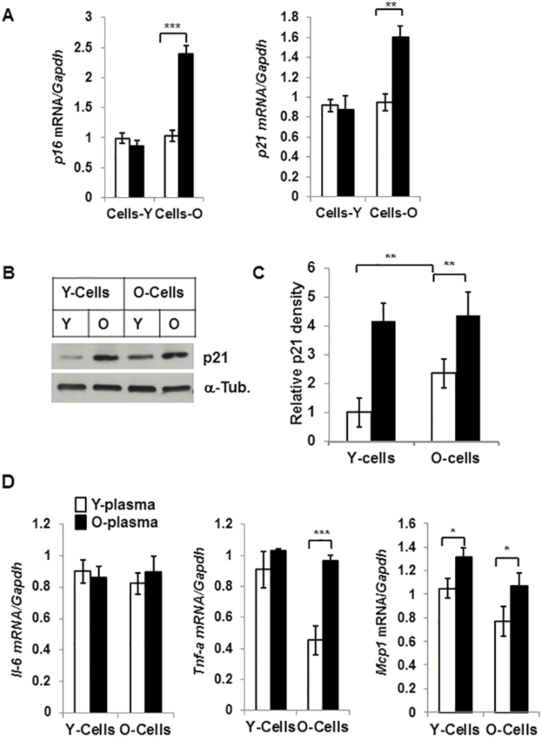
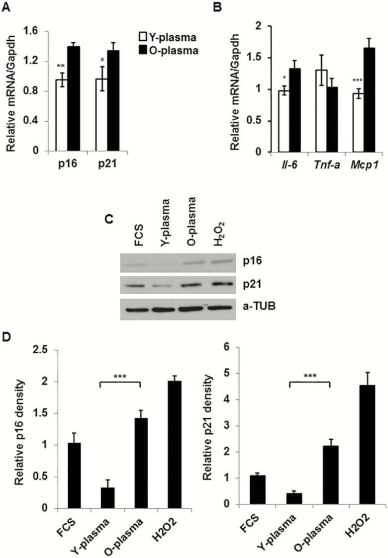
Visceral adipose tissue (VAT) inflammation plays a central role in longevity and multiple age-related disorders. Cellular senescence (SEN) is a fundamental aging mechanism that contributes to age-related chronic inflammation and organ dysfunction, including VAT. Recent studies using heterochronic parabiosis models strongly suggested that circulating factors in young plasma alter the aging phenotypes of old animals. Our study investigated if young plasma rescued SEN phenotypes in the VAT of aging mice. With heterochronic parabiosis model using young (3 months) and old (18 months) mice, we found significant reduction in the levels of pro-inflammatory cytokines and altered adipokine profile that are protective of SEN in the VAT of old mice. These data are indicative of protection from SEN of aging VAT by young blood circulation. Old parabionts also exhibited diminished expression of cyclin-dependent kinase inhibitors (CDKi) genes p16 (Cdkn2a) and p21 (Cdkn1a/Cip1) in the VAT. In addition, when exposed to young serum condition in an ex vivo culture system, aging adipose tissue-derived stromovascular fraction cells produced significantly lower amounts of pro-inflammatory cytokines (MCP-1 and IL-6) compared to old condition. Expressions of p16 and p21 genes were also diminished in the old stromovascular fraction cells under young serum condition. Finally, in 3T3-preadipocytes culture system, we found reduced pro-inflammatory cytokines (Mcp-1 and Il-6) and diminished expression of cyclin-dependent kinase inhibitor genes in the presence of young serum compared to old serum. In summary, this study demonstrates that young milieu is capable of protecting aging adipose tissue from SEN and thereby inflammation.

Keywords: Adipose tissue; Aging; Inflammation; Plasma factors; Senescence.


Figures

Figure 1.
Figure 1.
Increased body weight (BW) and visceral adipose tissue (VAT) mass in young but no change in old mice because of heterochronic parabiosis. (A) Photographs of heterochronic pairs both dorsal and ventral views. (B) Graphical representation of body weight and VAT weight and the ratio of BW:VAT of young (4 month) isochronic (Y-Iso), young heterchronic (Y-Het), old (20 month) heterochronic (O-Het), and old isochronic (O-Iso) pairs (n = 12 each) following 4 weeks of parabiosis. Statistical significance was determined by Tukey’s multiple comparison test followed by analysis of variance and indicated as *p < .05, **p < .001, ***p < .0001. (C) Relative mRNA abundance of Ppar-g, Cebp-a, and Ap2 in the parabiosis groups were also plotted in the graph after normalization (n is represented by no. of dots) with Gapdh expression. Statistical significance was determined by paired t test.
Figure 2.
Figure 2.
Young plasma diminishes pro-inflammatory and senescence phenotypes in old adipose tissue in heterochronic parabiosis system. Mouse (C57/Bl6: male) adipose tissue samples were collected from either isochronic young (Y-iso) or old (O-iso), and heterochronic young (Y-het) or old (O-het) groups following 4 weeks of parabiosis. (A) Levels of MCP-1 and (B) IL-6 in the adipose tissue lysate as quantified by ELISA. Statistical significance was determined by paired t test (n = 6 for each group) and expressed as * p < .05 or **p < .001.
Figure 3.
Figure 3.
Diminished abundance of cellular senescence–promoting adipokines (ADKs) protein in old adipose tissue following heterochronic parabiosis. (A) Representative image of three independent ADK arrays performed on visceral adipose tissue lysates from the parabionts. Proteins of interest are labeled with rectangles and adjacent numbers. The expression of each ADK band is normalized with internal reference spots and is plotted in (B). Statistical significances were determined by paired t test (n = 6 for each group) and expressed as *p < .05, **p < .001, ***p < .0001.
Figure 4.
Figure 4.
Reduced levels of p16 and p21 gene products in aging visceral adipose tissue following heterochronic parabiosis. (A) Graphical presentation of relative mRNA of p16 and p21 in different parabionts (n = 6 for each group). (B) Representative image of western blot analysis of p16 and p21 protein expression. (C) The relative protein abundance of p16 and p21 were expressed (n = 4) after the density was normalized with the respective α-Tubulin. Statistical significance was determined by paired t test and expressed as *p < .05, **p < .001.
Figure 5.
Figure 5.
Reduced expression of senescence-associated and pro-inflammatory genes in old stromovascular fractions (SVFs) cultured with young plasma. Epididymal adipose tissue–derived SVFs from young (4 months) and old (20 months) were cultured in presence of young or old serum (10%) for 3 days and were harvested for total RNA and protein lysates. mRNA expression of p16 and p21 was analyzed by quantitative reverse transcription-polymerase chain reaction and plotted in (A) and relative protein abundance (n = 3) was measured after the density was normalized with the respective α-Tubulin band (B and C). Relative mRNA abundance of Il-6, Tnf-α and Mcp-1 was also plotted in the graph (D). The significance levels *p < .05 or **p < .01 or ***p < .001 were determined by Student’s t test using means and standard error of the mean of three independent (n = 3) experiments.
Figure 6.
Figure 6.
Reduced expression of pro-inflammatory and cellular senescence genes in 3T3-preadipocytes and in primary stromovascular fractions (SVFs), cultured in the presence of young plasma. 3T3-preadipocytes or adipose tissue–derived old SVFs were cultured in the presence of young or old plasma (10%) for 3 days. Cells were harvested for total RNA and protein lysates. Expression of mRNA was analyzed by quantitative reverse transcription-polymerase chain reaction and protein analysis by western blotting. (A) Relative mRNA expressions of p16 (Cdkn2a) and p21 (Cdkn1a/Cip1) were evaluated after normalization with Gapdh (n = 4). (B) Relative expression of Il-6, Tnf-α, and Mcp-1 was expressed following normalization with Gapdh expression (n = 4). (C) Representative image of protein expressions of p16 and p21 in 3T3-preadipocyte lysates under different conditions. The relative protein abundance was shown in graph (D) after the density was normalized with the respective α-Tubulin band (n = 3). The significance levels *p < .05 or **p < .01 or ***p <.001 were determined by Student’s t test using means and standard error of the mean of three independent experiments.

References

    1. Cartwright MJ, Schlauch K, Lenburg ME, et al. Aging, depot origin, and preadipocyte gene expression. J Gerontol A Biol Sci Med Sci. 2010;65:242–251. doi: 10.1093/gerona/glp213 - DOI - PMC - PubMed
    1. van der Heijden RA, Sheedfar F, Morrison MC, et al. High-fat diet induced obesity primes inflammation in adipose tissue prior to liver in C57BL/6j mice. Aging (Albany NY). 2015;7:256–268. doi: 10.18632/aging.100738 - DOI - PMC - PubMed
    1. Barzilai N, Gupta G. Revisiting the role of fat mass in the life extension induced by caloric restriction. J Gerontol A Biol Sci Med Sci. 1999;54:B89–B96; discussion B97. - PubMed
    1. Masoro EJ. Caloric restriction and aging: controversial issues. J Gerontol A Biol Sci Med Sci. 2006;61:14–19. - PubMed
    1. Bluher M, Kahn BB, Kahn CR. Extended longevity in mice lacking the insulin receptor in adipose tissue. Science. 2003;299:572–574. doi: 10.1126/science.1078223299/5606/572 - DOI - PubMed

Publication types

MeSH terms