. 2019 Feb 20;47(3):1428-1439.
doi: 10.1093/nar/gky1275.

Structure and two-metal mechanism of fungal tRNA ligase

Affiliations

Structure and two-metal mechanism of fungal tRNA ligase

Ankan Banerjee et al. Nucleic Acids Res. .

Abstract

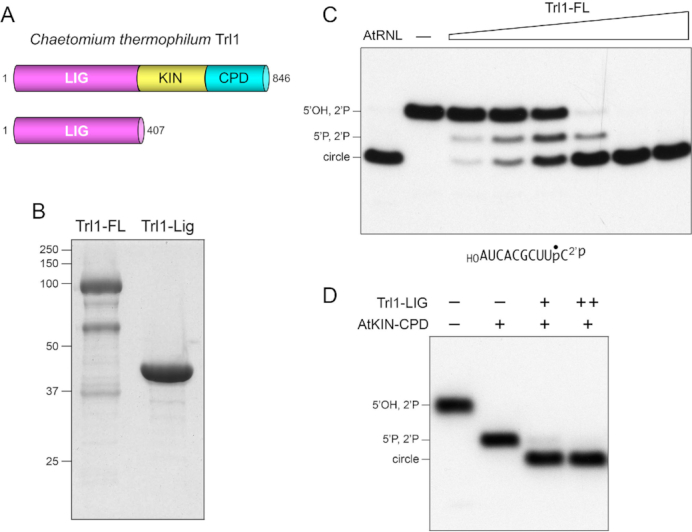
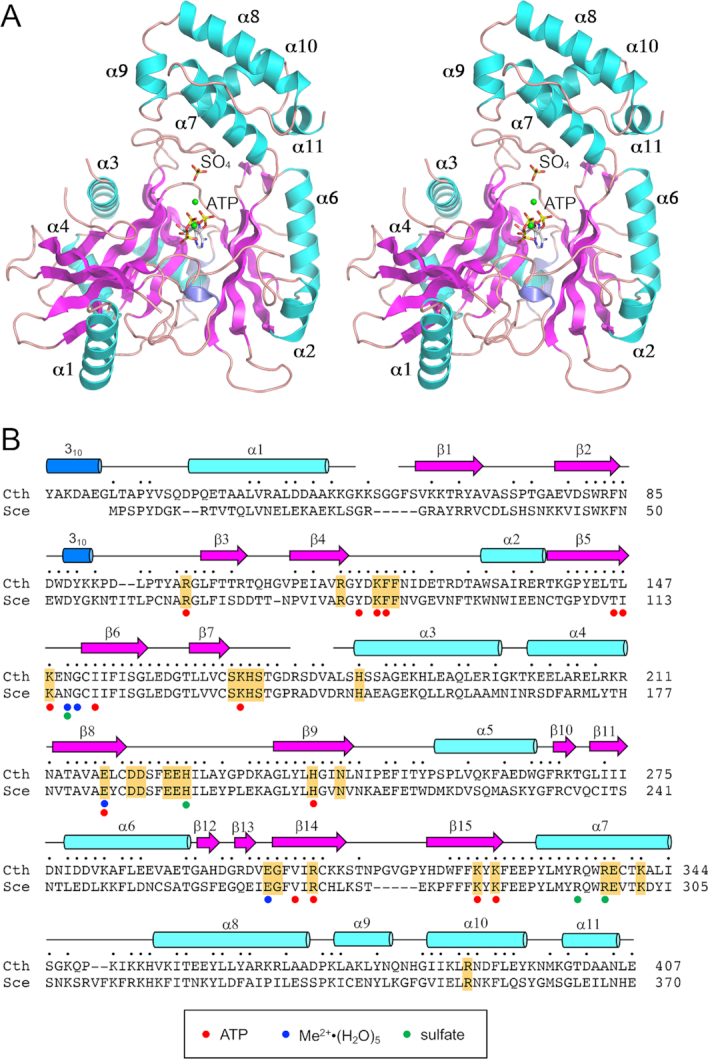
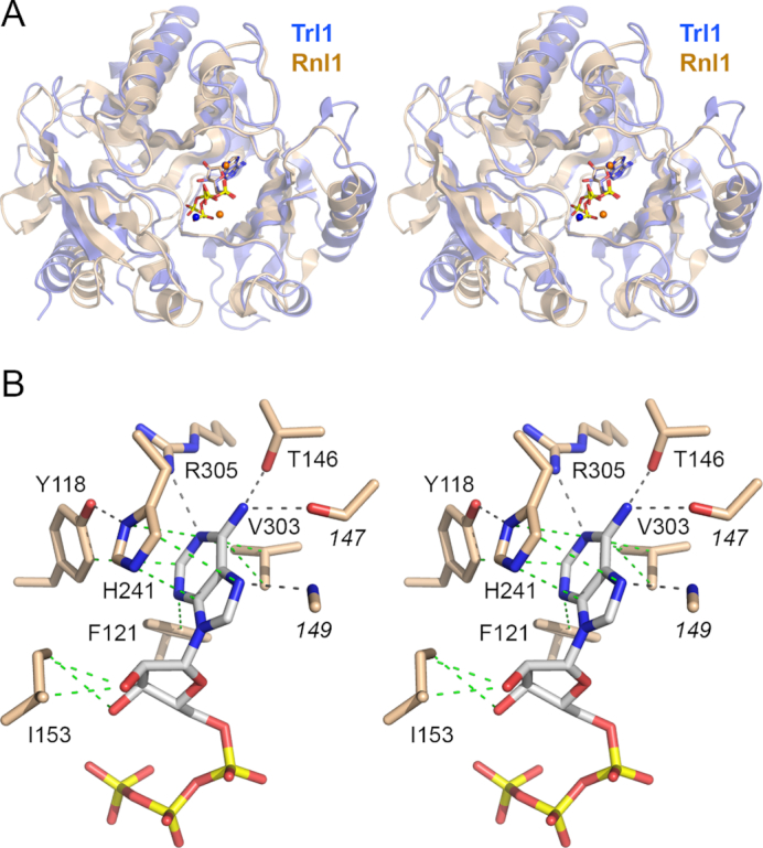
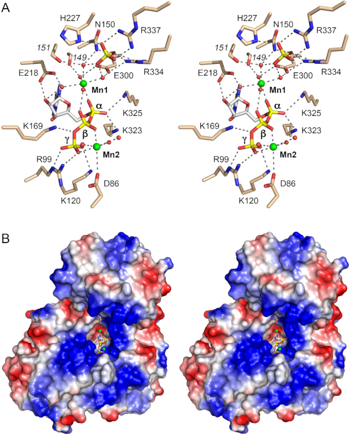
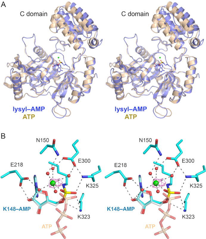
Fungal tRNA ligase (Trl1) is an essential enzyme that repairs RNA breaks with 2',3'-cyclic-PO4 and 5'-OH ends inflicted during tRNA splicing and non-canonical mRNA splicing in the fungal unfolded protein response. Trl1 is composed of C-terminal cyclic phosphodiesterase (CPD) and central GTP-dependent polynucleotide kinase (KIN) domains that heal the broken ends to generate the 3'-OH,2'-PO4 and 5'-PO4 termini required for sealing by an N-terminal ATP-dependent ligase domain (LIG). Here we report crystal structures of the Trl1-LIG domain from Chaetomium thermophilum at two discrete steps along the reaction pathway: the covalent LIG-(lysyl-Nζ)-AMP•Mn2+ intermediate and a LIG•ATP•(Mn2+)2 Michaelis complex. The structures highlight a two-metal mechanism whereby a penta-hydrated metal complex stabilizes the transition state of the ATP α phosphate and a second metal bridges the β and γ phosphates to help orient the pyrophosphate leaving group. A LIG-bound sulfate anion is a plausible mimetic of the essential RNA terminal 2'-PO4. Trl1-LIG has a distinctive C-terminal domain that instates fungal Trl1 as the founder of an Rnl6 clade of ATP-dependent RNA ligase. We discuss how the Trl1-LIG structure rationalizes the large body of in vivo structure-function data for Saccharomyces cerevisiae Trl1.


Figures

Figure 1.
Figure 1.
Chaetomium Trl1 ligase activity. (A) Cartoon depiction of the domain organization of full-length (FL) 846-aa C. thermophilum Trl1 consisting of N-terminal ligase (LIG), central kinase (KIN), and C-terminal cyclic phosphodiesterase (CPD) domains. An N-terminal 407-aa segment is shown here to comprise an autonomous LIG module. (B) Aliquots (10 μg) of the Superdex fractions of recombinant Trl1-FL and Trl1-LIG were analyzed by SDS-PAGE. The Coomassie blue-stained gel is shown. The positions and sizes (kDa) of marker polypeptides are indicated on the left. (C) RNA ligase reaction mixtures (10 μl) containing 50 mM Tris–HCl, pH 8.0, 50 mM NaCl, 2 mM DTT, 10 mM MgCl2, 100 μM GTP, 100 μM ATP, 0.5 pmol (50 nM) 32P-labeled 10-mer HORNA2′p (shown at the bottom with the 32P-label indicated by •), and either no enzyme (lane –), 1.25 μg plant tRNA ligase (AtRNL), or 0.625, 1.25, 2.5, 5, 10 or 25 ng Trl1-FL (from left to right in the titration series) were incubated at 37°C for 30 min. The labeled RNAs were resolved by urea-PAGE and visualized by autoradiography. The positions of the 5′-OH, 2′-PO4 RNA substrate, 5′-PO4, 2′-PO4 kinase reaction product, and the 10-mer circle product of intramolecular ligation are indicated on the left. (D) Reactions mixtures (10 μl) containing 50 mM Tris–HCl, pH 8.0, 50 mM NaCl, 2 mM DTT, 10 mM MgCl2, 100 μM GTP, 100 μM ATP, 0.5 pmol (50 nM) 32P-labeled 10-mer HORNA2′p, 250 ng AtKIN-CPD (where indicated by +), and 5 or 10 pg Trl1-LIG (where indicted by + and ++, respectively) were incubated at 37°C for 30 min. The labeled RNAs were resolved by urea-PAGE and visualized by autoradiography.
Figure 2.
Figure 2.
Structure of Trl1-LIG. (A) Stereo view of the LIG tertiary structure, depicted as a ribbon model with magenta β strands, cyan α helices (numbered sequentially), and blue 310 helices. The ATP in the active site and a nearby sulfate anion are rendered as stick models. Mn2+ ions are depicted as green spheres. (B) Secondary structure elements (colored as in panel A) are displayed above the C. thermophilum (Cth) Trl1-LIG primary structure, which is aligned to the primary structure of the LIG domain of S. cerevisiae (Sce) Trl1. Positions of amino acid side chain identity or similarity are indicated by dots above the Cth sequence. Gaps in the alignment are indicated by dashes. Amino acids in Trl1-LIG that make atomic contacts to ATP are denoted by red dots below the alignment. Amino acids that coordinate the hydrated metal complex are indicated by blue dots below the alignment. Amino acids that coordinate the sulfate anion are indicated by green dots below the alignment. The amino acids in S. cerevisiae Trl1-LIG that were identified by alanine scanning as essential for LIG activity in vivo (6) are highlighted in gold shading.
Figure 3.
Figure 3.
Homology to T4 Rnl1 and basis for adenine nucleotide specificity. (A) Stereo view of the superimposed structures of the adenylyltransferase domains of Trl1-LIG (blue) and T4 Rnl1 (beige) in their respective complexes with ATP (stick models) and divalent cations (shown as blue spheres for Trl1-LIG and beige spheres for Rnl1). (B) Stereo view of the adenylate binding pocket highlighting contacts to the adenine nucleobase and ribose sugar. Amino acids and ATP are shown as stick models with beige and gray carbons, respectively. Atomic contacts are indicated by black dashed lines (hydrogen bonds) or green dashed lines (van der Waals contacts).
Figure 4.
Figure 4.
LIG interactions with ATP•Mn2+ and surface electrostatics. (A) Stereo view of the active site highlighting contacts to the ATP phosphates, two associated manganese ions (green spheres), and a nearby sulfate anion (stick model). Amino acids and ATP are shown as stick models with beige and gray carbons, respectively. Waters are depicted as red spheres. Atomic contacts are indicated by black dashed lines. (B) Stereo view of a surface electrostatic model of Trl1-LIG (generated in Pymol). ATP in the active site and a nearby sulfate anion are depicted as a stick models. Manganese ions are green spheres.
Figure 5.
Figure 5.
Trl1-LIG–AMP covalent intermediate. (A) Stereo view of the tertiary structures of the Tr1LIG–AMP intermediate (in blue) and the LIG–ATP Michaelis complex (in beige) superimposed with respect to their N-terminal adenylyltransferase domains. The image highlights a rigid body movement of the C-terminal domain in the transitions from LIG•ATP to LIG–AMP. The lysyl-adenylate adduct in the LIG–AMP structure is shown as a stick model. The catalytic Mn2+ ion is rendered as a green sphere. (B) Stereo view of the active site of LIG–AMP. Amino acids and lysyl–AMP are shown as stick models with cyan carbons; the AMP phosphorus atom is colored yellow. A catalytic Mn2+ ion and associated waters are depicted as green and red spheres, respectively. The anomalous difference peak overlying the manganese ion, contoured at 4σ, is depicted in magenta mesh. Atomic contacts are indicated by dashed lines. The superimposed ATP from the Michaelis complex structure is shown as a semi-transparent stick model with gray carbons and beige phosphorus atoms (to highlight the stereochemical inversion of the α phosphorus center after lysine adenylylation).

References

    1. Abelson J., Trotta C.R., Li H.. tRNA splicing. J. Biol. Chem. 1998; 273:12685–12688. - PubMed
    1. Sidrauski C., Cox J.S., Walter P.. tRNA ligase is required for regulated mRNA splicing in the unfolded protein response. Cell. 1996; 87:405–413. - PubMed
    1. Greer C.L., Peebles C.L., Gegenheimer P., Abelson J.. Mechanism of action of a yeast RNA ligase in tRNA splicing. Cell. 1983; 32:537–546. - PubMed
    1. Apostol B.L., Westaway S.K., Abelson J., Greer C.L.. Deletion analysis of a multifunctional yeast tRNA ligase polypeptide: identification of essential and dispensable functional domains. J. Biol. Chem. 1991; 266:7445–7455. - PubMed
    1. Sawaya R., Schwer B., Shuman S.. Genetic and biochemical analysis of the functional domains of yeast tRNA ligase. J. Biol. Chem. 2003; 278:43298–43398. - PubMed

Publication types

MeSH terms