. 2019 Feb 15;124(4):511-525.
doi: 10.1161/CIRCRESAHA.118.313316.

Fine-Tuning of Sox17 and Canonical Wnt Coordinates the Permeability Properties of the Blood-Brain Barrier

Affiliations

Fine-Tuning of Sox17 and Canonical Wnt Coordinates the Permeability Properties of the Blood-Brain Barrier

Monica Corada et al. Circ Res. .

Abstract

Rationale: The microvasculature of the central nervous system includes the blood-brain barrier (BBB), which regulates the permeability to nutrients and restricts the passage of toxic agents and inflammatory cells. Canonical Wnt/β-catenin signaling is responsible for the early phases of brain vascularization and BBB differentiation. However, this signal declines after birth, and other signaling pathways able to maintain barrier integrity at postnatal stage are still unknown.

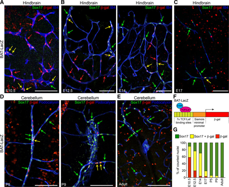
Objective: Sox17 (SRY [sex-determining region Y]-box 17) constitutes a major downstream target of Wnt/β-catenin in endothelial cells and regulates arterial differentiation. In the present article, we asked whether Sox17 may act downstream of Wnt/β-catenin in inducing BBB differentiation and maintenance.

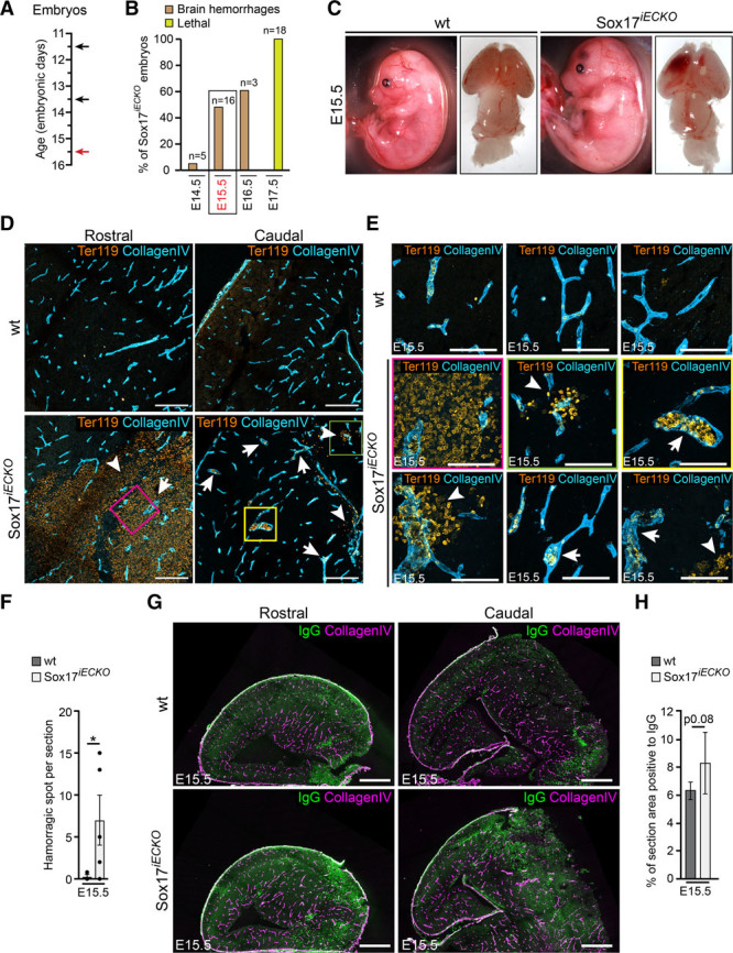
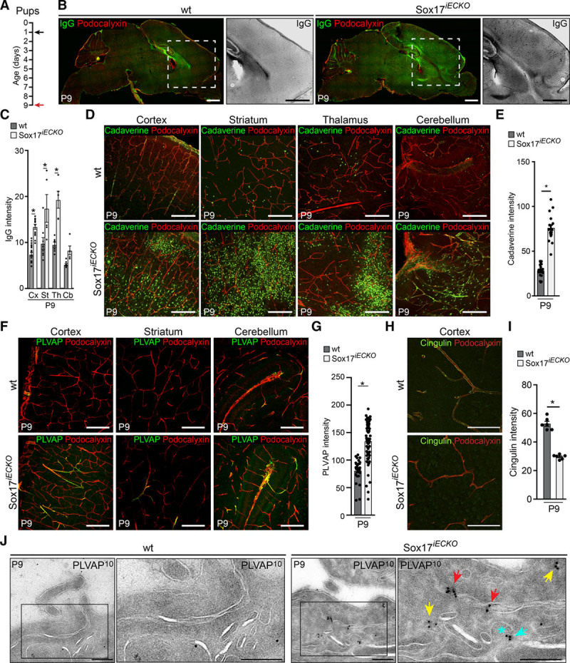
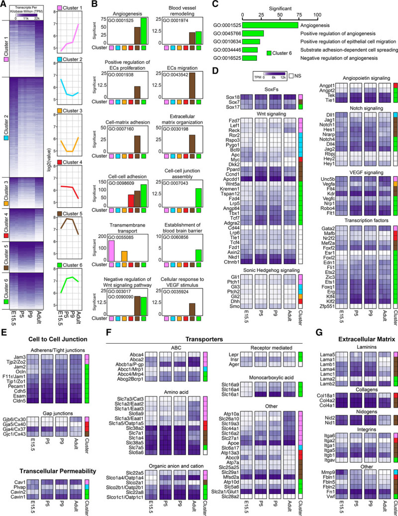
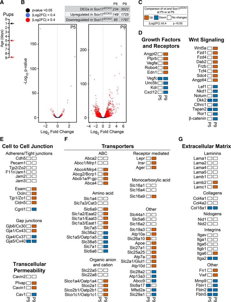
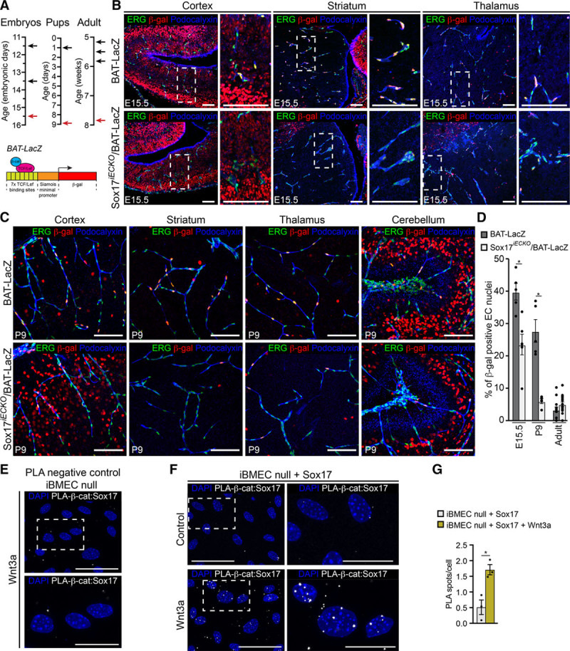
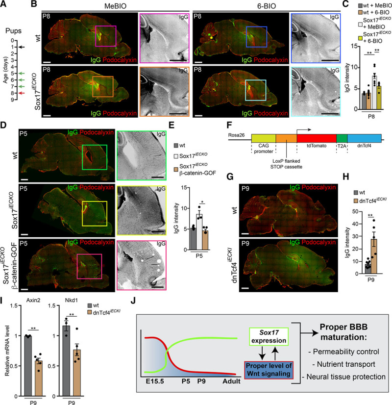
Methods and results: Using reporter mice and nuclear staining of Sox17 and β-catenin, we report that although β-catenin signaling declines after birth, Sox17 activation increases and remains high in the adult. Endothelial-specific inactivation of Sox17 leads to increase of permeability of the brain microcirculation. The severity of this effect depends on the degree of BBB maturation: it is strong in the embryo and progressively declines after birth. In search of Sox17 mechanism of action, RNA sequencing analysis of gene expression of brain endothelial cells has identified members of the Wnt/β-catenin signaling pathway as downstream targets of Sox17. Consistently, we found that Sox17 is a positive inducer of Wnt/β-catenin signaling, and it acts in concert with this pathway to induce and maintain BBB properties. In vivo, inhibition of the β-catenin destruction complex or expression of a degradation-resistant β-catenin mutant, prevent the increase in permeability and retina vascular malformations observed in the absence of Sox17.

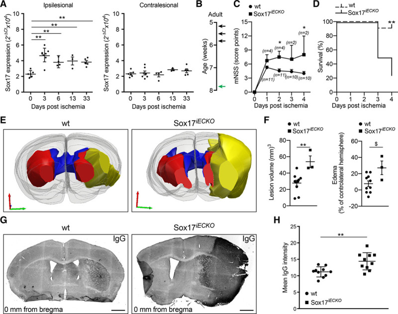
Conclusions: Our data highlight a novel role for Sox17 in the induction and maintenance of the BBB, and they underline the strict reciprocal tuning of this transcription factor and Wnt/β-catenin pathway. Modulation of Sox17 activity may be relevant to control BBB permeability in pathological conditions.

Keywords: Wnt/β-catenin; blood-brain barrier; endothelial cells; permeability; stroke.


Figures

Figure 1.
Figure 1.
Canonical Wnt/β-cat (β-catenin) signaling and Sox17 (SRY [sex-determining region Y]-box 17) expression show distinct time courses in endothelial cells (ECs) during brain angiogenesis and vessel maturation. AE, Confocal analysis of the brain microvasculature of BAT (β-catenin-activated transgene)-LacZ reporter embryos, pups, and adult mice. Whole-mount (A and B), vibratome sections (C), and cryosections (D and E) were stained for β-gal (β-galactosidase, red), to detect β-cat–induced expression of the Lac-Z reporter construct, Sox17 (green), and IB4 (isolectin B4, blue, AC) or Podocalyxin (blue, DE). Green, red, and yellow arrows indicate Sox17 staining, β-gal–positive nuclei, and double positive nuclei, respectively. Positive nuclei to β-gal outside the vascular system indicate active Wnt signaling in the brain parenchyma. Scale bar: 100 µm. F, Schematic representation of the BAT-LacZ reporter construct (see Methods). G, Quantification of nuclei positive for Sox17 only (green), β-gal only (red), or double-labeled (yellow; n=3–5 animals for each time point). These indicate different patterns of expression between Sox17 and β-cat signaling. LEF/TCF indicates lymphoid enhancer factor/T-cell factor.
Figure 2.
Figure 2.
Endothelial Sox17 (SRY [sex-determining region Y]-box 17) is required for correct brain vascular morphogenesis and barrier function. A, Scheme of tamoxifen administration at E11.5, E13.5 (black arrows), and embryo analysis at E15.5 (red arrow). See Methods section for details. B, Summary assessment of Sox17iECKO embryos presenting brain hemorrhages (brown columns) after tamoxifen treatment (as indicated in A). Data are presented as percentage of total number of embryos analyzed (n). E15.5 harvesting time was selected for subsequent experiments. At E17.5 (yellow column) none of the Sox17iECKO embryos was alive. C, Sox17iECKO embryos display brain hemorrhages. D and E, Sectioning of different regions of the embryonic brain showed enlarged vessels (arrows) stained with collagen IV (light blue) and extensive hemorrhages. Ter119 (orange) indicates red blood cell (arrowheads). Scale bar: 200 μm in C, 100 μm in D. F, Average count of hemorrhagic spots per section (n=3 wild-type [wt] and 5 Sox17iECKO, mean±SEM, *P<0.05, Mann-Whitney test). G, Confocal microscopy analysis of embryonic brain cryosections. Collagen IV (magenta) and IgG (green) of wt and Sox17iECKO revealed the presence of IgG leakage in Sox17iECKO embryos. Scale bar: 500 μm. H, Quantification of IgG leakage (left; n=3 wt and 5 Sox17iECKO, mean±SD, P=0.08, 2-tailed t test).
Figure 3.
Figure 3.
Endothelial Sox17 (SRY [sex-determining region Y]-box 17) deficiency affects blood-brain barrier integrity. A, Scheme for tamoxifen injections in the pups at P1 (black arrow), with analysis at P9 (red arrow). B, Confocal images of vibratome sections of wild-type (wt) and Sox17iECKO brains stained with Podocalyxin (red) and IgG (green). The presence of IgG leakage (black [magnified]) reveals high permeability in Sox17iECKO brains. Scale bar: 1 mm. C, Quantification of signal intensities for IgG extravasation indicated more prominent leakage in the cortex (Cx), striatum (St) and thalamus (Th) than in the cerebellum (Cb; n=5 wt and 8 Sox17iECKO, mean±SEM, *P<0.01, 1-way ANOVA and Bonferroni post hoc test). D, Confocal analysis of Alexa555-conjugated cadaverine leakage (green) in brain sections stained with Podocalyxin (red). Sox17iECKO present cadaverine accumulation in the brain parenchyma in all of the regions analyzed (lower). Scale bar: 200 μm. E, Quantification of cadaverine leakage (n=4 wt and 4 Sox17iECKO, mean±SEM, *P<0.01, Mann-Whitney test). FG, Confocal analysis of sections and quantification of PLVAP (plasmalemma vesicle–associated protein) expression (green) in the brain microvasculature (Podocalyxin in red) in wt and Sox17iECKO mouse. PLVAP expression is increased in Sox17iECKO mice. Scale bar: 200 μm. (n=6 wt and 6 Sox17iECKO, mean±SEM, *P<0.01, Mann-Whitney test). HI, Confocal images and quantification of Cingulin (green) in the microvasculature (Podocalyxin in red) of cortex in wt and Sox17iECKOmouse. Scale bar: 100 μm. (n=3 wt and 3 Sox17iECKO, mean±SEM, *P<0.01, Mann-Whitney test). J, Electron microscopy of PLVAP immunogold-labeled (10 nm gold, PLVAP) ultrathin sections of brain capillaries from wt and Sox17iECKO mice (P9). PLVAP is detectable only in the ECs of Sox17iECKO and localizes in the overlapping interendothelial contacts (red arrows) in the endosomal structures (yellow arrows) and in the neck of clathrin-coated buds (light blue arrow and asterisk).
Figure 4.
Figure 4.
Endothelial Sox17 (SRY [sex-determining region Y]-box 17) deficiency exacerbates stroke outcome. A, Real-time quantitative polymerase chain reaction analysis of Sox17 expression in the ischemic forebrain of wild-type (wt) animals. Expression was determined in the ischemic brain hemisphere (ipsilesional) and in the nonischemic controlateral hemisphere (contralesional) at different days postischemia (dpi; n=3–8 mice per time point, mean±CI95%, **P≤0.01, compared with 0 dpi, 1-way ANOVA, and Bonferroni post hoc test). B, Tamoxifen was injected in 5-wk-old mice (3 injections every other day; black arrows). Two weeks later, mice underwent left middle cerebral artery occlusion (MCAO; green arrow) and analysed at the time points indicated in the specific panels. C, Modified Neurological Severity Score in wt and Sox17iECKO mice (mean±SEM, *P≤0.05, 2-way ANOVA followed by Bonferroni post hoc test). D, Kaplan-Meier survival curve recorded in wt (n=11) and Sox17iECKO (n=4) mice subjected to MCAO up to 4 dpi (n=11 wt and 4 Sox17iECKO, **P≤0.01, Log-rank (Mantel-Cox) Test). E, Representative serial 3D reconstructions of the forebrains of a wt and a Sox17iECKO mouse at 4 dpi. Yellow represents the ischemic lesion tissue. Red represents the healthy striatum. Blue represents the ventricles. Gray represents the contours of the hemispheres. F, Graphs representing the lesion volume (left) and the edema of the ipsilesional hemisphere (right; n=10 wt and 3 Sox17iECKO, mean±SEM), (for lesion volume **P≤0.01, Mann-Whitney test), (for edema $P<0.05, 2-tailed t test and P=0.08, Mann-Whitney test). G, Confocal images of 1 representative cryosection from wt and Sox17iECKO at 4 dpi stained for IgG (black). The presence of IgG leakage reveals loss of BBB integrity in Sox17iECKO brains. Sections correspond to the bregma (0 mm). Scale bar: 1 mm. H, Quantification of IgG leakage from cryosections (n=5 wt and 3 Sox17iECKO, mean±SEM, **P<0.01, Mann-Whitney).
Figure 5.
Figure 5.
Key signaling pathways governing the maturation of a functional blood-brain barrier (BBB). A, Normalized expression of 10 783 differentially expressed genes (DEGs, obtained by Next-maSigPro analysis: P<0.05 and R2>0.6) during BBB development (E15.5), maturation (P5 and P9), and maintenance (adult; n=3 for each time point). Heat map shows gene expression levels (transcript per million [TPM]) in clusters (color-coded as indicated). Trajectory plots show the gene expression pattern of each cluster by displaying the average gene expression level at each time point in that cluster. B, Enriched gene ontology (GO) terms related to BBB development in clusters. Bar plot shows the number of genes that belong to enriched GO terms in each cluster. C, The top 5 most enriched GO terms in cluster 6. D, Heatmap shows expression levels over time of key signaling pathway endothelial cell (EC) genes during BBB development. EG, Heat maps are used to display the expression levels of genes relevant for key features of BBB formation and maturation (cell to cell junction [E], transcellular permeability [E], transporters [F] and extracellular matrix [G]) at different developmental stages. In DG, the genes are categorized based on time course analysis: DEGs (P<0.05 and R2>0.6) are grouped by the clusters that they belong to, and nondifferentially expressed genes are grouped into not significant (NS) category (R2<0.6 or P<0.05). VEGF indicates vascular endothelial growth factor.
Figure 6.
Figure 6.
Role of Sox17 (SRY [sex-determining region Y]-box 17) expression on blood-brain barrier (BBB)-related genes at P5 and P9. A, Scheme of tamoxifen injection in the pups at P1 (black arrow) and analysis of endothelial cells isolated from brains at P5 and P9 (red arrows). B, Volcano plots are used to display the magnitude of the differential expression between wild-type (wt) and Sox17iECKO either at P5 (left) or at P9 (right; n=3 wt and 3 Sox17iECKO for each time point). Each dot represents 1 gene that has detectable expression both in wt and Sox17iECKO cells. Black dots represent genes that are not significantly different (adjusted P>0.05). Significant differentially expressed genes (DEGs; adjusted P≤0.05) are colored either in red (Log2FC≥0.4) or in blue (Log2FC<0.4). The number of DEGs in Sox17iECKO are reported in the table on top of the Volcano plots. C, Description of thresholds and colors used in DG. D, Selected angiogenesis and Wnt/β-cat (β-catenin) related genes, differentially expressed, either up or down, in Sox17iECKO. EG, Regulation of the expression of gene relevant for key BBB features (cell to cell junction [E], transcellular permeability [E], transporters [F], and extracellular matrix [G]), as in Figure 5, modulated by the inactivation of Sox17 expression. The genes that do not have any expression changes are displayed by white box. FC indicates fold change.
Figure 7.
Figure 7.
Endothelial Sox17 (SRY [sex-determining region Y]-box 17) is required for Wnt/β-catenin signaling in the brain. A, Scheme of tamoxifen administration and analysis. Pregnant females were injected (E11.5 and E13.5; black arrows) and the embryos dissected and analyzed at E15.5 (red arrow); pups were injected at P1 (black arrow), and analyzed at P9 (red arrow); 5-wk-old mice received 3 injections every other day (black arrows) and were analyzed at 8-wk old (red arrow). See Methods for details. Schematic representation of the BAT (β-catenin-activated transgene)-LacZ reporter construct as in Figure 1F. B and C, Confocal analysis of wild-type and Sox17iECKO/BAT-LacZ mice (see Methods for details) at the indicated ages. Brains sections were stained for ERG (ETS transcription factor; green), β-gal (β-galactosidase; red) and Podocalyxin (blue). In the mutant embryos and pups (lower B and C, respectively), the number of β-gal–positive endothelial nuclei (ERG-positive) are significantly reduced in all of the brain regions. For the embryonic brain, higher magnification of the insets is also shown. Scale bar: 100 μm. D, Quantification of β-gal–positive nuclei with respect to total ERG-positive endothelial cells (ECs; percentages) as in B (embryos), C (pups), and Online Figure VIF (adult). β-gal–positive nuclei outside the vascular system indicate active Wnt signaling in the brain parenchyma (n=3–4 animals for each experimental condition, mean±SEM, *P<0.01, Mann-Whitney test). EF, In situ proximity ligation assay (PLA). iBMEC (immortalized brain microvascular endothelial cells) null for Sox17 was used as negative control (E). In iBMEC null transduced with Sox17, Sox17/β-cat (β-catenin) interaction (white) was observed. Nuclei were counterstained with DAPI (4’,6-diamidino-2-Phenylindole; blue). Magnification of boxed areas are shown. Note the increase in nuclear spot, induced by Wnt3a activation. G, Quantification of PLA nuclear signal expressed as the average number of PLA spots per cell; n=3 for each condition, mean±SEM, *P<0.01, 2-tailed t test. LEF/TCF indicates lymphoid enhancer factor/T-cell factor.
Figure 8.
Figure 8.
Stabilization of β-cat (β-catenin) signaling restores vascular defects induced by Sox17 (SRY [sex-determining region Y]-box 17) inactivation. A, Scheme of tamoxifen injections in the pups at P1 (black arrow), (2′Z,3′E)-6-bromoindirubin-3′-acetoxime (6-BIO; or 1-methyl- bromoindirubin [MeBIO]) injections at P5-7 (green arrows), and analysis of brain P8 (red arrow). B, Confocal images of sections stained with Podocalyxin (red) and IgG (green or black in magnification) of wild-type (wt) and Sox17iECKO pups revealed a complete rescue of IgG leakage in mutant brains treated with 6-BIO. Scale bar: 1 mm. C, Quantification of IgG leakage in brain as in B (n=3 for each experimental condition, mean±SEM, **P<0.01 ANOVA and Tukey post hoc analysis). D, β-cat stabilization rescue Sox17iECKO blood-brain barrier (BBB) defect. Confocal images of section stained with Podocalyxin (red) and IgG (green) from wt, Sox17iECKO, and Sox17iECKO/β-cat–gain of function (GOF) brains from P5 pups. The presence of IgG leakage (black [magnified]) reveals high permeability in Sox17iECKO brains. In the double mutant Sox17iECKO/β-cat GOF pups, IgG leakage is reduced to control levels. Scale bar 1 mm. E, Quantification of IgG extravasation indicates more prominent leakage in Sox17iECKO brains (n=6 wt and 3 Sox17iECKO and 5 Sox17iECKO/β-cat-GOF, mean±SEM, *P<0.05, Mann-Whitney test). F, Schematic representation of the td-Tomato-T2A–dominant-negative Tcf4 (transcription factor 7 like 2, T cell-specific, HMG box; dnTcf4) construct. See Methods for details (CAG, CMV immediate enhancer/β-actin). G, Confocal images of brain microvasculature. IgG (green) and Podocalyxin (red) staining of sections of wt and inducible endothelial-specific dominant-negative Tcf4 (dnTcf4iECKI) P9 pups. The presence of IgG reveals higher permeability in dnTcf4iECKI. Scale bar 1 mm. H, Quantification of IgG leakage indicates more prominent leakage in dnTcf4iECKI brains (n=8 wt and 5 dnTcf4iECKI, mean±SEM, **P<0.01, Mann-Whitney test). I, Real-time quantitative polymerase chain reaction analysis of 2 Wnt/β-cat target genes (Axin2 and Nkd1) expressed in brain endothelial cells isolated from dnTcf4iECKI P9 pups (n=3 wt and 4 dnTcf4iECKI, mean±SD **P<0.01, 2-tailed t test). J, Schematic model of the cross-talk between Wnt/β-cat and Sox17 signaling. Wnt signaling declines after birth and triggers activation of Sox17 that in turn control brain vascular permeability. A calibrated cross-talk of these 2 transcription pathways is necessary to prevent uncontrolled effect and pathogenic conditions.

References

    1. Zhao Z, Nelson AR, Betsholtz C, Zlokovic BV. Establishment and dysfunction of the blood-brain barrier. Cell. 2015;163:1064–1078. doi: 10.1016/j.cell.2015.10.067. - PMC - PubMed
    1. Augustin HG, Koh GY. Organotypic vasculature: from descriptive heterogeneity to functional pathophysiology. Science. 2017;357:eaal2379. doi: 10.1126/science.aal2379. - PubMed
    1. Armulik A, Genové G, Mäe M, Nisancioglu MH, Wallgard E, Niaudet C, He L, Norlin J, Lindblom P, Strittmatter K, Johansson BR, Betsholtz C. Pericytes regulate the blood-brain barrier. Nature. 2010;468:557–561. doi: 10.1038/nature09522. - PubMed
    1. Iadecola C. The neurovascular unit coming of age: a journey through neurovascular coupling in health and disease. Neuron. 2017;96:17–42. doi: 10.1016/j.neuron.2017.07.030. - PMC - PubMed
    1. Clevers H, Nusse R. Wnt/β-catenin signaling and disease. Cell. 2012;149:1192–1205. doi: 10.1016/j.cell.2012.05.012. - PubMed

Publication types