. 2018 Dec 27;6(1):145.
doi: 10.1186/s40478-018-0646-6.

Homozygous TBC1 domain-containing kinase (TBCK) mutation causes a novel lysosomal storage disease - a new type of neuronal ceroid lipofuscinosis (CLN15)?

Affiliations

Homozygous TBC1 domain-containing kinase (TBCK) mutation causes a novel lysosomal storage disease - a new type of neuronal ceroid lipofuscinosis (CLN15)?

Stefanie Beck-Wödl et al. Acta Neuropathol Commun. .

Abstract

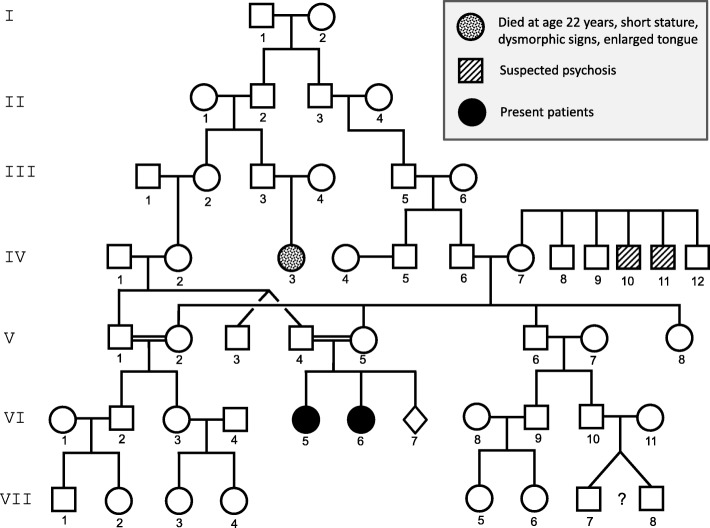
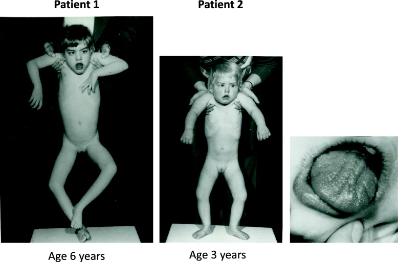
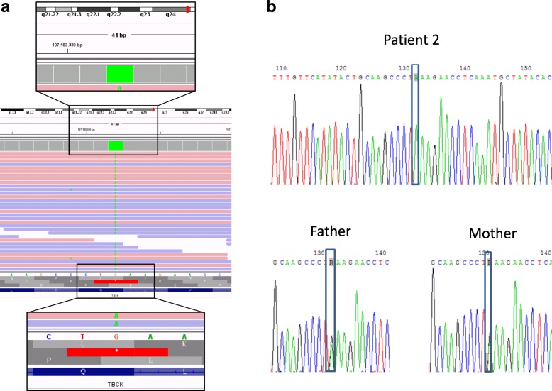
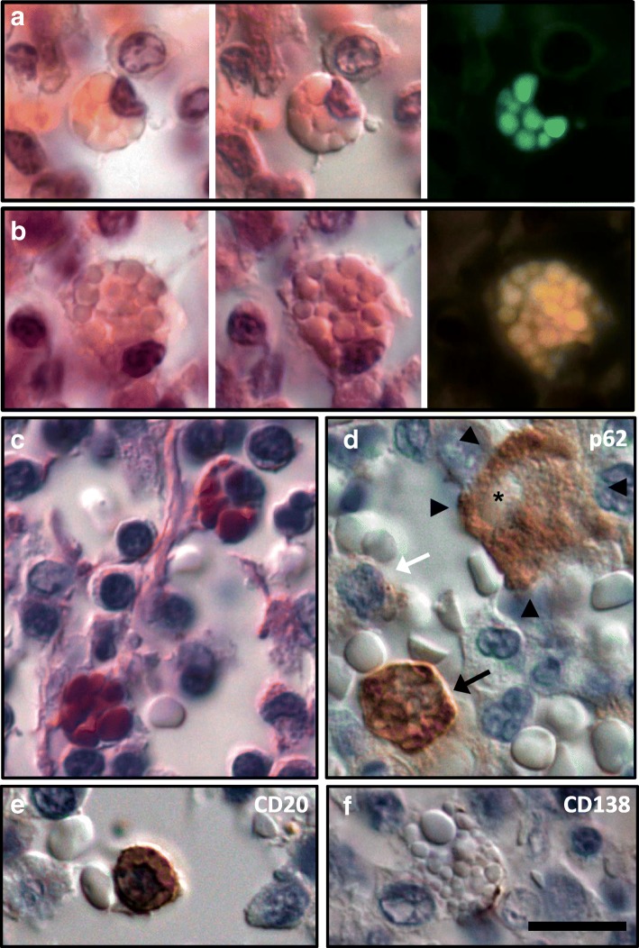
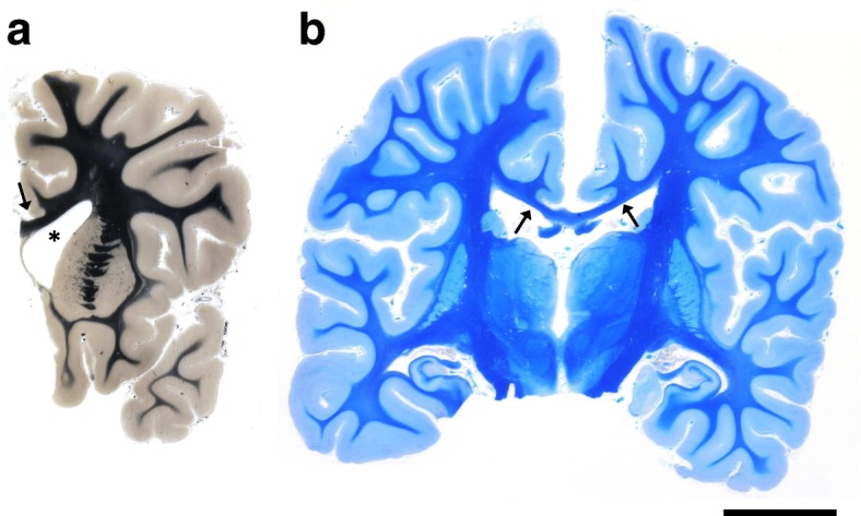
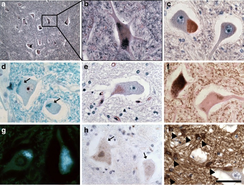
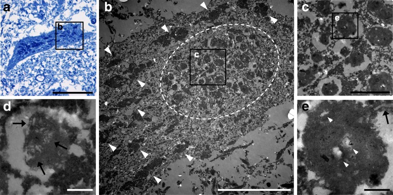
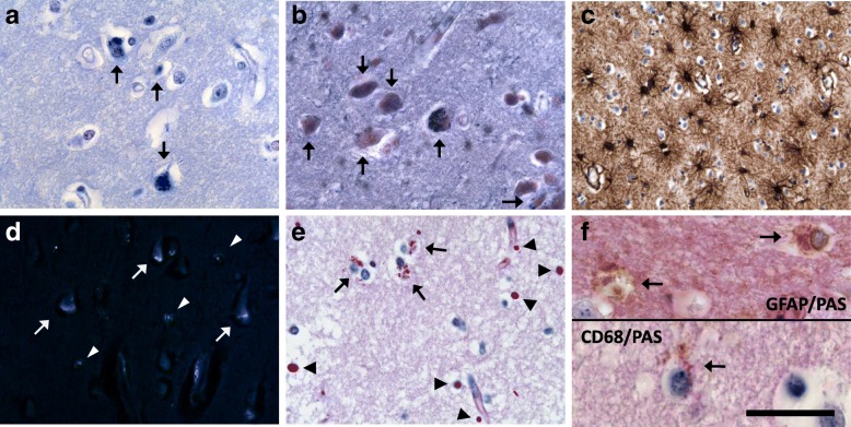
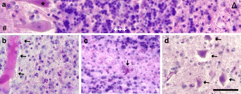
Homozygous mutation of TBC1 domain-containing kinase (TBCK) is the cause of a very recently defined severe childhood disorder, which is characterized by severe hypotonia, global developmental delay, intellectual disability, epilepsy, characteristic facies and premature death. The link between TBCK loss of function and symptoms in patients with TBCK deficiency disorder (TBCK-DD) remains elusive. Here we demonstrate for the first time the histopathological characteristics of TBCK deficiency consisting of 1) a widespread and massive accumulation of lipofuscin storage material in neurons of the central nervous system without notable neuronal degeneration, 2) storage deposits in few astrocytes, 3) carbohydrate-rich deposits in brain, spleen and liver and 4) vacuolated lymphocytes. Biochemical examinations ruled out more than 20 known lysosomal storage diseases. These investigations strikingly uncover TBCK-DD as a novel type of lysosomal storage disease which is characterized by different storage products rather than one specific type of accumulated material. Due to the clear predominance of intraneuronal lipofuscin storage material and the characteristic clinical presentation we propose to classify this disease as a new subtype of neuronal ceroid lipofuscinosis (CLN15). Our results and previous reports suggest an autophagosomal-lysosomal dysfunction caused by enhanced mTORC1-mediated autophagosome formation and reduced Rab-mediated autophagosome-lysosome fusion, thus disclosing potential novel targets for therapeutic approaches in TBCK-DD.

Keywords: Autophagy; Central nervous system (CNS); Infantile muscular hypotonia with psychomotor retardation and characteristic facies 3 (IHPRF3); Mammalian target of rapamycin (mTOR); Rab; Vacuolated lymphocytes.


Conflict of interest statement

Ethics approval and consent to participate

All procedures performed in this study were in accordance with the ethical standards of the institutional and/or national research committee and with the 1964 Helsinki declaration and its later amendments or comparable ethical standards.

Consent for publication

Informed consent was obtained from the parents of the siblings for whom identifying information is included in this article.

Competing interests

The authors declare that they have no competing interest.

Publisher’s Note

Springer Nature remains neutral with regard to jurisdictional claims in published maps and institutional affiliations.

Figures

Fig. 1
Fig. 1
Family tree of the patients. The siblings (VI.5 and VI.6, black circles) were born to Caucasian, distantly consanguineous parents, who did not suffer from the disease. ◊, pregnancy with induced abortion. ?, twins with unknown zygosity
Fig. 2
Fig. 2
External appearance of the two patients. Severe hypotonia, a short neck and mild facial dysmorphia with open mouth, tented upper lip vermilion, macroglossia, furrowed tongues and right esotropia are seen
Fig. 3
Fig. 3
The TBCK defect in the genome of patient 2 and her parents. a, Integrative Genomics Viewer presentation of the homozygous stop mutation in the TBCK gene of patient 2. Below the green square, green dots show the base exchange (c.304C > T) in the multiple reads. b, Sanger sequencing of the TBCK gene of patient 2 and her parents. Sequencing was performed on the reverse strand, showing the base exchange G > A in our patient (grey boxes). Both parents carry the same mutation heterozygously, as seen in the R (grey boxes) that resembles an A and a G in each allele
Fig. 4
Fig. 4
Vacuolated lymphocytes in lymph nodes of patient 1. A few lymphocytes with clear bold cytoplasmic vacuoles can be detected in the hematoxylin eosin staining in normal microscopy (a and b, first panel) and differential interference contrast (DIC) microscopy allowing for a three-dimensional illustration (a and b, second panel). The vacuoles exhibit a clear autofluorescence in different channels (a and b, right panel) and are strongly PAS-positive (c, DIC). An enhanced autophagosome formation is indicated by strong p62 immunoreactivity (d, black arrow), whereas normal lymphocytes are predominantly negative (white arrow). Note the positively stained larger macrophage (arrowheads) phagocytosing an erythrocyte (asterisk). Most of the vacuolated cells are CD20+ B-lymphocytes (e, DIC), and single cells show a partial CD138 expression, likely pointing at immature plasma cells (f, DIC). Scale bar: 10 μm
Fig. 5
Fig. 5
Whole mount coronar brain sections of patient 1 (a, Heidenhain-Woelcke stain) and patient 2 (b, luxol fast blue stain). The lateral ventricle is widened especially in patient 1 (a, asterisk). The brain of patient 2 shows narrowing of the white matter (b, blue staining of myelin). The corpus callosum is thinned in both patients (a and b, arrows). Scale bar: 2 cm
Fig. 6
Fig. 6
Neuronal inclusions in the spinal cord anterior horn of patient 2. Many neurons present with granular deposits in the perikaryon, which stain strongly with sudan black (a and b) and sudan red (c). Note the frequent localization adjacent to neuronal processes. The inclusions stain with luxol fast blue (d, arrows) and show a weak PAS-reaction (e). The storage material is moderately argyrophilic (f, Gallyas stain) and shows strong autofluorescence (g, unstained section). The storage material does not stain for p62 (h, arrows). Note the reactive GFAP+ gliosis (i, arrowheads). GFAP, glial fibrillary acidic protein. Scale bar: 250 μm in a, 50 μm in b-i
Fig. 7
Fig. 7
Ultrastructural morphology of neuronal storage material. Semi-thin section stained with toluidin blue of a neuron in the spinal cord anterior horn of patient 2 shows strongly stained storage material (a). Ultrastructural examination of the same neuron in a serial section (b-e) reveals a cluster of intracytoplasmic globular inclusions (delineated in b) consisting of amorphous osmiophilic material with high-density particles, lipid droplets (e; white arrowheads) and structures reminding of degraded membranous material (d and e; arrows). These inclusions correspond to lipofuscin granules in lysosomal residual bodies and remind of granular osmiophilic deposits (GRODs). Note the different polygonal shape of physiological Nissl substance (b; white arrowheads). Scale bar: 50 μm in a, 10 μm in b, 2 μm in c, 250 nm in d and e
Fig. 8
Fig. 8
Morphological changes in the cortex of patient 2. Many cortical neurons in lamina V and VI incorporate storage material, which stains strongly with luxol fast blue in the Klüver Barrera staining (a, arrows) and with sudan black (b, arrows). A massive reactive astrocytosis is seen in GFAP immunohistochemistry (c). The intraneuronal inclusions (arrows) and glial inclusions (arrowheads) exert a strong autofluorescence (d, unstained section). Several cortical glia cells show cytoplasmatic PAS-positive granula (e, arrows). Note the numerous PAS-positive corpora amylacea (e, arrowheads). The laden glia cells are mainly astrocytes, as shown in double staining for PAS and GFAP (f, upper panel, arrows) and very few microglia cells, as shown in double staining for PAS and CD68 (f, lower panel, arrow). GFAP, glial fibrillary acidic protein. Scale bar: 50 μm in a-e, 20 μm in f
Fig. 9
Fig. 9
Diffuse PAS-positive material in the cerebellum of patient 1. The PAS-positive deposits are abundantly present in the subcortical cerebellar white matter (a, Δ; b and c) with sparing of molecular layer (#), Purkinje cell layer (*) and granular cell layer (+++). The deposits often accumulate perivascular (b, arrows) and are sometimes seen in glia cells (c, arrow). The nucleus dentatus is also affected (d). Note the shrunken and hyperchromatic neurons (arrows) as a sign of previous hypoxia. Scale bar: 65 μm in a, 50 μm in b-d

References

    1. Alazami AM, Patel N, Shamseldin HE, Anazi S, Al-Dosari MS, Alzahrani F, Hijazi H, Alshammari M, Aldahmesh MA, Salih MA, Faqeih E, Alhashem A, Bashiri FA, Al-Owain M, Kentab AY, Sogaty S, Al Tala S, Temsah MH, Tulbah M, Aljelaify RF, Alshahwan SA, Seidahmed MZ, Alhadid AA, Aldhalaan H, AlQallaf F, Kurdi W, Alfadhel M, Babay Z, Alsogheer M, Kaya N, Al-Hassnan ZN, Abdel-Salam GM, Al-Sannaa N, Al Mutairi F, El Khashab HY, Bohlega S, Jia X, Nguyen HC, Hammami R, Adly N, Mohamed JY, Abdulwahab F, Ibrahim N, Naim EA, Al-Younes B, Meyer BF, Hashem M, Shaheen R, Xiong Y, Abouelhoda M, Aldeeri AA, Monies DM, Alkuraya FS. Accelerating novel candidate gene discovery in neurogenetic disorders via whole-exome sequencing of prescreened multiplex consanguineous families. Cell Rep. 2015;10:148–161. doi: 10.1016/j.celrep.2014.12.015. - DOI - PubMed
    1. Anderson G, Smith VV, Malone M, Sebire NJ. Blood film examination for vacuolated lymphocytes in the diagnosis of metabolic disorders; retrospective experience of more than 2,500 cases from a single Centre. J Clin Pathol. 2005;58:1305–1310. doi: 10.1136/jcp.2005.027045. - DOI - PMC - PubMed
    1. Baumann RJ, Markesbery WR. Juvenile amaurotic idiocy (neuronal ceroid lipofuscinosis) and lymphocyte fingerprint profiles. Ann Neurol. 1978;4:531–536. doi: 10.1002/ana.410040609. - DOI - PubMed
    1. Bhoj EJ, Li D, Harr M, Edvardson S, Elpeleg O, Chisholm E, Juusola J, Douglas G, Guillen Sacoto MJ, Siquier-Pernet K, Saadi A, Bole-Feysot C, Nitschke P, Narravula A, Walke M, Horner MB, Day-Salvatore DL, Jayakar P, Vergano SA, Tarnopolsky MA, Hegde M, Colleaux L, Crino P, Hakonarson H. Mutations in TBCK, encoding TBC1-domain-containing kinase, lead to a recognizable syndrome of intellectual disability and hypotonia. Am J Hum Genet. 2016;98:782–788. doi: 10.1016/j.ajhg.2016.03.016. - DOI - PMC - PubMed
    1. Bidinosti M, Botta P, Kruttner S, Proenca CC, Stoehr N, Bernhard M, Fruh I, Mueller M, Bonenfant D, Voshol H, Carbone W, Neal SJ, McTighe SM, Roma G, Dolmetsch RE, Porter JA, Caroni P, Bouwmeester T, Luthi A, Galimberti I. CLK2 inhibition ameliorates autistic features associated with SHANK3 deficiency. Science. 2016;351:1199–1203. doi: 10.1126/science.aad5487. - DOI - PubMed

MeSH terms