Review
. 2019 Jul;37(7):744-760.
doi: 10.1016/j.tibtech.2018.12.001. Epub 2018 Dec 24.

Primary Cell-Derived Intestinal Models: Recapitulating Physiology

Affiliations
Review

Primary Cell-Derived Intestinal Models: Recapitulating Physiology

Johanna S Dutton et al. Trends Biotechnol. 2019 Jul.

Abstract

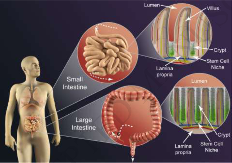
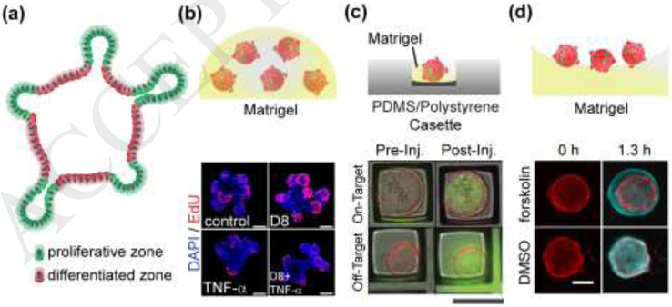
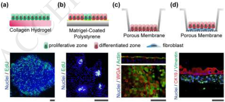
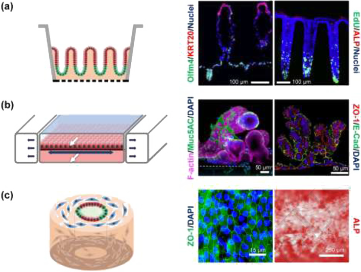
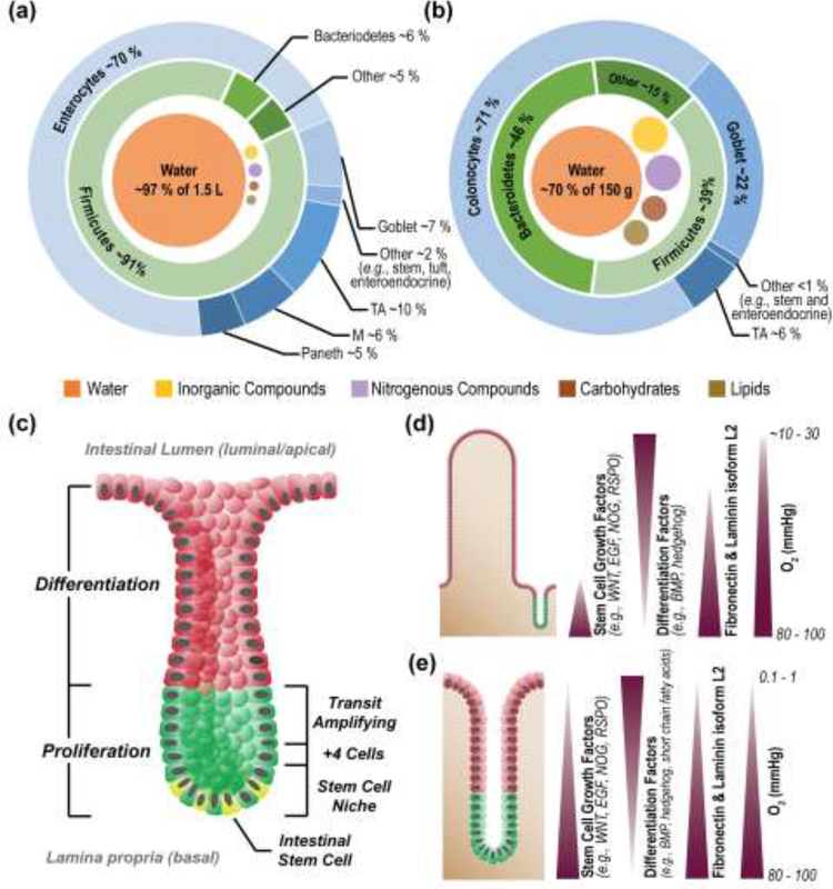
The development of physiologically relevant intestinal models fueled by breakthroughs in primary cell-culture methods has enabled successful recapitulation of key features of intestinal physiology. These advances, paired with engineering methods, for example incorporating chemical gradients or physical forces across the tissues, have yielded ever more sophisticated systems that enhance our understanding of the impact of the host microbiome on human physiology as well as on the genesis of intestinal diseases such as inflammatory bowel disease and colon cancer. In this review we highlight recent advances in the development and usage of primary cell-derived intestinal models incorporating monolayers, organoids, microengineered platforms, and macrostructured systems, and discuss the expected directions of the field.

Keywords: in vitro models; intestine; monolayers; organ-on-chips; organoids; stem cells.


Figures

Figure 1. Key Figure.
Figure 1. Key Figure.. Architectural characteristics of the human intestines.
The small intestinal epithelium hosts an array of repeating crypt/villus units (top right) to maximize absorptive surface area, while the large intestinal epithelium (bottom right) consists solely of crypts. The crypts of both organs harbor proliferative cells and a stem cell niche at each crypt base (green) on non-dividing, differentiated cells along the upper crypt (white), villus and luminal surface.
Figure 2.
Figure 2.. Organoid Systems.
(a) Schematic cross-section through an intestinal organoid. Stem cells (green) are enriched in buds, and differentiated cells (red) are enriched along the luminal aspect. (b) Organoids cultured within Matrigel. The top panel is a schematic of a patty with 5 organoids. The lower panel is a fluorescence image of four different organoids demonstrating that tumor necrosis factor α (TNF-α) decreases EdU incorporation or cell proliferation. Scale bars: 50 μm. Adapted with permission from [42]. (c) Organoids cultured within a microwell. The top panel is a schematic of a side view of a single organoid within a microwell on a microdevice. The lower panel is the top view of an overlaid fluorescence and brightfield image before and after microinjection of two organoids in microwells. The top organoid was successfully injected with a green fluorophore while the lower organoid was not. Scale bars: 200 μm. Reproduced from with permission from [38]. (d) Organoids on a Matrigel surface. The top panel is side-view schematic of 3 organoids on Matrigel. The lower panel is a top view image of two organoids before and after application of forskolin or a DMSO control. Red coloring indicates organoid size prior to stimulus addition while cyan indicates the organoid size after the stimuli. Scale bars: 150 μm. Adapted with permission from [39].
Figure 3.
Figure 3.. Intestinal monolayer systems.
(a) Self-renewing monolayers. The top panel is a schematic of the side view of the proliferative cells (green) on the hydrogel. The lower panel is a fluorescence image of a patch of cells showing EdU incorporation throughout the monolayer (original image used for illustration purposes). (b) Self-organizing monolayers. The top panel is a schematic of the side view showing the localized region of proliferative cells (green) amongst the larger area of differentiated cells (red). The lower panel is a fluorescence image (top view) of four proliferative zones EdU incorporation localized to these zones. Reproduced with permission from [63]. (c) Differentiated monolayers. The top panel is a schematic of the side view of the differentiated cells (red) on a porous membrane. The lower panel is a fluorescence image of cells showing actin staining which is found in the microvilli covering the differentiated cells, and wheat germ agglutinin (WGA) which binds glycoproteins found in mucus. Reproduced from [70] under a Creative Commons license. (d) Monolayer Co-cultures. The top panel is a schematic of the side view of the differentiated cells (green) overlying the myofibroblasts (blue). The lower panel is a cross section or side view of a fluorescence image demonstrating vimentin-expressing fibroblasts and cytokeratin 19 (CK19)-expressing intestinal epithelial cells. Reproduced from [68] under a Creative Commons license. All scale bars: 50 μm.
Figure 4.
Figure 4.. Shaped three-dimensional intestinal systems.
(a) Microstructured systems. Left: Side-view schematic of polarized intestinal crypts with a stem cell niche (green) and differentiated cell zone (red). Middle: Fluorescence image of cross section through an in vitro human small intestine epithelium showing a crypt and two villi. Reproduced with permission from [76]. Right: Fluorescence image of cross section through an in vitro human colon epithelium with three crypts. Stem/proliferative cells are marked by olfactomedin-4 (OLFM4) and differentiated cells by cytokeratin-20 (KRT20). Reproduced from [18] under a Creative Commons license. (b) Microfluidic systems. Left: Side-view schematic of differentiated epithelial cells (red) on a stretchable surface. White arrows mark fluid flow while dark arrows indicate the motion of the stretchable surface. Middle and right: Human small intestinal epithelial cells derived from the organoids of biopsied intestinal tissues (middle), reproduced from [77] under a Creative Commons license, and iPSC derived intestinal epithelial cells (right), reproduced from [78] under a Creative Commons license. Both tissues were grown in microfluidic devices with luminal and basal fluid flow leading to villi formation. (c) Macrostructured replica. Left: Schematic of an angled view of a silk scaffolding with fibroblasts (blue), proliferative epithelial cells (green) and differentiated epithelial cells (red). Middle and right: Fluorescence image of tight junction (ZO-1) staining and alkaline phosphatase (ALP) for human small intestinal cells grown in tubular silk scaffold embedded with myofibroblast. Reproduced from [80] under a Creative Commons license.
Figure I.
Figure I.. Comparison of human small and large intestine.
Luminal contents and epithelial cell types of the small (a) and large (b) intestine [, –105]. Structure of the crypts of the small and large intestines, with major zones and stem cell niche components labelled (c) Chemical gradients across the epithelium of the small and large (d) and large (e) intestine [106]. Images reproduced with permission from the indicated references.
Figure I.
Figure I.. Microfabrication methods.
(a) Photolithography creates patterns by exposing light onto a photoresist film through a patterned mask followed by removal of unpolymerized photoresist. (b) Soft lithography requires a master or starting pattern to form an elastomeric stamp or mold. A stamp transfers a two dimensional pattern of biomolecules (microcontact printing) or a mold forms three dimensional structures from a hydrogel or other material. (c) 3D printing can be used to create structures formed from hydrogels, often with cells embedded.

References

    1. Costello CM et al. (2014) 3-D intestinal scaffolds for evaluating the therapeutic potential of probiotics. Mol. Pharm 11, 2030–2039 - PMC - PubMed
    1. Kim HJ et al. (2012) Human gut-on-a-chip inhabited by microbial flora that experiences intestinal peristalsis-like motions and flow. Lab Chip 12, 2165–2174 - PubMed
    1. Kim HJ et al. (2016) Contributions of microbiome and mechanical deformation to intestinal bacterial overgrowth and inflammation in a human gut-on-a-chip. Proc. Natl. Acad. Sci. U. S. A 113, E7–E15 - PMC - PubMed
    1. Shah P et al. (2016) A microfluidics-based in vitro model of the gastrointestinal human– microbe interface. Nature communications 7, 11535 - PMC - PubMed
    1. Trietsch SJ et al. (2017) Membrane-free culture and real-time barrier integrity assessment of perfused intestinal epithelium tubes. Nature communications 8, 262 - PMC - PubMed

Publication types