

Bioactive cell-like hybrids from dendrimersomes with a human cell membrane and its components
- PMID: 30591566
- PMCID: PMC6338876
- DOI: 10.1073/pnas.1811307116
Bioactive cell-like hybrids from dendrimersomes with a human cell membrane and its components
Abstract
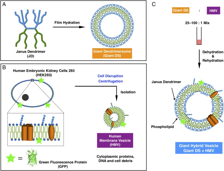
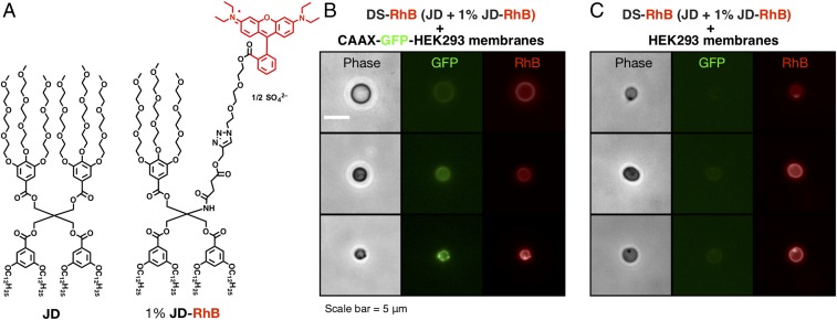
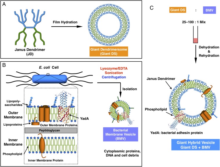
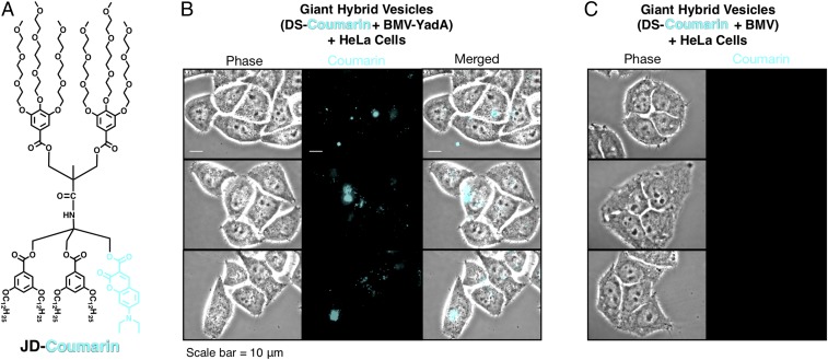
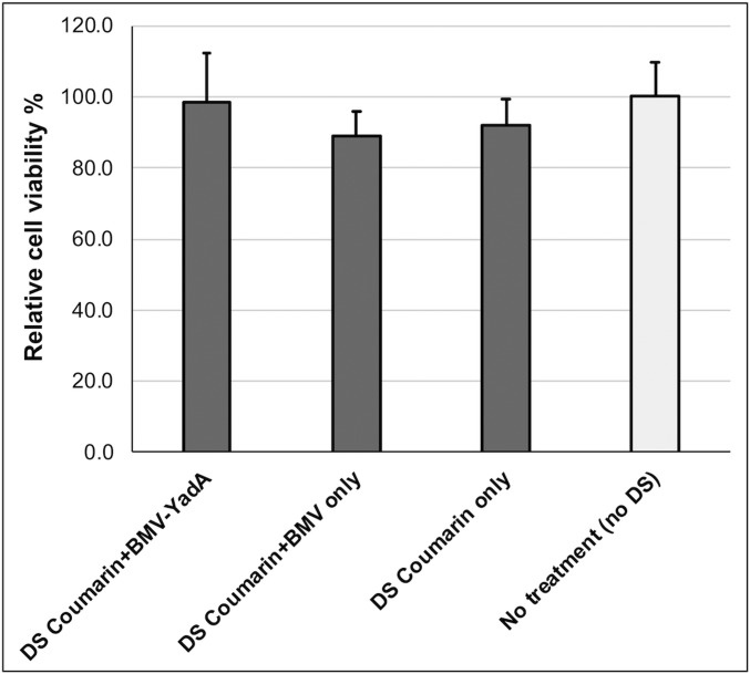
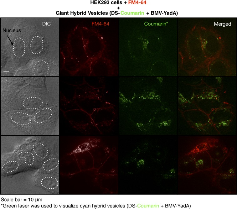
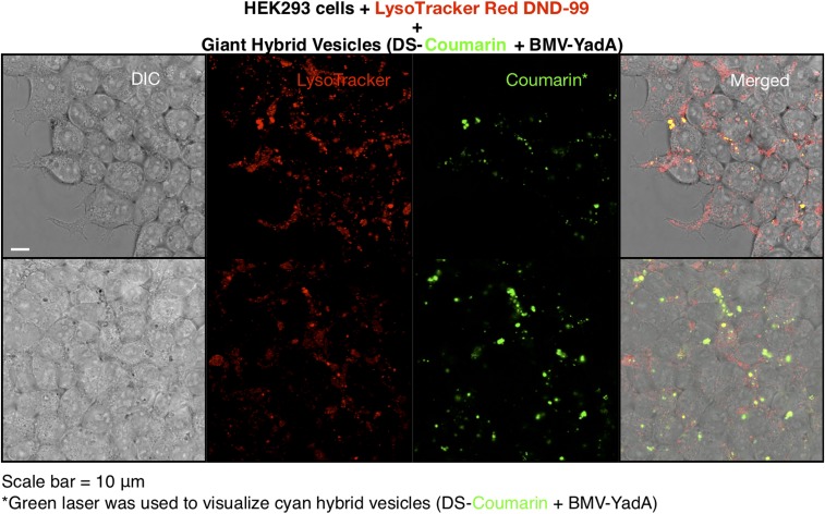
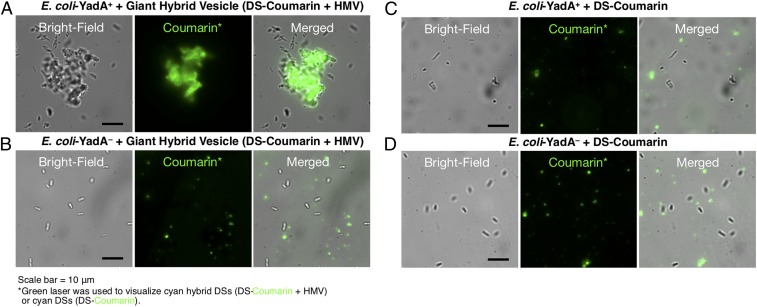
Cell-like hybrids from natural and synthetic amphiphiles provide a platform to engineer functions of synthetic cells and protocells. Cell membranes and vesicles prepared from human cell membranes are relatively unstable in vitro and therefore are difficult to study. The thicknesses of biological membranes and vesicles self-assembled from amphiphilic Janus dendrimers, known as dendrimersomes, are comparable. This feature facilitated the coassembly of functional cell-like hybrid vesicles from giant dendrimersomes and bacterial membrane vesicles generated from the very stable bacterial Escherichia coli cell after enzymatic degradation of its outer membrane. Human cells are fragile and require only mild centrifugation to be dismantled and subsequently reconstituted into vesicles. Here we report the coassembly of human membrane vesicles with dendrimersomes. The resulting giant hybrid vesicles containing human cell membranes, their components, and Janus dendrimers are stable for at least 1 y. To demonstrate the utility of cell-like hybrid vesicles, hybrids from dendrimersomes and bacterial membrane vesicles containing YadA, a bacterial adhesin protein, were prepared. The latter cell-like hybrids were recognized by human cells, allowing for adhesion and entry of the hybrid bacterial vesicles into human cells in vitro.
Keywords: bacterial adhesin; bacterial membrane; coassembly; hybrid vesicles; mammalian cell.
Copyright © 2019 the Author(s). Published by PNAS.
Conflict of interest statement
The authors declare no conflict of interest.
Figures
Similar articles
-
Bioactive cell-like hybrids coassembled from (glyco)dendrimersomes with bacterial membranes.Proc Natl Acad Sci U S A. 2016 Mar 1;113(9):E1134-41. doi: 10.1073/pnas.1525589113. Epub 2016 Feb 16. Proc Natl Acad Sci U S A. 2016. PMID: 26884210 Free PMC article.
-
Encapsulation of hydrophobic components in dendrimersomes and decoration of their surface with proteins and nucleic acids.Proc Natl Acad Sci U S A. 2019 Jul 30;116(31):15378-15385. doi: 10.1073/pnas.1904868116. Epub 2019 Jul 15. Proc Natl Acad Sci U S A. 2019. PMID: 31308223 Free PMC article.
-
Membrane-Mimetic Dendrimersomes Engulf Living Bacteria via Endocytosis.Nano Lett. 2019 Aug 14;19(8):5732-5738. doi: 10.1021/acs.nanolett.9b02349. Epub 2019 Jul 18. Nano Lett. 2019. PMID: 31306030
-
Mimicking Complex Biological Membranes and Their Programmable Glycan Ligands with Dendrimersomes and Glycodendrimersomes.Chem Rev. 2017 May 10;117(9):6538-6631. doi: 10.1021/acs.chemrev.7b00097. Epub 2017 Apr 18. Chem Rev. 2017. PMID: 28417638 Review.
-
Biopores/membrane proteins in synthetic polymer membranes.Biochim Biophys Acta Biomembr. 2017 Apr;1859(4):619-638. doi: 10.1016/j.bbamem.2016.10.015. Epub 2016 Oct 29. Biochim Biophys Acta Biomembr. 2017. PMID: 27984019 Review.
Cited by
-
Assembling Complex Macromolecules and Self-Organizations of Biological Relevance with Cu(I)-Catalyzed Azide-Alkyne, Thio-Bromo, and TERMINI Double "Click" Reactions.Polymers (Basel). 2023 Feb 21;15(5):1075. doi: 10.3390/polym15051075. Polymers (Basel). 2023. PMID: 36904317 Free PMC article.
-
Toward long-lasting artificial cells that better mimic natural living cells.Emerg Top Life Sci. 2019 Nov 11;3(5):597-607. doi: 10.1042/ETLS20190026. Emerg Top Life Sci. 2019. PMID: 33523164 Free PMC article.
-
Encoding biological recognition in a bicomponent cell-membrane mimic.Proc Natl Acad Sci U S A. 2019 Mar 19;116(12):5376-5382. doi: 10.1073/pnas.1821924116. Epub 2019 Feb 28. Proc Natl Acad Sci U S A. 2019. PMID: 30819900 Free PMC article.
-
Stimuli-Responsive Principles of Supramolecular Organizations Emerging from Self-Assembling and Self-Organizable Dendrons, Dendrimers, and Dendronized Polymers.Polymers (Basel). 2023 Apr 9;15(8):1832. doi: 10.3390/polym15081832. Polymers (Basel). 2023. PMID: 37111979 Free PMC article.
-
Screening Libraries to Discover Molecular Design Principles for the Targeted Delivery of mRNA with One-Component Ionizable Amphiphilic Janus Dendrimers Derived from Plant Phenolic Acids.Pharmaceutics. 2023 May 23;15(6):1572. doi: 10.3390/pharmaceutics15061572. Pharmaceutics. 2023. PMID: 37376020 Free PMC article.
References
-
- Mohandas N, Evans E. Mechanical properties of the red cell membrane in relation to molecular structure and genetic defects. Annu Rev Biophys Biomol Struct. 1994;23:787–818. - PubMed
-
- Bangham AD, Standish MM, Watkins JC. Diffusion of univalent ions across the lamellae of swollen phospholipids. J Mol Biol. 1965;13:238–252. - PubMed
-
- Ringsdorf H, Schlarb B, Venzmer J. Molecular architecture and function of polymeric oriented systems: Models for the study of organization, surface recognition, and dynamics of biomembranes. Angew Chem Int Ed Engl. 1988;27:113–158.
-
- Thomas JL, Tirrell DA. Polyelectrolyte-sensitized phospholipid vesicles. Acc Chem Res. 1992;25:336–342.
-
- Allen TM, Cullis PR. Drug delivery systems: Entering the mainstream. Science. 2004;303:1818–1822. - PubMed
Publication types
MeSH terms
Substances
Grants and funding
LinkOut - more resources
Full Text Sources
