Review
. 2018 Dec 27;20(1):95.
doi: 10.3390/ijms20010095.

Iron Metabolism in Cancer

Affiliations
Review

Iron Metabolism in Cancer

Yafang Wang et al. Int J Mol Sci. .

Abstract

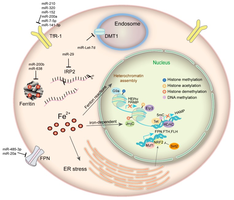
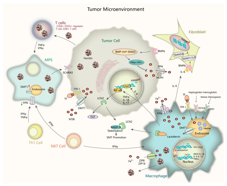
Demanded as an essential trace element that supports cell growth and basic functions, iron can be harmful and cancerogenic though. By exchanging between its different oxidized forms, iron overload induces free radical formation, lipid peroxidation, DNA, and protein damages, leading to carcinogenesis or ferroptosis. Iron also plays profound roles in modulating tumor microenvironment and metastasis, maintaining genomic stability and controlling epigenetics. in order to meet the high requirement of iron, neoplastic cells have remodeled iron metabolism pathways, including acquisition, storage, and efflux, which makes manipulating iron homeostasis a considerable approach for cancer therapy. Several iron chelators and iron oxide nanoparticles (IONPs) has recently been developed for cancer intervention and presented considerable effects. This review summarizes some latest findings about iron metabolism function and regulation mechanism in cancer and the application of iron chelators and IONPs in cancer diagnosis and therapy.

Keywords: cancer; epigenetic regulation; iron homeostasis; iron manipulating strategies; tumor microenvironment.


Conflict of interest statement

The authors declare no conflict of interest.

Figures

Figure 1
Figure 1
Iron and epigenetic regulation. Iron can modulate heterochromatin assembly mediated by Fenton reactions and induce global histone methylation changes through iron-dependent JmjC-domain-containing epigenetic modifying enzymes in cancer cells. Multiple miRNAs have been demonstrated to regulate iron metabolism-related proteins. DNA methylation, histone acetylation/methylation modification, and some transcription factors such as NRF2 and MZF-1 function corporately to maintain cellular iron metabolism in cancer. TfR, transferrin receptor 1; IRP2, iron regulatory protein; FPN, ferroportin; DMT1, divalentmetal transporter 1; ER, endoplasmic reticulum; HDAC, histone deacetylase; HEPH, hephaestin; Elp3, elongator complex protein 3; 5mC, 5-methylcytosine; TET, ten-eleven translocation protein.
Figure 2
Figure 2
Iron handling in the tumor microenvironment. Tumor microenvironment compartments play a critical role in controlling iron metabolism. Inflammatory cytokines upregulate Lcn2 via NF-κB pathway. After releasing out of the cell, Lcn2 sequesters iron and stabilize MMP-9, promoting cell survival and matrix degradation leading to EMT. M2 macrophages are major sites of taking up, metabolizing, storing, and exporting iron. They supply iron to accelerate tumor growth by multiple transport pathways. Tumor-associated fibroblasts contribute to hepcidin induction via paracrine IL-6/BMP/SMAD signaling. Circulating T cells has accumulated H-ferritin to maintain proper immune functions. Th1 cells and NKT cells can secret cytokines like IFN-γ and TNF to the environment, which increase DMT1 whereas decrease FPN level, thus resulting in iron sequestration in the MPS. Tumor-associated fibroblasts induce hepcidin expression via paracrine IL-6-BMP-SMAD signaling. Lcn2, Lipocalin 2; NF-κB, nuclear factor kappa-light-chain-enhancer of activated B cells; MMP-9, matrix metalloproteinases-9; EMT, epithelial-mesenchymal transition; IL-6, interleukin-6; NKT, natural killer T cells; IFN—γ, interferon-γ; TNF, tumor necrosis factor; FPN, ferroportin; MPS, mononuclear phagocyte system.

References

    1. Muckenthaler M.U., Rivella S., Hentze M.W., Galy B. A Red Carpet for Iron Metabolism. Cell. 2017;168:344–361. doi: 10.1016/j.cell.2016.12.034. - DOI - PMC - PubMed
    1. Recalcati S., Gammella E., Buratti P., Cairo G. Molecular regulation of cellular iron balance. IUBMB Life. 2017;69:389–398. doi: 10.1002/iub.1628. - DOI - PubMed
    1. Torti S.V., Torti F.M. Iron and cancer: More ore to be mined. Nat. Rev. Cancer. 2013;13:342–355. doi: 10.1038/nrc3495. - DOI - PMC - PubMed
    1. Martinkova P., Brtnicky M., Kynicky J., Pohanka M. Iron Oxide Nanoparticles: Innovative Tool in Cancer Diagnosis and Therapy. Adv. Healthc. Mater. 2018;7 doi: 10.1002/adhm.201700932. - DOI - PubMed
    1. Viktorinova A. Iron-mediated oxidative cell death is a potential contributor to neuronal dysfunction induced by neonatal hemolytic hyperbilirubinemia. Arch. Biochem. Biophys. 2018;654:185–193. doi: 10.1016/j.abb.2018.07.022. - DOI - PubMed