Review
. 2018 Dec 27;11(1):24.
doi: 10.3390/cancers11010024.

Obesity-Induced TNFα and IL-6 Signaling: The Missing Link between Obesity and Inflammation-Driven Liver and Colorectal Cancers

Affiliations
Review

Obesity-Induced TNFα and IL-6 Signaling: The Missing Link between Obesity and Inflammation-Driven Liver and Colorectal Cancers

Lara Kern et al. Cancers (Basel). .

Abstract

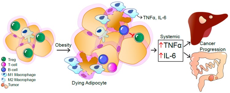
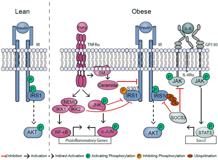
Obesity promotes the development of numerous cancers, such as liver and colorectal cancers, which is at least partly due to obesity-induced, chronic, low-grade inflammation. In particular, the recruitment and activation of immune cell subsets in the white adipose tissue systemically increase proinflammatory cytokines, such as tumor necrosis factor α (TNFα) and interleukin-6 (IL-6). These proinflammatory cytokines not only impair insulin action in metabolic tissues, but also favor cancer development. Here, we review the current state of knowledge on how obesity affects inflammatory TNFα and IL-6 signaling in hepatocellular carcinoma and colorectal cancers.

Keywords: IL-6 and TNF; Obesity; liver and colon cancer; low-grade inflammation; signaling.


Conflict of interest statement

The authors declare no conflict of interest.

Figures

Figure 1
Figure 1
The obesity-induced, systemic, low-grade inflammation contributes to the progression of hepatocellular carcinoma (HCC) and colorectal cancer (CRC). As a response to mechanical and endoplasmatic reticulum stress in obesity, immune cells are recruited to the white adipose tissue (WAT). Dying adipocytes are surrounded by macrophages visible as crown-like structures. In obese WAT, the number of M1-polarized macrophages is increased that release inflammatory cytokines like tumor necrosis factor α (TNFα) and Interleukin-6 (IL-6), resulting in a local and systemic low-grade inflammation. B and T cells are recruited to the obese WAT. Infiltrating B cells inhibit regulatory T cells, further contributing to systemic inflammation accompanied with elevated IL-6 and TNFα levels, causing HCC and CRC.
Figure 2
Figure 2
Obesity-induced inflammation causes insulin resistance.

References

    1. Hotamisligil G.S., Shargill N.S., Spiegelman B.M. Adipose expression of tumor necrosis factor-alpha: Direct role in obesity-linked insulin resistance. Science. 1993;259:87–91. doi: 10.1126/science.7678183. - DOI - PubMed
    1. Hotamisligil G.S., Peraldi P., Budavari A., Ellis R., White M.F., Spiegelman B.M. IRS-1-mediated inhibition of insulin receptor tyrosine kinase activity in TNF-alpha- and obesity-induced insulin resistance. Science. 1996;271:665–668. doi: 10.1126/science.271.5249.665. - DOI - PubMed
    1. Wellen K.E., Hotamisligil G.S. Inflammation, stress, and diabetes. J. Clin. Investig. 2005;115:1111–1119. doi: 10.1172/JCI25102. - DOI - PMC - PubMed
    1. Weisberg S.P., McCann D., Desai M., Rosenbaum M., Leibel R.L., Ferrante A.W., Jr. Obesity is associated with macrophage accumulation in adipose tissue. J. Clin. Investig. 2003;112:1796–1808. doi: 10.1172/JCI200319246. - DOI - PMC - PubMed
    1. Xu H., Barnes G.T., Yang Q., Tan G., Yang D., Chou C.J., Sole J., Nichols A., Ross J.S., Tartaglia L.A., et al. Chronic inflammation in fat plays a crucial role in the development of obesity-related insulin resistance. J. Clin. Investig. 2003;112:1821–1830. doi: 10.1172/JCI200319451. - DOI - PMC - PubMed

Grants and funding

LinkOut - more resources