. 2018 Dec 27;8(1):18083.
doi: 10.1038/s41598-018-35729-1.

Effects of Novel Calpain Inhibitors in Transgenic Animal Model of Parkinson's disease/dementia with Lewy bodies

Affiliations

Effects of Novel Calpain Inhibitors in Transgenic Animal Model of Parkinson's disease/dementia with Lewy bodies

Getaw Worku Hassen et al. Sci Rep. .

Abstract

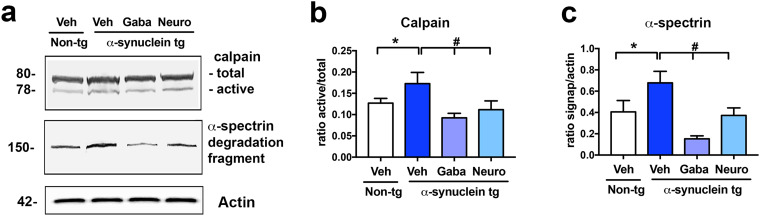
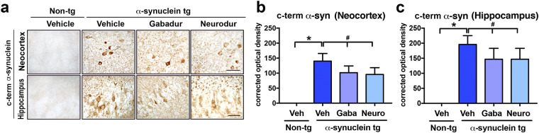
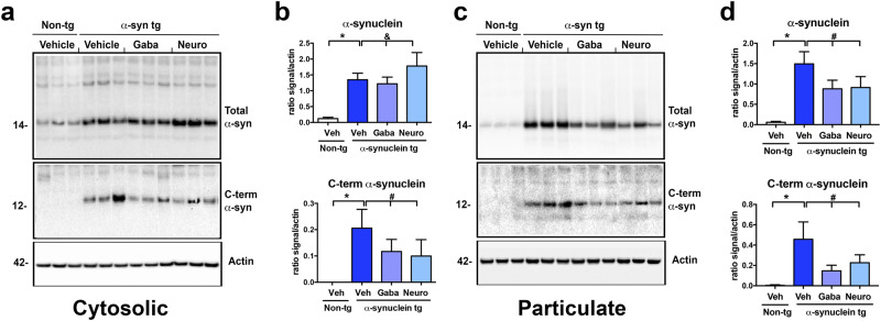
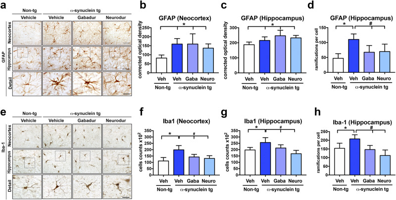
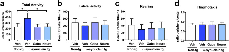
Parkinson's disease (PD) and dementia with Lewy bodies (DLB) are neurodegenerative disorders of the aging population characterized by the accumulation of α-synuclein (α-syn). The mechanisms triggering α-syn toxicity are not completely understood, however, c-terminus truncation of α-syn by proteases such as calpain may have a role. Therefore, inhibition of calpain may be of value. The main objective of this study was to evaluate the effects of systemically administered novel low molecular weight calpain inhibitors on α-syn pathology in a transgenic mouse model. For this purpose, non-tg and α-syn tg mice received the calpain inhibitors - Gabadur, Neurodur or a vehicle, twice a day for 30 days. Immunocytochemical analysis showed a 60% reduction in α-syn deposition using Gabadur and a 40% reduction using Neurodur with a concomitant reduction in c-terminus α-syn and improvements in neurodegeneration. Western blot analysis showed a 77% decrease in α-spectrin breakdown products (SBDPs) SBDPs with Gabadur and 63% reduction using Neurodur. There was a 65% reduction in the active calpain form with Gabadur and a 45% reduction with Neurodur. Moreover, treatment with calpain inhibitors improved activity performance of the α-syn tg mice. Taken together, this study suggests that calpain inhibition might be considered in the treatment of synucleinopathies.


Conflict of interest statement

The authors declare no competing interests.

Figures

Figure 1
Figure 1
Immunoblot analysis of the effects of calpain inhibitors on α-syn tg mice. α-Syn tg mice were injected ip twice a day for 30 days with either Gabadur (1 mg/mouse; N = 10), Neurodur (1 mg/mouse; N = 10), or PBS vehicle control (N = 10) and compared to vehicle-treated non-tg mice (N = 8). (a) Immunoblot and (b) quantification of calpain levels. Calpain levels were significantly increased in vehicle-treated α-syn tg mice compared to vehicle-treated non-tg mice. Treatment with either Gabadur or Neurodur significantly reduced calpain protein levels in α-syn tg mice compared to vehicle-treated α-syn tg mice. (c) Immunoblot and (d) quantification of α-spectrin breakdown products (α-SBDPs). α-SBDPs levels were significantly increased in vehicle-treated α-syn tg mice compared to vehicle-treated non-tg mice. Treatment with either Gabadur or Neurodur significantly reduced α-SBDPs protein levels in α-syn tg mice compared to vehicle-treated α-syn tg mice. Statistical analysis was conducted using one-way ANOVA post hoc Dunnett’s test for comparison with vehicle-treated non- tg mice (*p < 0.05) and Tukey–Kramer test for comparison with vehicle-treated α-syn tg mice (#p < 0.05).
Figure 2
Figure 2
Effects of Gabadur and Neurodur on total α-syn levels in the neocortex and hippocampus of α-syn tg mice. α-Syn tg mice were injected ip twice a day for 30 days with either Gabadur (1 mg/mouse; N = 10), Neurodur (1 mg/mouse; N = 10), or PBS vehicle control (N = 10) and compared to vehicle-treated non-tg mice (N = 8). (a) Representative photomicrographs and quantitation of total α-syn immunoreactivity in the (b) neocortex and (c) hippocampus. Immunoreactivity of total α-syn was significantly increased in vehicle-treated α-syn tg mice compared to vehicle-treated non-tg mice in both the neocortex and hippocampus. Treatment with either Gabadur or Neurodur significantly reduced total α-syn immunoreactivity in α-syn tg mice compared to vehicle-treated α-syn tg mice. Scale bar = 35 µm. Statistical analysis was conducted using one-way ANOVA post hoc Dunnett’s test for comparison with vehicle-treated non- tg mice (*p < 0.05) and Tukey–Kramer test for comparison with vehicle-treated α-syn tg mice (#p < 0.05).
Figure 3
Figure 3
Effects of Gabadur and Neurodur on c-terminus α-syn levels in the neocortex and hippocampus of α-syn tg mice. α-Syn tg mice were injected ip twice a day for 30 days with either Gabadur (1 mg/mouse; N = 10), Neurodur (1 mg/mouse; N = 10), or PBS vehicle control (N = 10) and compared to vehicle-treated non-tg mice (N = 8). (a) Representative photomicrographs and quantitation of c-terminus α-syn immunoreactivity in the (b) neocortex and (c) hippocampus. Immunoreactivity of c-terminus α-syn was significantly increased in vehicle-treated α-syn tg mice compared to vehicle-treated non-tg mice in both the neocortex and hippocampus. Treatment with either Gabadur or Neurodur significantly reduced c-terminus α-syn immunoreactivity in α-syn tg mice compared to vehicle-treated α-syn tg mice. Scale bar = 35 µm. Statistical analysis was conducted using one-way ANOVA post hoc Dunnett’s test for comparison with vehicle-treated non- tg mice (*p < 0.05) and Tukey–Kramer test for comparison with vehicle-treated α-syn tg mice (#p < 0.05).
Figure 4
Figure 4
Immunoblot analysis of the effects of Gabadur and Neurodur on total and C-terminus α-syn protein levels in α-syn tg mice. α-Syn tg mice were injected ip twice a day for 30 days with either Gabadur (1 mg/mouse; N = 10), Neurodur (1 mg/mouse; N = 10), or PBS vehicle control (N = 10) and compared to vehicle-treated non-tg mice (N = 8). Hemibrains were homogenized and divided into cytosolic (soluble) and particulate (membrane) fractions and analyzed by immunoblot. (a) Representative immunoblots of the cytosolic fraction analyzed for total and c-terminus α-syn protein levels. (b) Quantitative analysis of total α-syn and c-terminus α-syn protein levels. Total α-syn protein levels were significantly increased in vehicle-treated α-syn tg mice compared to vehicle-treated non-tg mice. Treatment with either Gabadur or Neurodur increased total and reduced c-terminus α-syn protein levels in α-syn tg mice compared to vehicle-treated α-syn tg mice. (c) Representative immunoblots of the particulate fraction analyzed for total and c-terminus α-syn protein levels. (d) Quantitative analysis of total α-syn and c-terminus α-syn protein levels. Treatment with either Gabadur or Neurodur decreased total and c-terminus α-syn protein levels in α-syn tg mice compared to vehicle-treated α-syn tg mice. Statistical analysis was conducted using one-way ANOVA post hoc Dunnett’s test for comparison with vehicle-treated non- tg mice (*p < 0.05) and Tukey–Kramer test for comparison with vehicle-treated α-syn tg mice (#p < 0.05).
Figure 5
Figure 5
Effects of Gabadur and Neurodur on neurodegeneration (NeuN) in the neocortex and hippocampus of α-syn tg mice. α-Syn tg mice were injected ip twice a day for 30 days with either Gabadur (1 mg/mouse; N = 10), Neurodur (1 mg/mouse; N = 10), or PBS vehicle control (N = 10) and compared to vehicle-treated non-tg mice (N = 8). (a) Representative photomicrographs and quantitation of NeuN-positive neurons in the (b) neocortex and (c) hippocampus. The number of NeuN-positive neurons was significantly reduced in vehicle-treated α-syn tg mice compared to vehicle-treated non-tg mice in both the neocortex and hippocampus. Treatment with either Gabadur or Neurodur significantly increased the number of NeuN-immunoreactive neurons in both the neocortex and hippocampus. Scale bar = 35 µm. Statistical analysis was conducted using one-way ANOVA post hoc Dunnett’s test for comparison with vehicle-treated non- tg mice (*p < 0.05) and Tukey–Kramer test for comparison with vehicle-treated α-syn tg mice (#p < 0.05).
Figure 6
Figure 6
Effects of Gabadur and Neurodur on astrogliosis and microgliosis in α-syn tg mice. α-Syn tg mice were injected ip twice a day for 30 days with either Gabadur (1 mg/mouse; N = 10), Neurodur (1 mg/mouse; N = 10), or PBS vehicle control (N = 10) and compared to vehicle-treated non-tg mice (N = 8). (a) Representative photomicrographs and quantitation of GFAP-positive astrocytes in the (b) neocortex and (c) hippocampus. The number of GFAP-positive neurons was significantly increased in vehicle-treated α-syn tg mice compared to vehicle-treated non-tg mice in both the neocortex and hippocampus. Neither treatment with Gabadur nor Neurodur significantly affected the number of GFAP-immunoreactive astrocytes in either the neocortex or hippocampus compared to vehicle-treated α-syn tg mice. (d) Representative photomicrographs and quantitation of Iba-1-positive microglia in the (e) neocortex and (f) hippocampus. The number of Iba-1-positive microglia was significantly increased in vehicle-treated α-syn tg mice compared to vehicle-treated non-tg mice in both the neocortex and hippocampus. Treatment with Gabadur or Neurodur significantly reduced the number of Iba-1-immunoreactive microglia in the neocortex and hippocampus. Scale bar = 35 µm. Statistical analysis was conducted using one-way ANOVA post hoc Dunnett’s test for comparison with vehicle-treated non- tg mice (*p < 0.05) and Tukey–Kramer test for comparison with vehicle-treated α-syn tg mice (#p < 0.05).
Figure 7
Figure 7
Effects of Gabadur and Neurodur on behavioral measures in α-syn tg mice. α-Syn tg mice were injected ip twice a day for 30 days with either Gabadur (1 mg/mouse; N = 10), Neurodur (1 mg/mouse; N = 10), or PBS vehicle control (N = 10) and compared to vehicle-treated non-tg mice (N = 8). Testing for (a) total spontaneous locomotor activity, (b) lateral activity, (c) rearing, and (d) thigmotaxis. Compared to vehicle-treated non-tg mice, vehicle-treated α-syn tg mice had a significant increase in total spontaneous activity. Gabadur and Neurodur significantly reduced total spontaneous locomotor compared to vehicle-treated α-syn tg mice. There were no significant differences between treatment groups in any of the other behavioral tests. Statistical analysis was conducted using one-way ANOVA post hoc Dunnett’s test for comparison with vehicle-treated non- tg mice (*p < 0.05) and Tukey–Kramer test for comparison with vehicle-treated α-syn tg mice (#p < 0.05).

References

    1. Alafuzoff, I. & Hartikainen, P. Alpha-synucleinopathies. Handb Clin Neurol145, 339–353, 10.1016/B978-0-12-802395-2.00024-9 (2017). - PubMed
    1. Jellinger, K. A. Dementia with Lewy bodies and Parkinson’s disease-dementia: current concepts and controversies. J Neural Transm (Vienna), 10.1007/s00702-017-1821-9 (2017). - PubMed
    1. Peng, C., Gathagan, R. J. & Lee, V. M. Distinct alpha-Synuclein strains and implications for heterogeneity among alpha-Synucleinopathies. Neurobiol Dis109, 209–218, 10.1016/j.nbd.2017.07.018 (2018). - PMC - PubMed
    1. Goedert, M., Jakes, R. & Spillantini, M. G. The Synucleinopathies: Twenty Years On. Journal of Parkinson’s disease7, S53–S71, 10.3233/JPD-179005 (2017). - PMC - PubMed
    1. Ono, K. The Oligomer Hypothesis in alpha-Synucleinopathy. Neurochem Res42, 3362–3371, 10.1007/s11064-017-2382-x (2017). - PubMed

Publication types