Review
. 2018 Dec 27;36(2):29.
doi: 10.1007/s11095-018-2554-7.

Pharmaceutical Development of AAV-Based Gene Therapy Products for the Eye

Affiliations
Review

Pharmaceutical Development of AAV-Based Gene Therapy Products for the Eye

Gerard A Rodrigues et al. Pharm Res. .

Abstract

A resurgence of interest and investment in the field of gene therapy, driven in large part by advances in viral vector technology, has recently culminated in United States Food and Drug Administration approval of the first gene therapy product targeting a disease caused by mutations in a single gene. This product, LUXTURNA™ (voretigene neparvovec-rzyl; Spark Therapeutics, Inc., Philadelphia, PA), delivers a normal copy of the RPE65 gene to retinal cells for the treatment of biallelic RPE65 mutation-associated retinal dystrophy, a blinding disease. Many additional gene therapy programs targeting both inherited retinal diseases and other ocular diseases are in development, owing to an improved understanding of the genetic basis of ocular disease and the unique properties of the ocular compartment that make it amenable to local gene therapy. Here we review the growing body of literature that describes both the design and development of ocular gene therapy products, with a particular emphasis on target and vector selection, and chemistry, manufacturing, and controls.

Keywords: adeno-associated virus (AAV) vector; formulation; gene therapy; ocular diseases; product development.


Figures
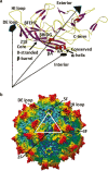
Fig. 1
Fig. 1
Adeno-associated virus serotype 1 (AAV1) structure. (a) Crystal structure of AAV1 capsid VP3 monomer (PDB ID, 3NG9). The β-strands are shown in purple ribbon, the conserved α-helix A is in red, and loops between the strands are in yellow. The dotted lines show the relative positions of the 5-fold (filled pentagon), 3-fold (filled triangle), and 2-fold (filled oval) interfaces of symmetry from the center of the capsid. An eight-stranded β-barrel (with β-sheets βCHEF and βBIDG), along with βA (labeled) and α-helix A (αA), forms the core of the VP monomer structure, flanked by large loop regions. The DE and HI loops (between β-strands D and E and between H and I, respectively) as well as the first ordered N-terminal residue (218), the C-terminus, and the interior and exterior capsid surfaces are labeled. (b) Radially color-cued (from capsid center to surface, blue to green to yellow to red) surface representation of the AAV1 capsid. The white triangle depicts a viral asymmetric unit bounded by one 5-fold axis and two 3-fold axes with a 2-fold axis between them. The approximate locations of the icosahedral 2-fold (2F), 3F, and 5F axes are indicated by the black arrows. The positions of the DE and HI loops are indicated by the dashed arrows. Reproduced from Venkatakrishnan et al. J Virol. 2013;87:4974–84. doi 10.1128/JVI.02524-12 (102) with permission from American Society for Microbiology. © 2013.
Fig. 2
Fig. 2
Adeno-associated virus (AAV) stability with respect to final frozen pH (correlation coefficient, r2 = 0.97). Lfu represents lac-forming unit. Reprinted by permission from Springer Nature: Springer Publishing Company, Gene Therapy. Croyle MA, Cheng X, Wilson JM. Development of formulations that enhance physical stability of viral vectors for gene therapy. © 2001. 2001 (101).
Fig. 3
Fig. 3
Dependence of adeno-associated virus serotype 2 (AAV2) vector aggregation on osmolarity (a), ionic strength (b), and purification method (c). The average particle radius of the AAV2-FIX vector was measured by dynamic light scattering following vector dilution in varying concentrations of excipients buffered with 10 mM sodium phosphate, pH 7.5. (a, b) Filled circles: sodium chloride; open circles: sodium citrate; filled squares: sodium phosphate; open squares: sodium sulfate; inverted filled squares: magnesium sulfate; open diamonds: glycerol. Vector was purified by method 3 (chromatography plus CsCl gradient). (c) The vector was purified by one of three different methods as the ionic strength was adjusted with NaCl. + symbols: method 1 (double CsCl gradient); open triangles: method 2 (cation exchange chromatography); filled triangles: method 2 plus nuclease digestion; x symbols: method 3. Republished from Wright et al. (93) under the terms of the CC BY-NC-ND 4.0 license (http://creativecommons.org/licenses/by-nc-nd/4.0/).
Fig. 4
Fig. 4
Recovery of adeno-associated virus serotype 2 (AAV2) following dilution and passage through the administration device. Stock AAV2-RPE65 vector diluted to 1×10¹¹ vg/mL in phosphate-buffered saline with (+PF68) or with (−PF68). Pluronic 68 0.001% was drawn into 1-mL syringes, and the vector was passed through device A, B, or C. Republished from Bennicelli et al. (105) under the terms of the CC BY-NC-ND 4.0 license (http://creativecommons.org/licenses/by-nc-nd/4.0/).
Fig. 5
Fig. 5
Potency of liquid formulation for an investigational protein drug product during storage at different temperatures (data from EY Shalaev, “Role of Ice in Destabilization of Proteins”, presented at the AAPS National Biotechnology Conference, Boston, MA, May 18, 2016).
Fig. 6
Fig. 6
Thermal profiles of rAAV serotypes 1 to 9 and rAAVrh.10 produced by DSF analysis. The DSF spectra display normalized RFUs (relative fluorescence units) vs. temperature. A representative DSF spectrum is shown for each rAAV serotype. Republished from Bennett et al. (124) under the terms of the CC BY-NC-ND 4.0 license (http://creativecommons.org/licenses/by-nc-nd/4.0/).
Fig. 7
Fig. 7
Example of circular dichroism (CD) spectra of AAV serotype 1 (AAV1) viral-linked proteins at different temperatures. A clear α-helical propensity can be seen for AAV1. This helical signal is lost as temperature increases. Reproduced from Venkatakrishnan et al. J Virol. 2013;87:4974–84. doi 10.1128/JVI.02524-12 (102) with permission from American Society for Microbiology. © 2013.

References

    1. Dunbar CE, High KA, Joung JK, Kohn DB, Ozawa K, Sadelain M. Gene therapy comes of age. Science. 2018;359(6372):eaan4672. doi: 10.1126/science.aan4672. - DOI - PubMed
    1. Bainbridge JW, Smith AJ, Barker SS, Robbie S, Henderson R, Balaggan K, Viswanathan A, Holder GE, Stockman A, Tyler N, Petersen-Jones S, Bhattacharya SS, Thrasher AJ, Fitzke FW, Carter BJ, Rubin GS, Moore AT, Ali RR. Effect of gene therapy on visual function in Leber's congenital amaurosis. N Engl J Med. 2008;358(21):2231–2239. doi: 10.1056/NEJMoa0802268. - DOI - PubMed
    1. Hauswirth WW, Aleman TS, Kaushal S, Cideciyan AV, Schwartz SB, Wang L, Conlon TJ, Boye SL, Flotte TR, Byrne BJ, Jacobson SG. Treatment of leber congenital amaurosis due to RPE65 mutations by ocular subretinal injection of adeno-associated virus gene vector: short-term results of a phase I trial. Hum Gene Ther. 2008;19(10):979–990. doi: 10.1089/hum.2008.107. - DOI - PMC - PubMed
    1. Maguire AM, Simonelli F, Pierce EA, Pugh EN, Jr, Mingozzi F, Bennicelli J, Banfi S, Marshall KA, Testa F, Surace EM, Rossi S, Lyubarsky A, Arruda VR, Konkle B, Stone E, Sun J, Jacobs J, Dell'Osso L, Hertle R, Ma JX, Redmond TM, Zhu X, Hauck B, Zelenaia O, Shindler KS, Maguire MG, Wright JF, Volpe NJ, McDonnell JW, Auricchio A, High KA, Bennett J. Safety and efficacy of gene transfer for Leber's congenital amaurosis. N Engl J Med. 2008;358(21):2240–2248. doi: 10.1056/NEJMoa0802315. - DOI - PMC - PubMed
    1. Naldini L. Medicine. A comeback for gene therapy. Science. 2009;326(5954):805–806. doi: 10.1126/science.1181937. - DOI - PubMed

MeSH terms