Review
. 2019 May;73(5):e22087.
doi: 10.1002/syn.22087. Epub 2019 Feb 18.

Current concepts in cochlear ribbon synapse formation

Affiliations
Review

Current concepts in cochlear ribbon synapse formation

Thomas M Coate et al. Synapse. 2019 May.

Abstract

In mammals, hair cells and spiral ganglion neurons (SGNs) in the cochlea together are sophisticated "sensorineural" structures that transduce auditory information from the outside world into the brain. Hair cells and SGNs are joined by glutamatergic ribbon-type synapses composed of a molecular machinery rivaling in complexity the mechanoelectric transduction components found at the apical side of the hair cell. The cochlear hair cell ribbon synapse has received much attention lately because of recent and important findings related to its damage (sometimes termed "synaptopathy") as a result of noise overexposure. During development, ribbon synapses between type I SGNs and inner hair cells form in the time window between birth and hearing onset and is a process coordinated with type I SGN myelination, spontaneous activity, synaptic pruning, and innervation by efferents. In this review, we highlight new findings regarding the diversity of type I SGNs and inner hair cell synapses, and the molecular mechanisms of selective hair cell targeting. Also discussed are cell adhesion molecules and protein constituents of the ribbon synapse, and how these factors participate in ribbon synapse formation. We also note interesting new insights into the morphological development of type II SGNs, and the potential for cochlear macrophages as important players in protecting SGNs. We also address recent studies demonstrating that the structural and physiological profiles of the type I SGNs do not reach full maturity until weeks after hearing onset, suggesting a protracted development that is likely modulated by activity.

Keywords: cochlea; hair cell; ribbon synapse; spiral ganglion neuron; synaptopathy.


Figures

Figure 1
Figure 1. Summary of cochlear hair cell afferent innervation and inner hair cell ribbon synapses.
(A) P0 mouse cochlear cross section with Tuj1 immunostaining (green) to reveal the spiral ganglion neurons (SGNs) and efferent fibers. Actin (phalloidin; magenta) was used to reveal the anatomy of the cochlea. sv, scala vestibuli; st, scala tympani; pa, peripheral axon; ca, central axon. (B) An E16.5 cochlear cross-section immunostained with Tuj1 antibodies to show the orientation of the nerve fibers within the organ of Corti, along with the inner and outer hair cells (IHCs and OHCs). (C) A representative whole-mount view of the cochlea at E17.5. Sox2 immunostaining reveals the organ of Corti (oC), and is also apparent in the spiral ganglion, but at lower levels in this image. (D) Cartoon schematic of the different classes of nerve fibers that innervate the organ of Corti. The high-magnification view of the inner hair cell shows the arrangement of ribbon bodies and postsynaptic densities of different sizes. TM, tectorial membrane; LOC, Lateral Olivary Complex; MOC, Medial Olivary Complex; SGN, spiral ganglion neuron; SR, spontaneous rate; m, modiolar; p, pillar; c, cuticular; h, habenular. Parts of this schematic were published previously (Zhang & Coate 2017) and reproduced here with permission from Elsevier. (E-G) Whole-mount immunostaining of the P12 cochlea using anti-Ctbp2 (red) and anti-Shank (green) antibodies reveals the pre- and postsynaptic components of the inner hair cell ribbon synapses. Scale bar in G: A = approximately 300 μm; B = 25 μm; C = 150 μm, E-G = 8 μm.
Figure 2
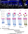
Figure 2. Highlights of Ribbon Synapse Development in the Mouse Cochlea.
(A) A whole-mount confocal image of a P4 mouse cochlea showing sparse numbers of genetically labeled SGNs. The cochlea came from a mouse carrying Neurog1CreERT2 (Koundakjian et al. 2007) and R26R-tdTomato and its mother was not exposed to tamoxifen (so “background” labeling is shown). The hair cells are immunostained with anti-myosin VI antibodies (blue) and tdTomato was immunolabeled using anti-dsRed antibodies (white). The arrowheads point to small branches from the type I SGNs not yet refined at this stage. IHC, inner hair cell; OHC, outer hair cell. (B) Similar preparation as in A, but at P10. The micrograph illustrates how the type I SGNs show unramified bouton endings at this stage. (C and D) High magnification images from the boxed regions in B. 3D reconstructions were generated (using Imaris software) from the original confocal z-stacks and then rotated to show SGN fibers contacting the “pillar” or “modiolar” side of the inner hair cell. C is an image in the XY plane that was rotated slightly toward the Z axis. D is an image that was rotated 90° to show the YZ plane. Scale bar in D: A and B = 25 μm; C and D = 8 μm. (E) A schematic of the developmental time line of ribbon synapse formation. See section 3.1 for additional details. Around P0, the SGNs show elaborate branching and the ribbon synapses (red) are small and immature. The small branches refine through P10 (~hearing onset). By P30, the ribbon bodies and their apposed postsynaptic densities (blue) have fully matured, and the three type I subdivisions (1a, 1b, and 1c) are apparent. m, modiolar; p, pillar; c, cuticular; h, habenular. (F) Approximations of the timing of different important events in SGN development with respect to the time points illustrated in E.

References

    1. Adamson CL, Reid M a, & Davis RL (2002). Opposite actions of brain-derived neurotrophic factor and neurotrophin-3 on firing features and ion channel composition of murine spiral ganglion neurons. The Journal of Neuroscience : The Official Journal of the Society for Neuroscience doi:22/4/1385 [pii] - PMC - PubMed
    1. Appler J, & Lu C (2013). Gata3 is a critical regulator of cochlear wiring. The Journal of …, 33, 3679–3691. - PMC - PubMed
    1. Babola TA, Li S, Gribizis A, … Bergles DE (2018). Homeostatic Control of Spontaneous Activity in the Developing Auditory System. Neuron, 99(3), 511–524.e5. - PMC - PubMed
    1. Barclay M, Constable R, James NR, Thorne PR, & Montgomery JM (2016). Reduced sensory stimulation alters the molecular make-up of glutamatergic hair cell synapses in the developing cochlea. Neuroscience doi:10.1016/j.neuroscience.2016.03.043 - DOI - PubMed
    1. Battisti AC, Fantetti KN, Moyers BA, & Fekete DM (2014). A subset of chicken statoacoustic ganglion neurites are repelled by Slit1 and Slit2. Hear Res, 310, 1–12. - PMC - PubMed

Publication types