

Multiple myeloma with crystal-storing histiocytosis, crystalline podocytopathy, and light chain proximal tubulopathy, revealed by retinal abnormalities: A case report
- PMID: 30593133
- PMCID: PMC6314660
- DOI: 10.1097/MD.0000000000013638
Multiple myeloma with crystal-storing histiocytosis, crystalline podocytopathy, and light chain proximal tubulopathy, revealed by retinal abnormalities: A case report
Abstract
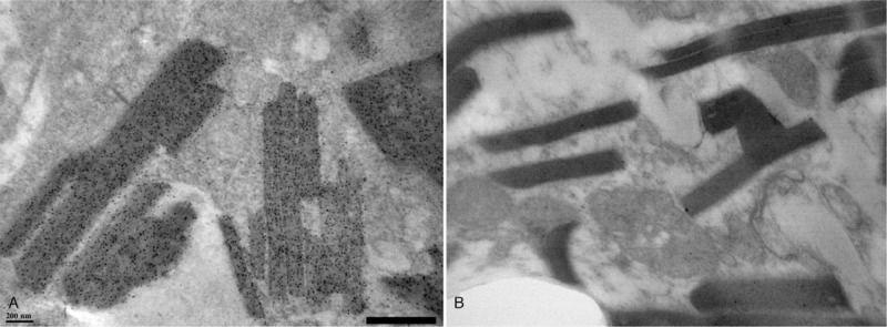
Rationale: Crystal sorting histiocytosis (CSH) is a rare disorder that is morphologically characterized by the accumulation of monoclonal immunoglobulin crystals, predominantly of a kappa light chain type, within lysosomes of macrophages. CSH may result in a variety of clinical manifestations depending on the involved organs. In this case report, we aim to describe a patient with ophthalmic manifestations which lead to the diagnosis of multiple myeloma with crystal-storing histiocytosis, crystalline podocytopathy, and light chain proximal tubulopathy.
Patient concerns: A 60-year-old male patient presented with progressive bilateral decreased vision for 2 years.
Diagnosis: Ophthalmic explorations showed bilateral macular and papillary edema, and multiple crystalline deposits in the anterior stromal cornea and in the retina. Laboratory tests showed nephrotic syndrome and renal dysfunction. Further work-up revealed IgG kappa multiple myeloma, with biopsy-proven combined crystalline podocytopathy and tubulopathy.
Interventions: The patient received chemotherapy (bortezomib, cyclophosphamide, and dexamethasone for 3 cycles, then bortezomib, lenalidomide, and dexamethasone).
Outcomes: Despite partial hematologic response and improvement of the papilledema and macular edema, the patient developed dialysis-dependent end-stage renal failure.
Lessons: This report, highlighting the protean presentation of paraprotein-mediated injuries, provides additional information on the ocular anomalies not previously described that may be associated with crystal-storing histiocytosis.
Conflict of interest statement
The authors report no conflict of interest.
Figures


Similar articles
-
Combined crystal-storing histiocytosis, light chain proximal tubulopathy, and light chain crystalline podocytopathy in a patient with multiple myeloma: a case report and literature review.Ren Fail. 2023 Dec;45(1):2145970. doi: 10.1080/0886022X.2022.2145970. Ren Fail. 2023. PMID: 36632756 Free PMC article. Review.
-
Combined light chain crystalline tubulopathy, podocytopathy, and histiocytosis associated with Bence-Jones κ protein diagnosed via immuno-electron microscopy.CEN Case Rep. 2021 Aug;10(3):453-458. doi: 10.1007/s13730-021-00588-9. Epub 2021 Mar 5. CEN Case Rep. 2021. PMID: 33675012 Free PMC article.
-
Combined proximal tubulopathy, crystal-storing histiocytosis, and cast nephropathy in a patient with light chain multiple myeloma.BMC Nephrol. 2017 May 25;18(1):170. doi: 10.1186/s12882-017-0584-8. BMC Nephrol. 2017. PMID: 28545410 Free PMC article.
-
Crystals, crystals everywhere but not a clue till late… Light chain crystalline proximal tubulopathy with concomitant myeloma cast nephropathy.Indian J Pathol Microbiol. 2020 Jul-Sep;63(3):463-466. doi: 10.4103/IJPM.IJPM_756_18. Indian J Pathol Microbiol. 2020. PMID: 32769341
-
Monoclonal light chain crystalline podocytopathy and tubulopathy associated with monoclonal gammopathy of renal significance: a case report and literature review.BMC Nephrol. 2018 Nov 12;19(1):322. doi: 10.1186/s12882-018-1108-x. BMC Nephrol. 2018. PMID: 30419839 Free PMC article. Review.
Cited by
-
Immunoglobulin-Storing Histiocytosis: A Case Based Systemic Review.J Clin Med. 2021 Apr 23;10(9):1834. doi: 10.3390/jcm10091834. J Clin Med. 2021. PMID: 33922555 Free PMC article. Review.
-
Crystal-Storing Histiocytosis: The Iceberg of More Serious Conditions.Diagnostics (Basel). 2023 Jan 11;13(2):271. doi: 10.3390/diagnostics13020271. Diagnostics (Basel). 2023. PMID: 36673081 Free PMC article. Review.
-
Combined crystal-storing histiocytosis, light chain proximal tubulopathy, and light chain crystalline podocytopathy in a patient with multiple myeloma: a case report and literature review.Ren Fail. 2023 Dec;45(1):2145970. doi: 10.1080/0886022X.2022.2145970. Ren Fail. 2023. PMID: 36632756 Free PMC article. Review.
-
Crystalline deposits in the cornea and various areas of the kidney as symptoms of an underlying monoclonal gammopathy: a case report.BMC Nephrol. 2021 Apr 6;22(1):117. doi: 10.1186/s12882-021-02309-x. BMC Nephrol. 2021. PMID: 33823814 Free PMC article.
-
Combined light chain crystalline tubulopathy, podocytopathy, and histiocytosis associated with Bence-Jones κ protein diagnosed via immuno-electron microscopy.CEN Case Rep. 2021 Aug;10(3):453-458. doi: 10.1007/s13730-021-00588-9. Epub 2021 Mar 5. CEN Case Rep. 2021. PMID: 33675012 Free PMC article.
References
-
- Merlini G, Stone MJ. Dangerous small B-cell clones. Blood 2006;108:2520–30. - PubMed
-
- Leung N, Bridoux F, Hutchison CA, et al. Monoclonal gammopathy of renal significance: when MGUS is no longer undetermined or insignificant. Blood 2012;120:4292–5. - PubMed
-
- Vignon M, Javaugue V, Alexander MP, et al. Current anti-myeloma therapies in renal manifestations of monoclonal light chain-associated Fanconi syndrome: a retrospective series of 49 patients. Leukemia 2017;31:123–9. - PubMed
Publication types
MeSH terms
LinkOut - more resources
Full Text Sources
Medical
