

Imaging features of spinal atypical teratoid rhabdoid tumors in children
- PMID: 30593171
- PMCID: PMC6314652
- DOI: 10.1097/MD.0000000000013808
Imaging features of spinal atypical teratoid rhabdoid tumors in children
Abstract
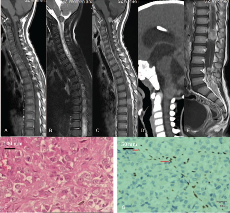
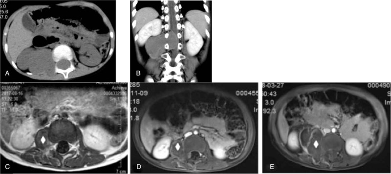
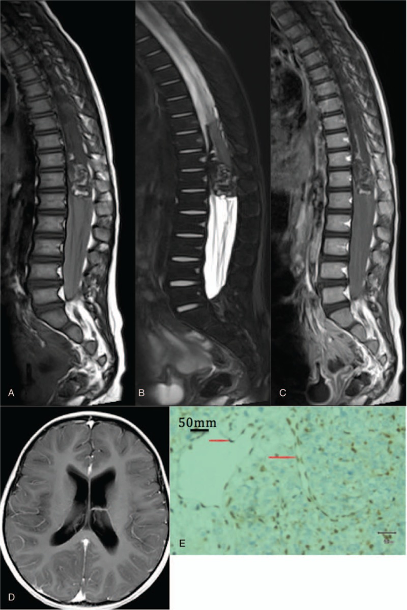
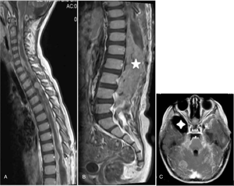
This study aims to analyze and summarize the imaging features of spinal atypical teratoid/rhabdoid tumors (AT/RT) in children.Imaging features in 8 children with spinal AT/RT confirmed by surgical pathology were retrospectively analyzed. All patients had underwent total spine 3.0 T magnetic resonance imaging (MRI) and 64-slice spiral computed tomography (CT). Among these 8 patients, head MR non-enhanced and spinal enhanced scanning was applied to 5 patients, while CT examination was applied to 3 patients.All 8 patients were characterized by cauda equina syndrome. The lesions of 7 patients were in the thoracolumbar spinal junction, while the lesion of the remaining patient was in the lumbar spine. Furthermore, among these patients, the lesions of 5 patients were limited to the intraspinal canal (1 lesion in the epidural space, and 4 lesions in the subdural space), while the lesions of 3 patients invaded the paravertebra (2 lesions in the epidural space and 1 lesion in the subdural space). Three or more spinal segments were invaded by tumors in 7 patients, while sacral canal was affected in 5 patients. All 8 patients experienced bleeding in the tumors. Enhanced MRI revealed meningeal enhancement in 6 patients, and bilateral nerve root enhancement in 4 patients. The masses in 3 patients brought damages to the intervertebral foramen or sacral pore. The lesion of 1 patient was featured by skip growth. One patient had total spinal metastasis and 3 had hydrocephalus. The masses in 2 patients had a slightly low density when detected by CT, and enhanced scanning revealed a mild to moderate enhancement.Spinal AR/TR had the following characteristics: children were characterized by cauda equina syndrome; the mass that invaded the thoracolumbar spinal junction and the extramedullary space of multiple segments grew along the spinal longitudinal axis; bleeding mass was revealed in MRI imaging; meninges, nerve root, and sacral canal metastases occurred. The gold standard for the definite diagnosis of AT/RT is biopsy combined with immunohistochemistry.
Conflict of interest statement
All authors have contributed significantly to the manuscript and declare that the work is original and has not been submitted or published elsewhere. None of the authors have any financial disclosure or conflict of interest.
Figures
References
-
- Tulla M, Berthold F, Graf N, et al. Incidence, trends, and survival of children with embryonal tumors. Pediatrics 2015;136:e623–32. - PubMed
MeSH terms
Supplementary concepts
LinkOut - more resources
Full Text Sources
Medical
Research Materials
