. 2019 Mar 1;294(9):3207-3218.
doi: 10.1074/jbc.RA118.006682. Epub 2018 Dec 28.

Bladder cancer extracellular vesicles drive tumorigenesis by inducing the unfolded protein response in endoplasmic reticulum of nonmalignant cells

Affiliations

Bladder cancer extracellular vesicles drive tumorigenesis by inducing the unfolded protein response in endoplasmic reticulum of nonmalignant cells

Chia-Hao Wu et al. J Biol Chem. .

Abstract

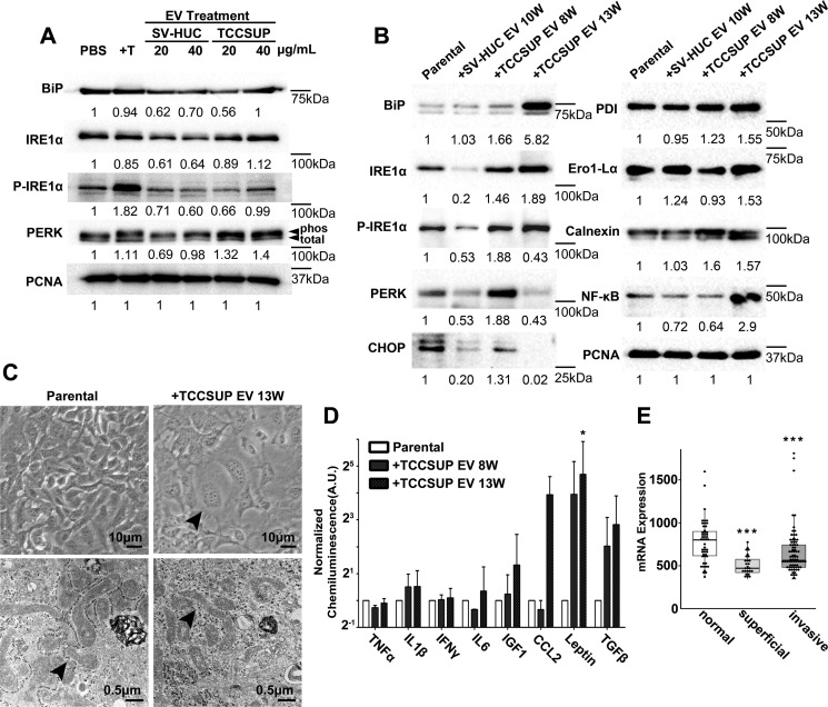
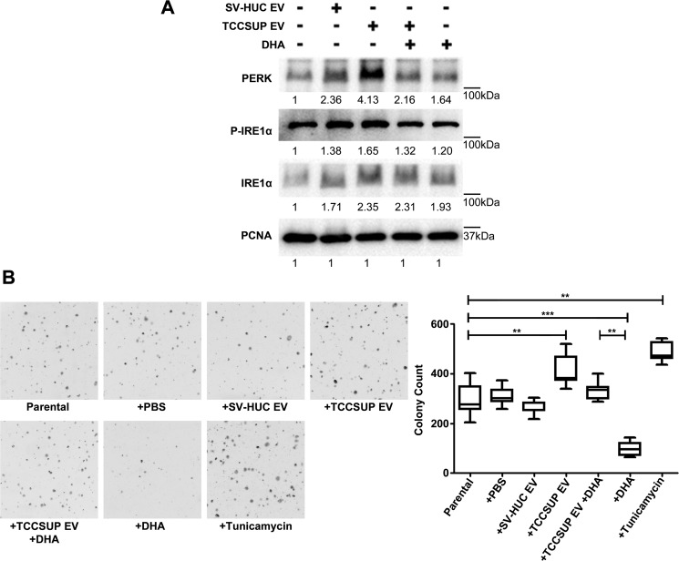
The field cancerization effect has been proposed to explain bladder cancer's multifocal and recurrent nature, yet the mechanisms of this effect remain unknown. In this work, using cell biology, flow cytometry, and qPCR analyses, along with a xenograft mouse tumor model, we show that chronic exposure to tumor-derived extracellular vesicles (TEVs) results in the neoplastic transformation of nonmalignant human SV-HUC urothelial cells. Inhibition of EV uptake prevented this transformation. Transformed cells not only possessed several oncogenic properties, such as increased genome instability, loss of cell-cell contact inhibition, and invasiveness, but also displayed altered morphology and cell structures, such as an enlarged cytoplasm with disrupted endoplasmic reticulum (ER) alignment and the accumulation of smaller mitochondria. Exposure of SV-HUC cells to TEVs provoked the unfolded protein response in the endoplasmic reticulum (UPRER). Prolonged induction of UPRER signaling activated the survival branch of the UPRER pathway, in which cells had elevated expression of inositol-requiring enzyme 1 (IRE1), NF-κB, and the inflammatory cytokine leptin, and incurred loss of the pro-apoptotic protein C/EBP homologous protein (CHOP). More importantly, inhibition of ER stress by docosahexaenoic acid prevented TEV-induced transformation. We propose that TEVs promote malignant transformation of predisposed cells by inhibiting pro-apoptotic signals and activating tumor-promoting ER stress-induced unfolded protein response and inflammation. This study provides detailed insight into the mechanisms underlying the bladder cancer field effect and tumor recurrence.

Keywords: bladder cancer; cell stress; cytokine; endoplasmic reticulum stress (ER stress); exosome (vesicle); extracellular vesicles; filed cancerization; inflammation; unfolded protein response (UPR); urothelial carcinoma.


Conflict of interest statement

The authors declare that they have no conflicts of interest with the contents of this article

Figures

Figure 1.
Figure 1.
Chronic exposure to cancer EVs leads to malignant transformation of recipient urothelial cells. A, cell cycle profiles of parental and TCCSUP EV 13-week treated SV-HUC cells as determined by flow cytometric measurement of PI staining. The cells were measured on day 6 of culture when they had reached 100% confluence. Shaded regions represent G0/G1 phase (blue) and G2/M phase (red). B, in vitro colony formation by SV-HUC cells following the indicated EV exposures and at least 5 weeks of maintenance in normal culture. C, quantification of colony formation in B. Asterisks represent statistical significance evaluated by Student's t test; p values of <0.05 (*), <0.01 (**), and <0.001 (***) are indicated. D, subcutaneous xenografts of 5-weeks recovered SV-HUC cells that had received cancer EV exposure for 8 weeks (left flank) and 13 weeks (right flank). E, mass measurements of resected xenografts from the experiment in D. F, hematoxylin and eosin staining of a representative TCCSUP EV 13-week xenograft section. W, week(s).
Figure 2.
Figure 2.
Characterization of cancer EV-transformed urothelial cells. A, quantification of gene expression in respond to oxidative stress and DNA damage. B, cellular reactive oxygen species levels are represented by geometric mean of the DCFDA fluorescent intensity as quantified by flow cytometric measurement. C, DNA damage level examination by γH2AX staining. Quantification of the γH2AX foci within the cell nucleus was done by ImageJ analysis. D, representative photographs of SV-HUC cell invasion in a Transwell invasion assay. E, quantification of the total area of invaded cells was done by ImageJ analysis of the photographs in A. n = 3 biological repeats. Normalized relative mRNA expression of invasion-related genes in EV-treated SV-HUC cells. Error bars represent standard deviations. Asterisks represent statistically significant difference from parental cell expression as evaluated by CFX Manager software; p values of <0.05 (*), <0.01 (**), and <0.001 (***) are indicated. W, week(s).
Figure 3.
Figure 3.
Inhibition of EV internalization by urothelial cells reduces cancer EV-driven colony forming ability. A, evaluation of EV internalization inhibitor efficacy in reducing uptake of PKH67-labeled TCCSUP EVs (green) by recipient SV-HUC cells. Inhibitors Dynasore and NSC23766 were used alone or in combination (D/N). SV-HUC nuclei were labeled with DAPI (blue). Quantification of EV uptake in the images. The integrated density (IntDen) as measured by Image J of the PKH67 signal in a given field is normalized by cell number. B, effect of the EV internalization inhibitor mixture (D/N) on SV-HUC in vitro colony forming ability during the final 5 weeks of TCCSUP EV exposure. Error bars represent standard deviations. Asterisks represent statistical significance evaluated by Student's t test; p values of <0.05 (*), <0.01 (**), and <0.001 (***) are indicated. ns, not significant; W, week(s).
Figure 4.
Figure 4.
Cancer EV-transformed urothelial cells show altered cellular ER stress signals and abnormal cellular ultra-structure. A and B, Western blotting showing ER stress-related protein expression after short-term (72 h, A) and long-term (up to 13 weeks, B) EV exposures. Normalized densitometry values are given below each band. Measurement of PERK includes both total and phosphorylated (PHOS) bands. C, parental and TCCSUP EV 13-week transformed SV-HUC cells shown in phase contrast micrographs (top panels) and by transmission EM (bottom panels). The black arrow at the upper right indicates an enlarged cell. In the bottom right panel, the disrupted mitochondrial-endoplasmic reticulum alignment is indicated by the black arrow. D, inflammation-related cytokines released from EV-transformed cells as determined by ELISA analysis. Statistically significant difference from parental cell expression is indicated. E, oligonucleotide array expression of CHOP mRNA in normal, superficial, and invasive bladder cancer patient tissue samples as published by Sanchez-Carbayo et al. (38) (normal, n = 48; superficial, n = 28; invasive, n = 81). Asterisks represent statistical significance evaluated by Student's t test; p values of <0.05 (*), <0.01 (**), and <0.001 (***) are indicated. W, week(s).
Figure 5.
Figure 5.
Administration of DHA inhibits cellular ER stress and tumorigenicity. A, Western blotting analysis of ER stress–related signals in SV-HUC cells after 72 h of treatment. B, in vitro colony formation of 8-week TCCSUP EV-treated SV-HUC cells exposed another 5 weeks of treatments as labeled. PCNA, proliferating cell nuclear antigen.

Comment in

References

    1. Abdollah F., Gandaglia G., Thuret R., Schmitges J., Tian Z., Jeldres C., Passoni N. M., Briganti A., Shariat S. F., Perrotte P., Montorsi F., Karakiewicz P. I., and Sun M. (2013) Incidence, survival and mortality rates of stage-specific bladder cancer in United States: a trend analysis. Cancer Epidemiol. 37, 219–225 10.1016/j.canep.2013.02.002 - DOI - PubMed
    1. Slaughter D. P., Southwick H. W., and Smejkal W. (1953) Field cancerization in oral stratified squamous epithelium: clinical implications of multicentric origin. Cancer 6, 963–968 10.1002/1097-0142(195309)6:5<963::AID-CNCR2820060515>3.0.CO;2-Q - DOI - PubMed
    1. Cianciulli A. M., Leonardo C., Guadagni F., Marzano R., Iori F., De Nunzio C., Franco G., Merola R., and Laurenti C. (2003) Genetic instability in superficial bladder cancer and adjacent mucosa: an interphase cytogenetic study. Hum. Pathol. 34, 214–221 10.1053/hupa.2003.30 - DOI - PubMed
    1. Jones T. D., Wang M., Eble J. N., MacLennan G. T., Lopez-Beltran A., Zhang S., Cocco A., and Cheng L. (2005) Molecular evidence supporting field effect in urothelial carcinogenesis. Clin. Cancer Res. 11, 6512–6519 10.1158/1078-0432.CCR-05-0891 - DOI - PubMed
    1. Kanada M., Bachmann M. H., and Contag C. H. (2016) Signaling by extracellular vesicles advances cancer hallmarks. Trends Cancer 2, 84–94 10.1016/j.trecan.2015.12.005 - DOI - PubMed

Publication types