. 2019 Mar;189(3):604-618.
doi: 10.1016/j.ajpath.2018.11.015. Epub 2018 Dec 27.

Blocking NF-κB Activation in Ly6c+ Monocytes Attenuates Necrotizing Enterocolitis

Affiliations

Blocking NF-κB Activation in Ly6c+ Monocytes Attenuates Necrotizing Enterocolitis

Elizabeth Managlia et al. Am J Pathol. 2019 Mar.

Abstract

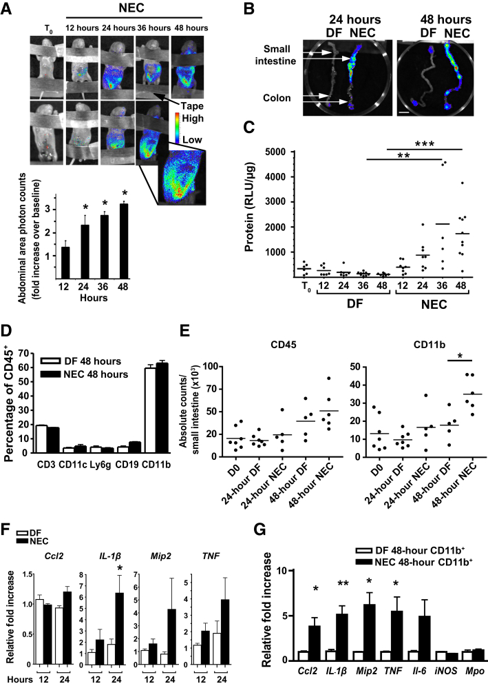
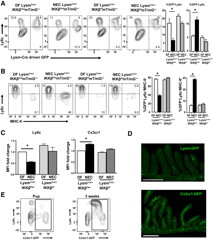
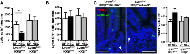
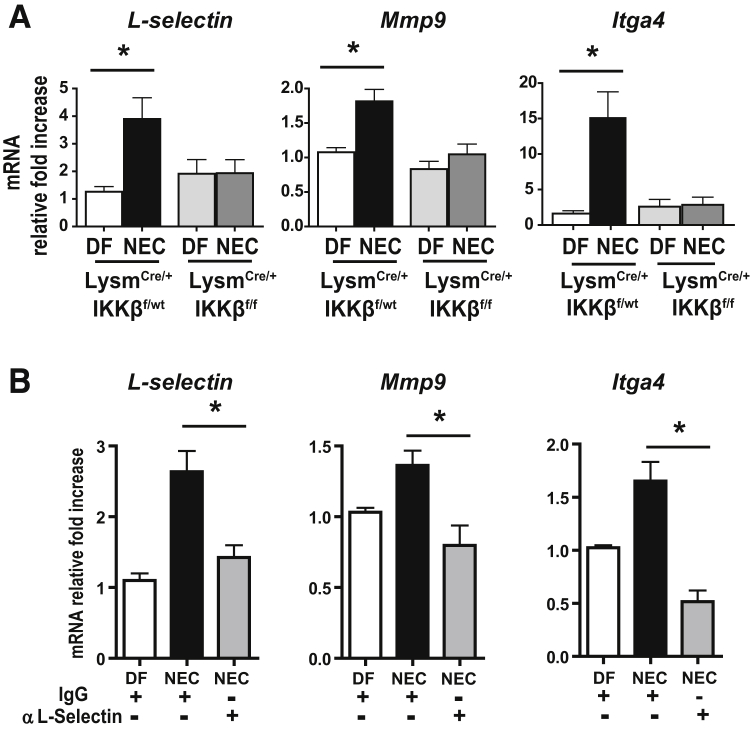
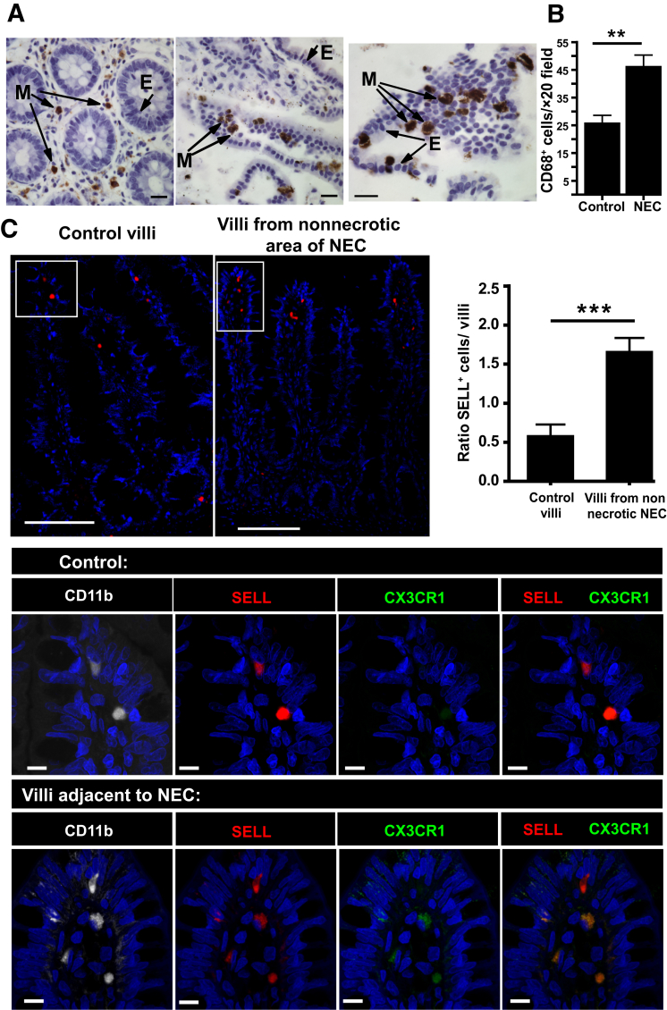
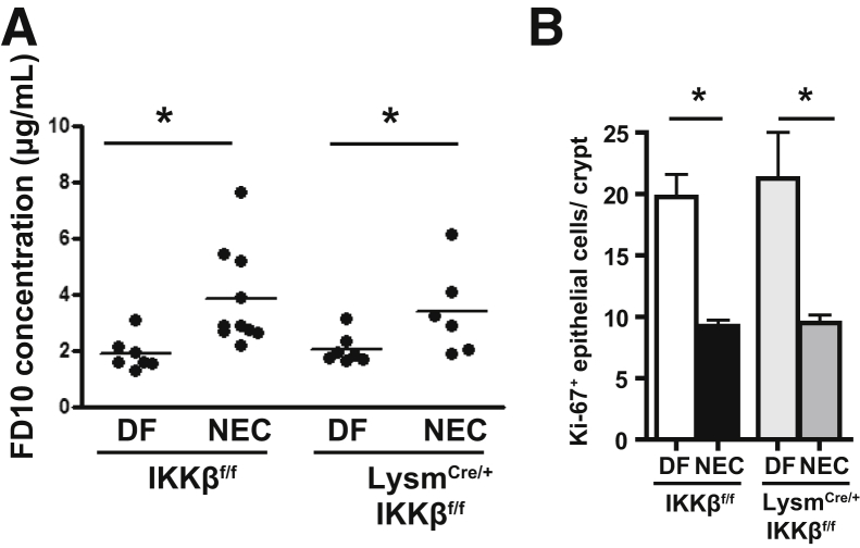
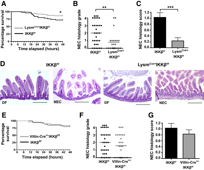
Necrotizing enterocolitis (NEC) is a devastating disease affecting premature infants with intestinal inflammation and necrosis. The neonatal intestinal inflammatory response is rich in macrophages, and blood monocyte count is low in human NEC. We previously found that NF-κB mediates the intestinal injury in experimental NEC. However, the role of NF-κB in myeloid cells during NEC remains unclear. Herein, inhibitor of kappaB kinase β (IKKβ), a critical kinase mediating NF-κB activation, was deleted in lysozyme M (Lysm)-expressing cells, which were found to be Cd11b+Ly6c+ monocytes but not Cd11b+Ly6c- macrophages in the dam-fed neonatal mouse intestine. NEC induced differentiation of monocytes into intestinal macrophages and up-regulation of monocyte recruitment genes (eg, L-selectin) in the macrophage compartment in wild-type mice, but not in pups with IKKβ deletion in Lysm+ cells. Thus, NF-κB is required for NEC-induced monocyte activation, recruitment, and differentiation in neonatal intestines. Furthermore, pups with Lysm-IKKβ deletion had improved survival and decreased incidence of severe NEC compared with littermate controls. Decreased NEC severity was not associated with an improved intestinal barrier. In contrast, NEC was unabated in mice with IKKβ deletion in intestinal epithelial cells. Together, these data suggest that recruitment of Ly6c+ monocytes into the intestine, NF-κB activation in these cells, and differentiation of Ly6c+ monocytes into macrophages are critical cellular and molecular events in NEC development to promote disease.


Figures

Figure 1
Figure 1
NF-κB is activated in the intestine of mice within 12 hours of exposure to a necrotizing enterocolitis (NEC) protocol, and myeloid cells express NF-κB–target genes on NEC induction. A: Less than 12-hour–old heterozygous NF-κB–reporter mice were subjected to NEC protocol and imaged every 12 hours with an IVIS camera. Luciferin (0.1 mg, intraperitoneally) was administered 10 minutes before each image acquisition. Top panel: Representative bioluminescence images from two mice obtained at time 0 (T0) and then every 12 hours during exposure to the NEC protocol are shown. Bottom panel: Abdominal area bioluminescence images were quantified, and results are presented as count-fold increase over baseline. Three to five pups were used per time point. B: Bioluminescence of NEC or dam-fed (DF) control intestines obtained at 24 and 48 hours. DF and NEC pups were euthanized simultaneously, and intestines were immediately dissected by two different investigators and placed on a petri dish to be immediately imaged. White arrows indicate the small intestine and the colon. C: Heterozygous NF-κB–reporter pups were placed on NEC protocol or allowed to be DF for different time periods, and luciferase activity of homogenized small-intestinal tissues was measured by in vitro assay. Luciferase activity normalized to protein concentration [relative light units (RLU)/μg] is presented. D: Percentage of specific leukocyte subsets from intestinal lamina propria of 48-hour DF control (open bars) and NEC pups (closed bars) determined by flow cytometry. Cells were gated on live, single, CD45+ cells. Cd11c+ refers to Cd11c+MHC-II+. Cd11b+ refers to Cd11b+Ly6g. E: Absolute counts determined by flow cytometry. Gates were made on live, single cells and evaluated for CD45 expression or further gated on CD45+Ly6gSSClo Cd11b+ cells. F: Inflammatory gene expression of isolated Cd11b+ cells from 12- to 24-hour NEC (closed bars) or control (open bars) pups, as measured by real-time PCR. G: Inflammatory gene expression of isolated Cd11b+ cells from either 48-hour NEC (closed bars) or control (open bars) pups, as measured by real-time PCR. Data are expressed as means ± SEM. n = 3 per group (D); n = 6 per group (F); n = 5 per group (G). P ≤ 0.05, ∗∗P ≤ 0.01, and ∗∗∗P ≤ 0.001. Scale bar = 1 cm (B). D0, day 0.
Figure 2
Figure 2
Lysozyme M (Lysm) expression is predominantly limited to monocytes in dam-fed (DF) pups but is detected in both monocytes and newly differentiated macrophages during necrotizing enterocolitis (NEC) in an inhibitor of kappaB kinase β (IKKβ)–dependent manner. A: Lamina propria (LP) cells were gated on live, single, CD45+Cd11b+Ly6g SSClo to select the monocyte/macrophage population (total Cd11b+ cells). To identify intestinal LP Cd11b+ cells undergoing Lysm-dependent Cre-recombination, LP cells from NEC (24 hours) or DF control IKKβ sufficient (LysmCre/+-IKKβf/wt-mT/mG+/−) and IKKβ deleted (LysmCre/+-IKKβf/f-mT/mG+/−) mice were analyzed for Ly6c and for Lysm-Cre–dependent green fluorescent protein (GFP) expression on total Cd11b+Ly6g cells by flow cytometry. Bar graphs show the percentage of Cd11b+Ly6g cells that were GFP+Ly6c+ (left panel) and those that were GFP+Ly6c (right panel). B: Cells were further gated on the Lysm-GFP+ population and analyzed for Ly6c and major histocompatibility complex (MHC)-II. Bar graphs show the percentage of GFP+ cells that were Ly6c+MHC-II (left panel) and those that were Ly6cMHC-II+ (right panel). C: The mean fluorescence intensity (MFI) of Ly6c and Cx3cr1 in Lysm-GFP+ cells was assessed. Bar graph represents the fold-increase in Ly6c or Cx3cr1 MFI over DF LysmCre/+-IKKβf/wt pup value. All experiments were repeated at least twice, and cells were fixed before data acquisition. D: Representative images of immunofluorescence staining for Lysm-GFP (top panel) from a DF 24-hour LysmCre/+IKKβf/wtmT/mG+/− (IKKβ-sufficient) pup or Cx3cr1-GFP (bottom panel) from a DF 24-hour Cx3cr1GFP/+ pup on formalin-fixed, paraffin-embedded tissue sections (16 μm thick). Z-stack confocal images are displayed as maximum projection. E: Representative flow cytometry plots from 24-hour (left panel) or 3-week–old (right panel) Cx3cr1GFP/+ mice showing lack of Cx3cr1 expression on Ly6c+ cells. Data are expressed as means ± SEM. n = 3 per group (C). P ≤ 0.05. Scale bar = 200 μm (D). Original magnification, ×10 (D).
Figure 3
Figure 3
Necrotizing enterocolitis (NEC) induces a decrease in intestinal Ly6c+ cells in inhibitor of kappaB kinase β (IKKβ)–sufficient mice, but not in lysozyme M (Lysm)–IKKβ–deleted pups. A and B: Absolute counts of Ly6c+ (A) and Lysm–green fluorescent protein–positive (GFP+) (B) expressing cells per small intestine were determined from at least three mice per group. C: Representative LysmCre/+IKKβf/wtmT/mG+/− and LysmCre/+IKKβf/fmT/mG+/− formalin-fixed, paraffin-embedded tissue sections from NEC pups stained for GFP (green), terminal deoxynucleotidyl transferase-mediated dUTP nick-end labeling (TUNEL; red), and DAPI (blue); white arrowheads indicate TUNEL+ cells. Bar graph indicates TUNEL+ cells/high-power field (HPF) on four mice per group and four HPFs per slide. Data are expressed as means ± SEM. P ≤ 0.05. Scale bar = 50 μm (C). DF, dam fed.
Figure 4
Figure 4
Monocyte-specific genes involved in recruitment and tissue infiltration are up-regulated in Cx3cr1+ lamina propria (LP) macrophages during necrotizing enterocolitis (NEC) in an inhibitor of kappaB kinase β (IKKβ)–dependent manner. A:L-selectin, Mmp9, and Itga4 mRNA expression from Cx3cr1+ cells isolated from LP cells of 24-hour dam-fed (DF) or NEC littermates with or without lysozyme M (Lysm)–dependent IKKβ deletion. B: Wild-type mice were injected with a neutralizing antibody against L-selectin 2 hours before NEC induction, and L-selectin, Mmp9, and Itga4 mRNA expression levels from LP Cx3cr1+ cells were analyzed. Bar graphs representing mRNA relative fold-increases compared with DF are presented. Data are expressed as means ± SEM. n ≥ 7 per group (A); n = 5 per group (B). P ≤ 0.05.
Figure 5
Figure 5
Selectin-L (SELL)+ cell numbers are increased in the villous lamina propria (LP) of human necrotizing enterocolitis (NEC). A: Tissue specimens collected from the small intestine of patients undergoing resection for ileal atresia (left panel) or NEC (middle and right panels) were fixed in formalin and stained for the monocyte/macrophage marker CD68 by immunohistochemistry [myeloid cell (M) is brown; epithelial cell (E)]. B: Bar graph showing CD68+ cells/×20 field of control slides or from patients with NEC. C: Representative immunofluorescence images of intestinal tissues stained for SELL (top left panel) and SELL, CD11b, and CX3CR1 [bottom panels (magnified areas from the white boxed areas of the top panels)]. Top right panel: Bar graph shows the ratio of SELL cells per LP villi in NEC patients in areas adjacent to necrotic areas versus villi of non-NEC patient controls. Data are expressed as means ± SEM. n = 4 (B, patients per group with at least five ×20 fields per slide, and C, SELL+ cells per LP villi of non-NEC patient controls); n = 7 (C, SELL+ cells per LP villi in NEC patients in areas adjacent to necrotic areas). ∗∗P ≤ 0.01, ∗∗∗P ≤ 0.001. Scale bars: 20 μm (A); 100 μm (C, top panels); 10 μm (C, bottom panels). Original magnifications: ×10 (C, top panels); ×100 (C, bottom panels).
Figure 6
Figure 6
Inhibitor of kappaB kinase β (IKKβ) deletion in lysozyme M–positive (Lysm+) monocytes does not abrogate the increase in intestinal permeability associated with necrotizing enterocolitis (NEC). A: Intestinal permeability expressed as fluorescein isothiocyanate (FITC)–dextran plasma concentration 4 hours after administration of FITC-dextran by gavage in LysmCre/+-IKKβf/f and IKKβf/f mice exposed to the NEC protocol for 24 hours and dam-fed (DF) controls. B: Quantification of epithelial cell proliferation on formalin-fixed, paraffin-emebedded tissue sections stained for Ki-67 from 24-hour DF or NEC pups with or without Lysm-Cre expression. Counts represent the number of Ki-67+ epithelial cells per crypt in ≥20 well-oriented crypts from at least three mice per group. Data are expressed as means ± SEM. P ≤ 0.05.
Figure 7
Figure 7
Mice with inhibitor of kappaB kinase β (IKKβ)–deleted monocytes have increased survival and decreased intestinal disease severity when exposed to a necrotizing enterocolitis (NEC) protocol. A: Survival curve demonstrating decreased mortality in lysozyme M (Lysm)Cre/+-IKKβf/f mice when exposed to the NEC model. B and C: NEC histologic grades (χ2 = 8.89; B) and NEC histologic scores (C) of LysmCre/+-IKKf/f mice and controls (IKKβf/f). D: Representative hematoxylin and eosin staining of dam-fed (DF) and NEC LysmCre/+-IKKf/f mice and littermate controls (IKKβf/f) 72 hours into NEC induction. E: Survival curves demonstrating no change in mortality in Villin-Cre+/−-IKKβf/f mice when exposed to the NEC model. F and G: NEC histologic grades (F) and NEC histologic scores (G) of Villin-Cre+/−-IKKf/f and controls. Data are expressed as means ± SEM. n = 73 (A, LysmCre/+-IKKβf/f); n = 50 (A, IKKβf/f); n = 24 (E, Villin-Cre+/−-IKKβf/f); n = 34 (E, IKKβf/f). P ≤ 0.05, ∗∗P ≤ 0.01, and ∗∗∗P ≤ 0.001. Scale bar = 200 μm (D).

Similar articles

Cited by

References

    1. Sharma R., Tepas J.J., 3rd, Hudak M.L., Mollitt D.L., Wludyka P.S., Teng R.J., Premachandra B.R. Neonatal gut barrier and multiple organ failure: role of endotoxin and proinflammatory cytokines in sepsis and necrotizing enterocolitis. J Pediatr Surg. 2007;42:454–461. - PubMed
    1. Bergmann K.R., Liu S.X., Tian R., Kushnir A., Turner J.R., Li H.L., Chou P.M., Weber C.R., De Plaen I.G. Bifidobacteria stabilize claudins at tight junctions and prevent intestinal barrier dysfunction in mouse necrotizing enterocolitis. Am J Pathol. 2013;182:1595–1606. - PMC - PubMed
    1. MohanKumar K., Kaza N., Jagadeeswaran R., Garzon S.A., Bansal A., Kurundkar A.R., Namachivayam K., Remon J.I., Bandepalli C.R., Feng X., Weitkamp J.H., Maheshwari A. Gut mucosal injury in neonates is marked by macrophage infiltration in contrast to pleomorphic infiltrates in adult: evidence from an animal model. Am J Physiol Gastrointest Liver Physiol. 2012;303:G93–G102. - PMC - PubMed
    1. He Y.M., Li X., Perego M., Nefedova Y., Kossenkov A.V., Jensen E.A., Kagan V., Liu Y.F., Fu S.Y., Ye Q.J., Zhou Y.H., Wei L., Gabrilovich D.I., Zhou J. Transitory presence of myeloid-derived suppressor cells in neonates is critical for control of inflammation. Nat Med. 2018;24:224–231. - PMC - PubMed
    1. Pang Y., Du X., Xu X., Wang M., Li Z. Monocyte activation and inflammation can exacerbate Treg/Th17 imbalance in infants with neonatal necrotizing enterocolitis. Int Immunopharmacol. 2018;59:354–360. - PubMed

Publication types

MeSH terms