

doi: 10.1016/j.ymeth.2018.12.008.
Epub 2018 Dec 26.
Selective profiling of ribosomes associated with yeast Upf proteins
Affiliations
- PMID: 30593864
- PMCID: PMC6387845
- DOI: 10.1016/j.ymeth.2018.12.008
Item in Clipboard
Selective profiling of ribosomes associated with yeast Upf proteins
Methods.
.
Abstract
Ribosomes associated with nonsense-mediated decay factors Upf1, Upf2, or Upf3 were purified by immunoprecipitation, and enrichment and stoichiometry of Upf factors and ribosomal proteins were analyzed by western blot and mass spectrometry. Using a small RNA library preparation protocol that eliminates in-gel RNA and cDNA size selection and incorporates four random nucleotides on each side of the ribosome-protected RNA fragment allowed recovery, detection, and analysis of all size classes of protected fragments from a sample simultaneously.
Keywords: NMD; Selective ribosome profiling; Upf proteins.
Copyright © 2018 Elsevier Inc. All rights reserved.
Figures
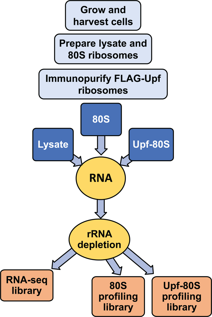
Diagram of protocol workflow.
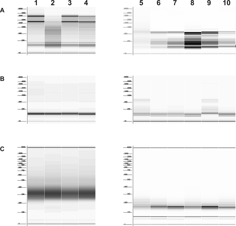
Starting material lanes 1–4, yeast lysate, untreated with RNase I; lanes 5–6, immunopurified ribosomes; lanes 7–10, total ribosomes. Numbers to the left of each figure indicate marker sizes in nt. A. RNA before removal of ribosomal RNA. Samples in lanes 2 and 5 are degraded and should be re-prepared; if the problem persists, it is necessary to make another preparation of lysate and ribosomes. Samples in lanes 5–10 have fragmented ribosomal RNA due to incubation of the lysate from which they were prepared with RNase I; this is expected. B. RNA after removal of ribosomal RNA. Samples in lanes 5 and 9 have incomplete removal of ribosomal RNA; these RNAs should be subjected to another round of ribosomal RNA removal. C. Finished libraries after PCR and cleanup. Sample in lane 5 is a library with a broad size range. This library should be prepared again if sequencing results indicate poor quality.
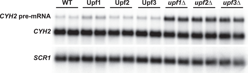
Northern blot of total RNA from yeast strains expressing: empty vector (WT); high-copy FLAG-tagged Upf1, Upf2, or Upf3 in the respective deletion strains; or bearing a deletion of the UPF1, UPF2 or UPF3 coding region (designated upf1Δ, upf2Δ, or upf3Δ). NMD phenotype was determined by hybridization with a random-primed labeled probe for CYH2. SCR1 was used as a loading control.
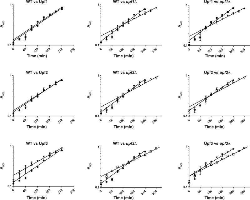
Left panel, between WT and FLAG-tagged Upf strains; center panel, between WT and deletion strains; right panel, between FLAG-tagged Upf strains and corresponding deletion strains. WT, ●; FLAG-Upf1, ■; FLAG-Upf2, ▲; FLAG-Upf3, ▼; upf1Δ, ♦; upf2Δ, ○; upf3Δ, □. Timepoints as mean A600 from cultures of two independent isolates. Error bars=range of values.
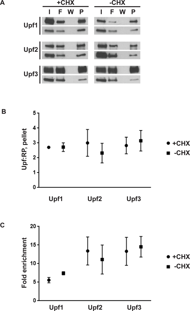
A. Western blot of input (I), flowthrough (F), final wash (W), or pelleted ribosomes (P) from anti-FLAG immunoprecipitation reactions. Cultures were cycloheximide treated (+CHX) or untreated (-CHX) prior to cell collection. Western blots were probed with anti-FLAG (top panel per set) or anti-TCM1/RPL3 antibodies (bottom panel per set). B. Relative abundance of Upf protein per average relative abundance of core ribosomal proteins present in a sample (Upf:RP) in pellet as determined by mass spectrometry; mean and range of two biological replicates per strain and condition. C. Fold enrichment of Upf protein in pellet vs input; mean and range of two biological replicates per strain and condition. FLAG-tagged proteins are indicated, all panels.
Similar articles
-
Ribosome-bound Upf1 forms distinct 80S complexes and conducts mRNA surveillance.RNA. 2022 Dec;28(12):1621-1642. doi: 10.1261/rna.079416.122. Epub 2022 Oct 3. RNA. 2022. PMID: 36192133 Free PMC article.
-
Nonsense-mediated mRNA decay involves two distinct Upf1-bound complexes.EMBO J. 2018 Nov 2;37(21):e99278. doi: 10.15252/embj.201899278. Epub 2018 Oct 1. EMBO J. 2018. PMID: 30275269 Free PMC article.
-
NMD monitors translational fidelity 24/7.Curr Genet. 2017 Dec;63(6):1007-1010. doi: 10.1007/s00294-017-0709-4. Epub 2017 May 23. Curr Genet. 2017. PMID: 28536849 Free PMC article. Review.
-
Yeast Upf1 CH domain interacts with Rps26 of the 40S ribosomal subunit.RNA. 2013 Aug;19(8):1105-15. doi: 10.1261/rna.039396.113. Epub 2013 Jun 25. RNA. 2013. PMID: 23801788 Free PMC article.
-
Yeast and human RNA helicases involved in ribosome biogenesis: current status and perspectives.Biochim Biophys Acta. 2013 Aug;1829(8):775-90. doi: 10.1016/j.bbagrm.2013.01.007. Epub 2013 Jan 26. Biochim Biophys Acta. 2013. PMID: 23357782 Review.
Cited by
-
Features and factors that dictate if terminating ribosomes cause or counteract nonsense-mediated mRNA decay.J Biol Chem. 2022 Nov;298(11):102592. doi: 10.1016/j.jbc.2022.102592. Epub 2022 Oct 13. J Biol Chem. 2022. PMID: 36244451 Free PMC article. Review.
-
Transcriptome-wide investigation of stop codon readthrough in Saccharomyces cerevisiae.PLoS Genet. 2021 Apr 20;17(4):e1009538. doi: 10.1371/journal.pgen.1009538. eCollection 2021 Apr. PLoS Genet. 2021. PMID: 33878104 Free PMC article.
-
Identification of the hyaluronic acid pathway as a therapeutic target for facioscapulohumeral muscular dystrophy.Sci Adv. 2019 Dec 11;5(12):eaaw7099. doi: 10.1126/sciadv.aaw7099. eCollection 2019 Dec. Sci Adv. 2019. PMID: 31844661 Free PMC article.
-
Ribosome-bound Upf1 forms distinct 80S complexes and conducts mRNA surveillance.RNA. 2022 Dec;28(12):1621-1642. doi: 10.1261/rna.079416.122. Epub 2022 Oct 3. RNA. 2022. PMID: 36192133 Free PMC article.
References
-
- Nicholson P, Joncourt R, Muhlemann O, Analysis of nonsense-mediated mRNA decay in mammalian cells, Current protocols in cell biology editorial board, Juan S. Bonifacino [et al.] Chapter 27 (2012) Unit27 4. - PubMed
Publication types
MeSH terms
Substances
Grants and funding
LinkOut - more resources
Full Text Sources
Other Literature Sources
Molecular Biology Databases
Research Materials
