. 2019 Mar 1:14:131-141.
doi: 10.1016/j.omtn.2018.11.012. Epub 2018 Dec 1.

An RNA Aptamer Targeting the Receptor Tyrosine Kinase PDGFRα Induces Anti-tumor Effects through STAT3 and p53 in Glioblastoma

Affiliations

An RNA Aptamer Targeting the Receptor Tyrosine Kinase PDGFRα Induces Anti-tumor Effects through STAT3 and p53 in Glioblastoma

Sorah Yoon et al. Mol Ther Nucleic Acids. .

Abstract

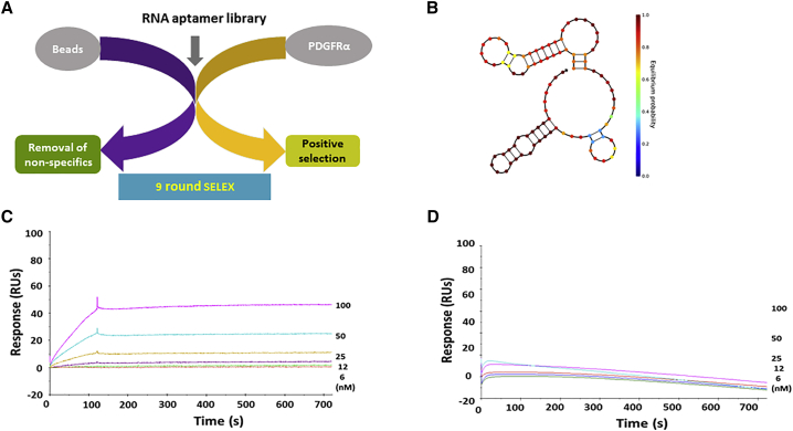
Human glioblastoma (GBM) is the most aggressive malignancy of the CNS, with less than 5% survival. Despite great efforts to find effective therapeutics, current options remain very limited. To develop a targeted cancer therapeutic, we selected RNA aptamers against platelet-derived growth factor receptor α (PDGFRα), which is a receptor tyrosine kinase. One RNA aptamer (PDR3) with high affinity (0.25 nM) showed PDGFRα specificity and was internalized in U251-MG cells. Following treatment with the PDR3 aptamer, expression of the transcription factor STAT3 (signal transducer and activator of transcription 3) was inhibited, whereas the expression of the histone demethylase JMJD3 and the tumor suppressor p53 were upregulated. PDR3 also upregulated serine phosphorylation of p53, which subsequently mediated apoptosis through the death receptors: tumor necrosis factor (TNF)-related apoptosis-inducing ligand receptors 1/2 (TRAIL-R1/R2), Fas-associated via death domain (FADD), and Fas. PDR3 significantly decreased cell viability in a dose-dependent manner. Furthermore, translocation of PDR3 into the nucleus induced hypomethylation at the promoters of cyclin D2. To assess the feasibility of targeted delivery, we conjugated PDR3 aptamer with STAT3-siRNA for a chimera. The PDR3-siSTAT3 chimera successfully inhibited the expression of target genes and showed significant inhibition of cell viability. In summary, our results show that well-tailored RNA aptamers targeting the PDGFRα-STAT3 axis have the potential to act as anti-cancer therapeutics in GBM.

Keywords: GBM; PDGFRα aptamer; STAT3; apoptosis; p53.


Figures

Figure 1
Figure 1
Secondary Structure and Biosensor Assays (A) A protein-based SELEX method was used to isolate anti-PDGFRα RNA aptamers from a randomized 40-nt RNA library. After nine rounds of SELEX, the amplified aptamers were cloned, and individual clones were identified by DNA sequencing. (B) The secondary structure of the anti-PDGFRα aptamer, PDR3, was predicted using NUPACK software. (C) Surface plasmon resonance (SPR) sensorgrams of binding between PDR3 and recombinant PDGFRα. To assess binding, we used a Biacore T100 to monitor label-free interactions and to measure the increase in response units (RUs) from baseline. BIAevaluation software was used to calculate binding affinity (KD). (D) The binding of PDR3 to recombinant PDGFRβ was measured using SPR. Binding affinity was not measurable.
Figure 2
Figure 2
Internalization Assays of PDR3 in Live Cells and Relative Target Gene Expression Analysis (A) Cy3-labeled PDR3 aptamer and aptamer library as negative control (NC) were assessed for binding efficiency by flow cytometry in U251-MG cells. The data show measurements of positively stained cells and are representative of duplicates. (B) Cy3-labeled PDR3 aptamer was assessed for intracellular uptake in live cells. Human GBM U251-MG cells were treated with 200 nM Cy3-labeled PDR3 aptamer or aptamer library for 2 hr, and internalization was visualized using confocal microscopy. Cells show punctate regions of Cy3 intracellular labeling. Red: Cy3-labeled RNAs; blue: Hoechst 33342 for nuclear staining. Scale bars: 5 μm. NC, Cy3-labeled initial aptamer library; PDR3, Cy3-labeled anti-PDGFRα aptamer. (C) Quantification of internalization. Images were quantified to yield mean intensity per cell of the red fluorophore (Cy3) using Zen Blue (v2.3 Carl Zeiss Microimaging). Intensity level (12-bit) is presented on the y axis. Data are presented as the mean ± SD. NC, Cy3 labeled initial aptamer library; PDR3, Cy3 labeled anti-PDGFRα aptamers. (D) Relative expression of STAT3 mRNA transcripts was quantified using real-time PCR (qPCR) in U251-MG cells treated with 200 nM PDR3 or irrelevant aptamer (IRRE) over time. Data are presented as the mean ± SD. (E) Protein expression of STAT3 after treatment was measured by western blot. Cells were treated with PDR3 (200 nM) at 24-hr intervals, and total protein was extracted at 72 hr. For loading control, GAPDH was used. Each group is shown in duplicate. (F) Pixel intensity was quantified using Image-pro premier. Data are presented as the mean ± SD. (G) Relative expression of JMJD3 mRNA transcripts was quantified using qPCR in U251-MG cells treated with 200 nM PDR3 or IRRE for 48 hr. Data are presented as the mean ± SD. (H) Relative expression of p53 mRNA transcripts was quantified using qPCR on U251-MG cells treated with 200 nM PDR3 or IRRE for 48 hr. Data are presented as the mean ± SD. One-way ANOVA was used to assess statistical significance; *p ≤ 0.05; **p ≤ 0.01. CC, untreated control cells; PDR3, anti-PDGFRα aptamer.
Figure 3
Figure 3
Apoptosis Assays after PDR3 Treatment (A) U251-MG cells were treated with 500 nM PDR3 aptamer for 24 hr. The cells were stained with Annexin V-Alexa Fluor 488, and fluorescent images were taken using confocal microscopy. Green: Annexin V-Alexa Fluor 488; blue: Hoechst 33342. Scale bars: 10 μm. (B) Cells were stained with Annexin V and analyzed using flow cytometry after treatment with PDR3 for 24 hr. Propidium iodide (PI) was used to discriminate dead cells. (C) Apoptosis-related molecules were determined using protein array. Total proteins (200 μg) after treatment with PDR3 aptamer for 72 hr were used for apoptosis assays. Upregulated serine phosphorylation of p53 and death receptors TRAIL-R1, TRAIL-R2, FADD, and Fas were observed. (D and E) Pixel intensity of phospho-p53 (D) and death receptors (E) were quantified using ImageJ. Data are presented as the mean ± SD. (F) The EC50 of PDR3 was calculated, using PDGFRα in vitro kinase assay, to be 55 ng. (G) Cell viability was measured in U251-MG cells treated twice with various concentrations of PDR3 or IRRE at 24-hr intervals and harvested at a final incubation time of 48 hr. Cell viability was measured using MTS assay. Data were normalized to untreated control cells. Data are presented as the mean ± SD. (H) Inhibition of cell proliferation after PDGFRα activation with PDGF-AA ligands was measured using MTS assay. After pretreatment with PDR3 aptamer, PDGF-AA ligands were incubated with cells. Cell viability was measured using MTS assay. Cell proliferation was normalized to untreated control cells. Data are presented as the mean ± SD. One-way ANOVA was used to assess statistical significance; *p ≤ 0.05; **p ≤ 0.01. CC, untreated control cells; PDR3, anti-PDGFRα aptamer.
Figure 4
Figure 4
DNA Methylation by PDR3 Aptamer (A) Nuclear translocation of PDR3 was observed in live U215-MG cells using confocal microscopy. The nuclear region is indicated with a line. Nuclear translocation of PDR3 is indicated with an arrow. Red: Cy3-labeled RNAs; blue: Hoechst 33342 for nuclear staining. Scale bars: 10 μm. (B) The diagram summarizes the relative percentages of hypomethylated regions. Changes in DNA methylation were measured using the bisulfite method. (C) Hierarchical heatmap of hypomethylation changes induced by PDR3 treatment. Heatmap shows promoter methylation levels. Each group is shown in duplicate. (D) Chromosomal views of methylation differences in the CCND2 gene between control and PDR3 treatment. (E) Schematic working model of biological pathways affected by PDR3. Inhibition of STAT3 by binding of PDR3 to PDGFRα upregulates the expression of JMJD3 and its downstream effector p53. Activated p53 induces apoptosis-related genes such as TRAIL R1/R2, FADD, and Fas to promote cell death. Along with apoptosis, translocation of PDR3 into the nucleus induces methylation changes. NC, Cy3-labeled initial aptamer library; PDR3, anti-PDGFRα aptamer; CC, untreated cell control.
Figure 5
Figure 5
The Inhibition of STAT3 Expression by PDR3-siSTAT3 chimeras (A) Schematic of cell surface receptor-mediated siRNA delivery. An RNA aptamer targeting PDGFRα, which is overexpressed on GBM, allows specific delivery of therapeutic siSTAT3 into target cells. (B) Intracellular uptake of chimeras. U251-MG cells were treated with 200 nM Cy3-labeled chimeric aptamer-siRNAs and analyzed using confocal microscopy, which shows punctate regions of Cy3 labeling in the cytoplasm. Red: Cy3-labeled RNA; blue: Hoechst 33342. Scale bars: 10 μm. (C) Relative expression of STAT3 mRNA transcripts was quantified using qPCR over time in U251-MG cells treated with 200 nM PDR3 chimeras. Data are presented as the mean ± SD. (D) U251-MG cells were treated with various concentrations of chimeras twice at 24-hr intervals and harvested at a final incubation time of 48 hr. Cell viability was measured using MTS assay, normalized to untreated control cells. One-way ANOVA was used to assess statistical significance; *p ≤ 0.05; **p ≤ 0.01. IRRE-STAT3, irrelevant aptamer with siSTAT3 sequence; PDR3-scr, PDR3 aptamer conjugated to scrambled siRNA control; PDR3-STAT3, PDR3 aptamer conjugated to siSTAT3 sequence.

Similar articles

Cited by

References

    1. Tuerk C. Using the SELEX combinatorial chemistry process to find high affinity nucleic acid ligands to target molecules. Methods Mol. Biol. 1997;67:219–230. - PubMed
    1. Yoon S., Armstrong B., Habib N., Rossi J.J. Blind SELEX approach identifies RNA aptamers that regulate EMT and inhibit metastasis. Mol. Cancer Res. 2017;15:811–820. - PMC - PubMed
    1. Jia W., Ren C., Wang L., Zhu B., Jia W., Gao M., Zeng F., Zeng L., Xia X., Zhang X. CD109 is identified as a potential nasopharyngeal carcinoma biomarker using aptamer selected by cell-SELEX. Oncotarget. 2016;7:55328–55342. - PMC - PubMed
    1. Kruspe S., Giangrande P.H. Aptamer-siRNA chimeras: discovery, progress, and future prospects. Biomedicines. 2017;5:E45. - PMC - PubMed
    1. Yoon S., Huang K.W., Reebye V., Mintz P., Tien Y.W., Lai H.S., Sætrom P., Reccia I., Swiderski P., Armstrong B. Targeted delivery of C/EBPalpha -saRNA by pancreatic ductal adenocarcinoma-specific RNA aptamers inhibits tumor growth in vivo. Mol. Ther. 2016;24:1106–1116. - PMC - PubMed