. 2019 Mar 1:14:171-183.
doi: 10.1016/j.omtn.2018.11.010. Epub 2018 Nov 24.

miR675 Accelerates Malignant Transformation of Mesenchymal Stem Cells by Blocking DNA Mismatch Repair

Affiliations

miR675 Accelerates Malignant Transformation of Mesenchymal Stem Cells by Blocking DNA Mismatch Repair

Yanan Lu et al. Mol Ther Nucleic Acids. .

Abstract

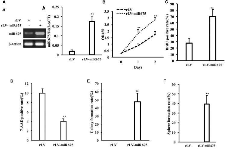
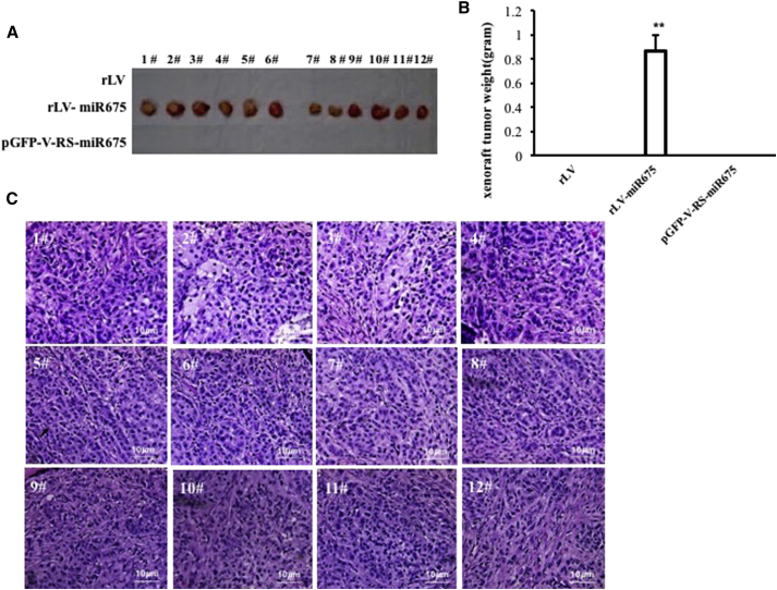
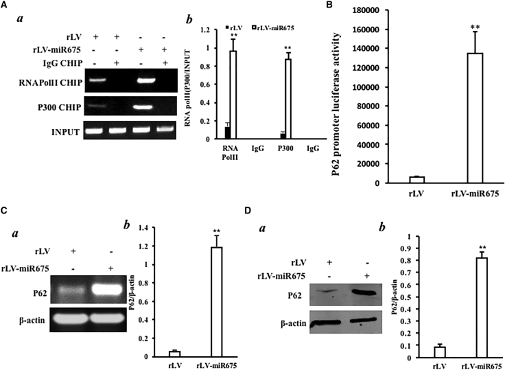
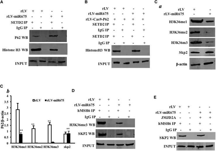
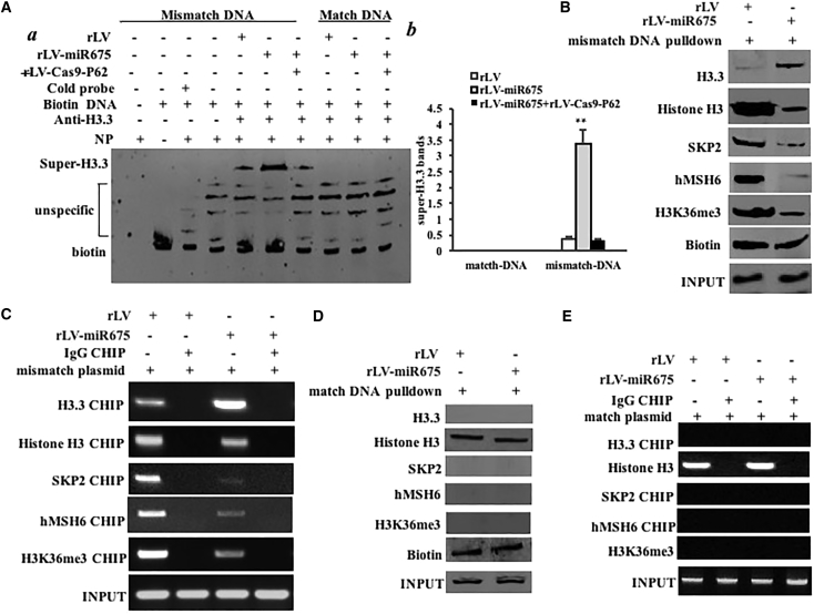
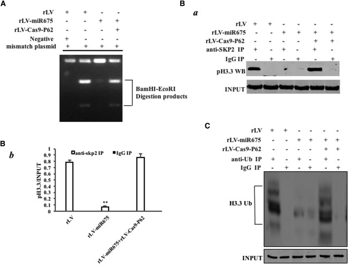
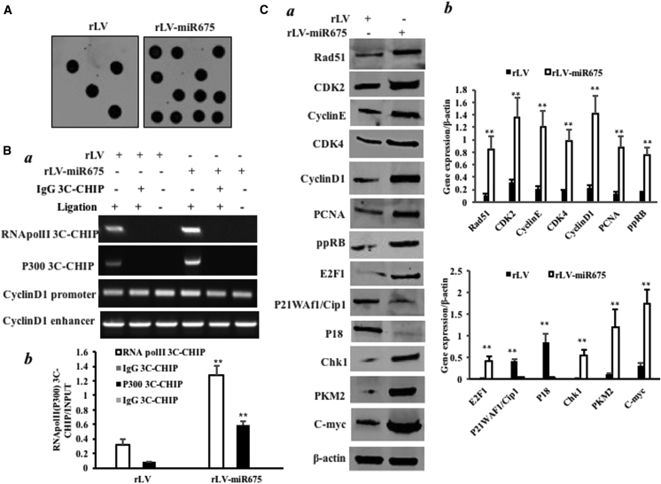
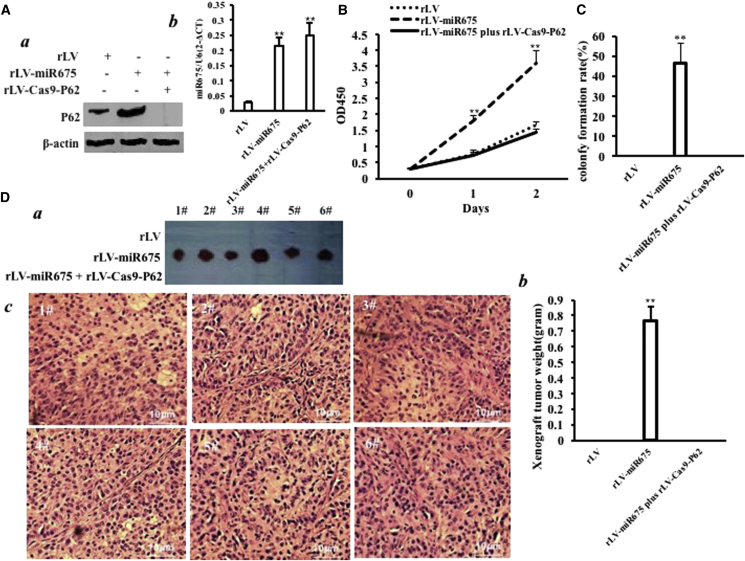
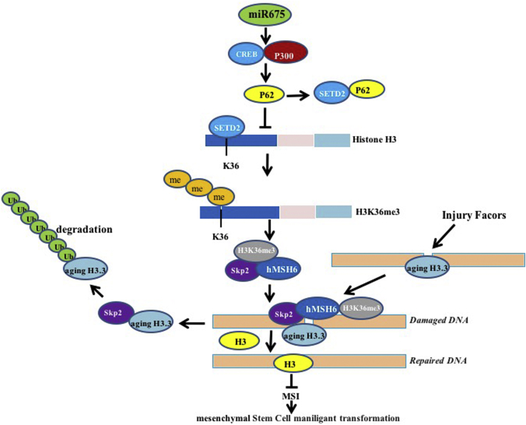
miR675 is highly expressed in several human tumor tissues and positively regulates cell progression. Herein, we demonstrate that miR675 promotes malignant transformation of human mesenchymal stem cells. Mechanistically, we reveal that miR675 enhances the expression of the polyubiquitin-binding protein p62. Intriguingly, P62 competes with SETD2 to bind histone H3 and then significantly reduces SETD2-binding capacity to substrate histone H3, triggering drastically the reduction of three methylation on histone H3 36th lysine (H3K36me3). Thereby, the H3K36me3-hMSH6-SKP2 triplex complex is significantly decreased. Notably, the ternary complex's occupancy capacity on chromosome is absolutely reduced, preventing it from DNA damage repair. By virtue of the reductive degradation ability of SKP2 for aging histone H3.3 bound to mismatch DNA, the aging histone H3.3 repair is delayed. Therefore, the mismatch DNA escapes from repair, triggering the abnormal expression of several cell cycle-related genes and causing the malignant transformation of mesenchymal stem cells. These observations strongly suggest understanding the novel functions of miR675 will help in the development of novel therapeutic approaches in a broad range of cancer types.

Keywords: P62; human mesenchymal stem cell; malignant transformation; miR675.


Figures

Figure 1
Figure 1
miR675 Accelerates Malignant Growth of Human Mesenchymal Stem Cells In Vitro (A) (a) The RT-PCR analysis of pre-miR675 in HBMMSCs infected with rLV and rLV-miR675, respectively. β-actin was the internal control. (b) The real-time RT-PCR analysis of mature miR675 in the HBMMSCs infected with rLV and rLV-miR675, respectively. U6 was the internal control. (B) Cell growth assay using CCK8. Each value was presented as mean ± SEM (n = 3). (C) S phase cell assay using BrdU. Each value was presented as mean ± SEM (n = 3). (D) The assay of flow cytometry using 7-AAD. Each value was presented as mean ± SEM (n = 3). (E) Cell soft agar colony formation assay. Each value was presented as mean ± SEM (n = 3). (F) Cell sphere formation ability. Each value was presented as mean ± SEM (n = 3).
Figure 2
Figure 2
Tumorigenesis Test In Vivo (A) The mice were stratified and the tumors were recovered; the photography shows xenograft tumor in the three groups (indicated at left, n = 12). (B) The wet weight of each tumor was determined for each mouse. Each value was presented as mean ± SEM. (C) A portion of each tumor was fixed in 4% paraformaldehyde and embedded in paraffin for histological H&E staining (original magnification ×100).
Figure 3
Figure 3
miR675 Enhances the Expression of P62 in the Human Mesenchymal Stem Cells Infected with rLV and rLV-miR675, Respectively (A) (a) Chromatin immunoprecipitation (ChIP) with anti-P300 and Pol II followed by PCR with P62 promoter primers. IgG ChIP was the negative control. P62 promoter DNA was the INPUT. (b) The quantitative analysis of ChIP. (B) The activity assay of P62 promoter luciferase reporter gene. (C) (a) P62 expression analysis by RT-PCR P62 in the human mesenchymal stem cells infected with rLV and rLV-miR675, respectively. β-actin was the internal control. (b) The quantitative analysis of RT-PCR. (D) (a) P62 expression analysis by western blotting with anti-P62 in the human mesenchymal stem cells infected with rLV and rLV-miR675, respectively. β-actin was the internal control. (b) The gray scan analysis of positive bands of western blotting.
Figure 4
Figure 4
miR675 Affects the Interaction among hMSH6, H3k36me3, and SKP2 in the Human Mesenchymal Stem Cells Infected with rLV and rLV-miR675, Respectively (A) Anti-STED2 co-immunoprecipitation (coIP) followed by western blotting with anti-P62 and anti-histone 3. IgG IP was the negative control. INPUT refers to western blotting with anti-STED2. (B) Anti-SETD2 coIP followed by western blotting with anti-histone 3. IgG IP was the negative control. INPUT refers to western blotting with anti-SETD2. (C) (a) Western blotting with anti-H3K36me1, anti-H3K36me2, and anti-H3K36me1. β-actin was the internal control. (b) The gray scan analysis of positive bands of western blotting. (D) Anti-hMSH6 coIP followed by western blotting with anti-H3K36me3 and anti-SKP2. IgG IP was the negative control. INPUT refers to western blotting with anti-hMSH6. (E) Anti-hMSH6 coIP followed by western blotting with anti-SKP2 in the human mesenchymal stem cells, including the rLV and rLV-miR675 plus pCMV6-AC-GFP-JMJD2A groups. IgG IP was the negative control. INPUT refers to western blotting with anti-hMSH6.
Figure 5
Figure 5
miR675 Delays the hMSH6-H3k36me3-Skp2 Ternary Complex Occupancy on the Mismatch DNA in the Human Mesenchymal Stem Cells Infected with rLV and rLV-miR675, Respectively (A) (a) Super-EMSA (gel shift) with biotin-DNA probe (including mismatch and match DNA double strands) and anti-histone H3.3 antibody. The intensity of the band was examined by western blotting with anti-biotin. (b) The gray scan analysis of positive bands of super-EMSA. (B) Biotin-mismatch probe pull-down followed by western blotting with anti-histone 3.3, anti-histone 3, anti-SKP2, anti-hMSH6, and anti-H3K36me3. Biotin was the INPUT and histone was the internal control. (C) Chromatin immunoprecipitation (ChIP) with anti-histone 3.3, anti-histone H3, anti-SKP2, anti-hMSH6, and anti-H3K36me3 followed by PCR with damaged DNA primers. IgG ChIP was the negative control and damaged DNA was the INPUT. (D) Biotin-match double DNA probe pull-down followed by western blotting with anti-histone H3.3, anti-histone H3, anti-SKP2, anti-hMSH6, and anti-H3K36me3. Biotin was the INPUT and histone was the internal control. (E) ChIP with anti-histone 3.3, anti-histone H3, anti-SKP2, anti-hMSH6, and anti-H3K36me3 followed by PCR with match double DNA primers. IgG ChIP was the negative control and match double DNA was the INPUT.
Figure 6
Figure 6
miR675 Inhibits DNA Damage Repair and Decreases Aging Histone H3.3 Degradation in the Human Mesenchymal Stem Cells Infected with rLV, rLV-miR675, and rLV-miR675 Plus rLV-Cas9-P62, Respectively (A) Restriction endonuclease analysis with BamHI and EcoRI for plasmid DNA injury repair. (B) (a) Anti-SKP2 coIP followed by western blotting with anti-histone 3.3. IgG IP was the negative control. INPUT refers to western blotting with anti-histone H3.3. (b) The gray scan analysis of positive bands. (C) Anti-Ub coIP followed by western blotting with anti-histone H3.3. IgG IP was the negative control. INPUT refers to western blotting with β-actin.
Figure 7
Figure 7
miR675 Triggers MSI and Abnormal Gene Expression in the Human Mesenchymal Stem Cells Infected with rLV and rLV-miR675, Respectively (A) Microsatellite instability (MSI) analysis through dot blot (slot blot) using various biotin-labeling MSI probes (biotin-MSIs). (B) (a) Chromosome conformation capture (3C)-chromatin immunoprecipitation (ChIP) with anti-P300 and anti-Pol II. The chromatin was cross-linked, digested with restriction enzymes, and ligated under conditions that favor intramolecular ligation. Immediately after ligation, the chromatin was immunoprecipitated using an antibody (anti-P300, anti-Pol II) against the protein of interest. Thereafter, the cross-links were reversed and the DNA was purified further. The PCR anlysis was applied for detecting CyclinD1 promoter-enhancer coupling product using CyclinD1 promoter and enhancer primers. The CyclinD1 promoter and enhancer was the INPUT. (b) The quantitative analysis of ChIP-3C. (C) (a) Western blotting with anti-Rad51, anti-CDK2, anti-CyclinE, anti-CDK4, anti-CyclinD1, anti-PCNA, anti-ppRB, anti-E2F1, anti-P18, anti-P21, anti-PKM2, anti-c-Myc, and anti-Chk1. β-actin was the internal control. (b) The gray scan analysis of positive bands of western blotting.
Figure 8
Figure 8
The Rescue Experiment of the Carcinogenesis Effect of miR675 P62 knockdown abrogated the oncogenic function of miR675 in the human mesenchymal stem cells infected with rLV, rLV-miR675, and rLV-miR675 plus rLV-Cas9-P62, respectively. (A) (a) The western blotting analysis with anti-P62. β-actin was the internal control. (b) The real-time RT-PCR analysis for mature miR675. U6 was the internal control. (B) Cell growth assay using CCK8. Each value was presented as mean ± SEM. (C) Cell soft agar colony formation assay. (D) Tumorigenesis test in vivo. (a) The mice were stratified and the tumors were recovered. (b) The wet weight of each tumor was determined for each mouse. Each value was presented as mean ± SEM. (c) A portion of each tumor was fixed in 4% paraformaldehyde and embedded in paraffin for histological H&E staining.
Figure 9
Figure 9
The Schematic Illustrates a Model of the Differentiation of Human Mesenchymal Stem Cells into Cancer Cells via miR675 Oncogenic miR675 promotes the interaction between CREB and P300, which leads to the high expression of P62. That P62 competes with STED2 to bind histone H3 greatly reduces the STED2-binding capacity with substrate histone H3, triggering a reduction of three methylations on histone H3 36th lysine (H3K36me3); thereby, the H3K36me3-hMSH6-SKP2 tri-complex is decreased. Meanwhile, the ternary complex occupancy capacity on chromosome is absolutely reduced, preventing normal DNA repair. By virtue of the reductive degradation ability of SKP2 for aging histone H3.3 bound to damaged DNA, the aging histone H3.3 repair is delayed and eliminated. That the damaged DNA escaped repair can lead to the abnormal expression of some cell cycle- and metabolism-related genes, causing the human mesenchymal stem cell malignant transformation.

Similar articles

Cited by

References

    1. Lu Y., Liu J., Liu Y., Qin Y., Luo Q., Wang Q., Duan H. TLR4 plays a crucial role in MSC-induced inhibition of NK cell function. Biochem. Biophys. Res. Commun. 2015;464:541–547. - PubMed
    1. Li X., Zhang P., Liu X., Lv P. Expression of interleukin-12 by adipose-derived mesenchymal stem cells for treatment of lung adenocarcinoma. Thorac. Cancer. 2015;6:80–84. - PMC - PubMed
    1. Keniry A., Oxley D., Monnier P., Kyba M., Dandolo L., Smits G., Reik W. The H19 lincRNA is a developmental reservoir of miR-675 that suppresses growth and Igf1r. Nat. Cell Biol. 2012;14:659–665. - PMC - PubMed
    1. Wang R., Zhou S., Wu P., Li M., Ding X., Sun L., Xu X., Zhou X., Zhou L., Cao C., Fei G. Identifying Involvement of H19-miR-675-3p-IGF1R and H19-miR-200a-PDCD4 in Treating Pulmonary Hypertension with Melatonin. Mol. Ther. Nucleic Acids. 2018;13:44–54. - PMC - PubMed
    1. Goodell M.A. Parental permissions: H19 and keeping the stem cell progeny under control. Cell Stem Cell. 2013;13:137–138. - PubMed

LinkOut - more resources