

miR-18a reactivates the Epstein-Barr virus through defective DNA damage response and promotes genomic instability in EBV-associated lymphomas
- PMID: 30594162
- PMCID: PMC6311029
- DOI: 10.1186/s12885-018-5205-9
miR-18a reactivates the Epstein-Barr virus through defective DNA damage response and promotes genomic instability in EBV-associated lymphomas
Erratum in
-
Correction to: miR-18a reactivates the Epstein-Barr virus through defective DNA damage response and promotes genomic instability in EBV-associated lymphomas.BMC Cancer. 2019 Mar 1;19(1):189. doi: 10.1186/s12885-019-5378-x. BMC Cancer. 2019. PMID: 30823908 Free PMC article.
Abstract
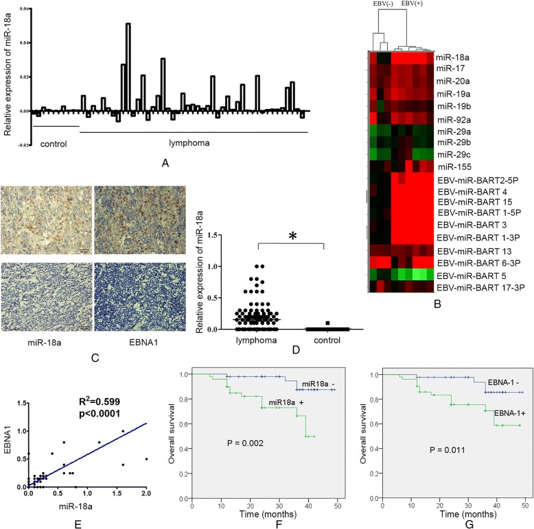
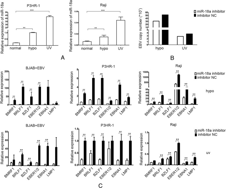
Background: The Epstein-Barr virus (EBV) is closely associated with several types of malignancies. EBV is normally present in the latent state in the peripheral blood B cell compartment. The EBV latent-to-lytic switch is required for virus spread and virus-induced carinogenesis. Immunosuppression or DNA damage can induce the reactivation of EBV replication. EBV alone is rarely sufficient to cause cancer. In this study, we investigated the roles of host microRNAs and environmental factors, such as DNA-damage agents, in EBV reactivation and its association with lymphomagenesis.
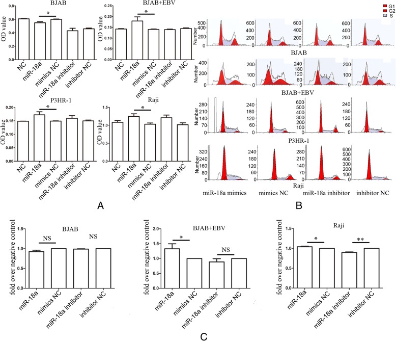
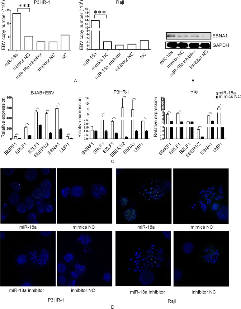
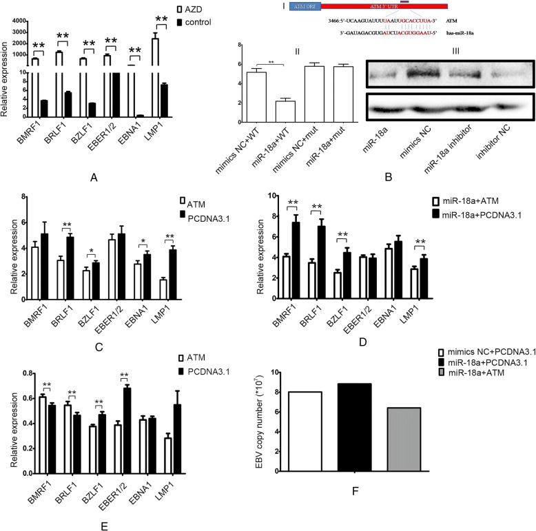
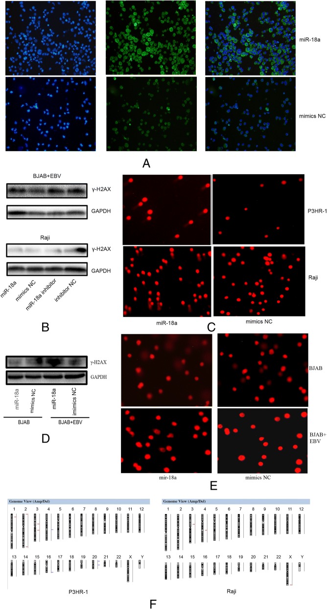
Methods: We first analyzed the publicly available microRNA array data containing 45 diffuse large B-cell lymphoma patients and 10 control lymph nodes or B cells with or without EBV infection. In situ hybridization for miR-18a and immunohistochemitry were performed to evaluate the correlation between the expression of miR-18a and nuclear EBV protein EBNA1 in lymphoid neoplasm. The proliferative effects of miR-18a were investigated in EBV-positive or -negative lymphoid neoplasm cell lines. EBV viral load was measured by a quantitative real-time EBV PCR and FISH assay. The genomic instability was evaluated by CGH-array.
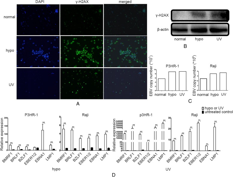
Results: In this study, we analyzed the publicly available microRNA array data and observed that the expression of the miR-17-92 cluster was associated with EBV status. In situ hybridization for miR-18a, which is a member of the miR-17-92 cluster, showed a significant upregulation in lymphoma samples. miR-18a, which shares the homolog sequence with EBV-encoded BART-5, promoted the proliferation of lymphoma cells in an EBV status-dependent manner. The DNA-damaging agent UV or hypoxia stress induced EBV activation, and miR-18a contributed to DNA damaging-induced EBV reactivation. In contrast to the promoting effect of ATM on the lytic EBV reactivation in normoxia, ATM inhibited lytic EBV gene expression and decreased the EBV viral load in the prescence of hypoxia-induced DNA damage. miR-18a reactivated EBV through inhibiting the ATM-mediated DNA damage response (DDR) and caused genomic instability.
Conclusions: Taken together, these results indicate that DNA-damaging agents and host microRNAs play roles in EBV reactivation. Our study supported the interplay between host cell DDR, environmental genotoxic stress and EBV.
Keywords: DNA damage response; EBV reactivation; Genomic instability; miR-18a.
Conflict of interest statement
Ethics approval and consent to participate
The research presented here has been performed in accordance with the Declaration of Helsinki and has been approved by the ethics committee of Xiangya Hospital, Central South University, China (reference number 201312484). The patients were informed about the sample collection and had signed informed consent forms.
Consent for publication
Not applicable.
Competing interest
The authors declare that they have no competing interests.
Publisher’s Note
Springer Nature remains neutral with regard to jurisdictional claims in published maps and institutional affiliations.
Figures
Similar articles
-
DNA Damage Signaling Is Induced in the Absence of Epstein-Barr Virus (EBV) Lytic DNA Replication and in Response to Expression of ZEBRA.PLoS One. 2015 May 7;10(5):e0126088. doi: 10.1371/journal.pone.0126088. eCollection 2015. PLoS One. 2015. PMID: 25950714 Free PMC article.
-
Role of ATM in the formation of the replication compartment during lytic replication of Epstein-Barr virus in nasopharyngeal epithelial cells.J Virol. 2015 Jan;89(1):652-68. doi: 10.1128/JVI.01437-14. Epub 2014 Oct 29. J Virol. 2015. PMID: 25355892 Free PMC article.
-
Epstein-Barr Virus Hijacks DNA Damage Response Transducers to Orchestrate Its Life Cycle.Viruses. 2017 Nov 16;9(11):341. doi: 10.3390/v9110341. Viruses. 2017. PMID: 29144413 Free PMC article. Review.
-
Epstein-Barr Virus Infection of Cell Lines Derived from Diffuse Large B-Cell Lymphomas Alters MicroRNA Loading of the Ago2 Complex.J Virol. 2019 Jan 17;93(3):e01297-18. doi: 10.1128/JVI.01297-18. Print 2019 Feb 1. J Virol. 2019. PMID: 30429351 Free PMC article.
-
Epstein-Barr Virus (EBV)-Related Lymphoproliferative Disorders in Ataxia Telangiectasia: Does ATM Regulate EBV Life Cycle?Front Immunol. 2019 Jan 4;9:3060. doi: 10.3389/fimmu.2018.03060. eCollection 2018. Front Immunol. 2019. PMID: 30662441 Free PMC article. Review.
Cited by
-
A review on EBV encoded and EBV-induced host microRNAs expression profile in different lymphoma types.Mol Biol Rep. 2021 Feb;48(2):1801-1817. doi: 10.1007/s11033-021-06152-z. Epub 2021 Feb 1. Mol Biol Rep. 2021. PMID: 33523370 Review.
-
Stress-Induced Epstein-Barr Virus Reactivation.Biomolecules. 2021 Sep 18;11(9):1380. doi: 10.3390/biom11091380. Biomolecules. 2021. PMID: 34572593 Free PMC article. Review.
-
EBV and 1q Gains Affect Gene and miRNA Expression in Burkitt Lymphoma.Viruses. 2023 Aug 25;15(9):1808. doi: 10.3390/v15091808. Viruses. 2023. PMID: 37766215 Free PMC article.
-
Hydroa Vacciniforme and Hydroa Vacciniforme-Like Lymphoproliferative Disorder: A Spectrum of Disease Phenotypes Associated with Ultraviolet Irradiation and Chronic Epstein-Barr Virus Infection.Int J Mol Sci. 2020 Dec 7;21(23):9314. doi: 10.3390/ijms21239314. Int J Mol Sci. 2020. PMID: 33297336 Free PMC article. Review.
-
A comprehensive overview on the crosstalk between microRNAs and viral pathogenesis and infection.Med Res Rev. 2025 Mar;45(2):349-425. doi: 10.1002/med.22073. Epub 2024 Aug 26. Med Res Rev. 2025. PMID: 39185567 Free PMC article. Review.
References
MeSH terms
Substances
Grants and funding
- 81272255,81472695,81773147/National Natural Science Foundation of China
- 81402249/National Natural Science Foundation of China
- 2015JJ2181/Natural Science Foundation of Hunan Province
- 2013JJ3039/Natural Science Foundation of Hunan Province
- 2012TT2013/Hunan provincial scientific research innovation special key project
LinkOut - more resources
Full Text Sources
Research Materials
Miscellaneous
