. 2019 Feb:40:276-289.
doi: 10.1016/j.ebiom.2018.12.044. Epub 2018 Dec 26.

Targeting cancer stem cell signature gene SMOC-2 Overcomes chemoresistance and inhibits cell proliferation of endometrial carcinoma

Affiliations

Targeting cancer stem cell signature gene SMOC-2 Overcomes chemoresistance and inhibits cell proliferation of endometrial carcinoma

Huan Lu et al. EBioMedicine. 2019 Feb.

Abstract

Background: Endometrial cancer is one of the most common gynecological malignancies and has exhibited an increasing incidence rate in recent years. Cancer stem cells (CSCs), which are responsible for tumor growth and chemoresistance, have been confirmed in endometrial cancer. However, it is still challenging to identify endometrial cancer stem cells to then target for therapy.

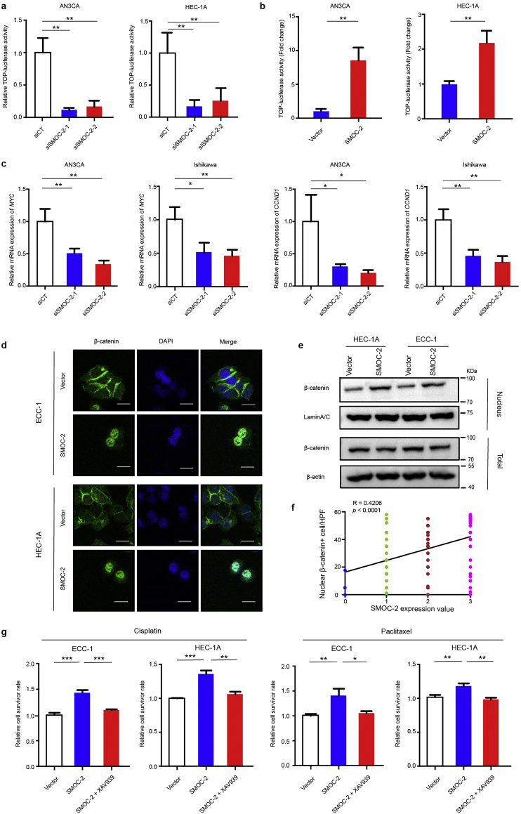
Methods: Flow cytometry was used to identify the endometrial cancer stem cells. Sphere formation assay, western blotting, qRT-PCR assay, cell viability assay, xenograft assay and immunohistochemistry staining analysis were utilized to evaluate the effect of SPARC-related modular calcium binding 2 (SMOC-2) on the cells proliferation and drug resistance. Cell viability assay, qRT-PCR assay, immunofluorescence staining, Co-IP assay and luciferase reporter gene assay were performed to explore the possible molecular mechanism by which SMOC-2 activates WNT/β-catenin pathway.

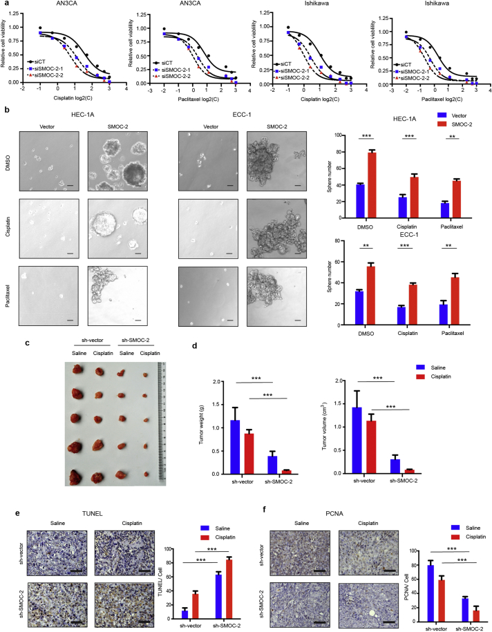
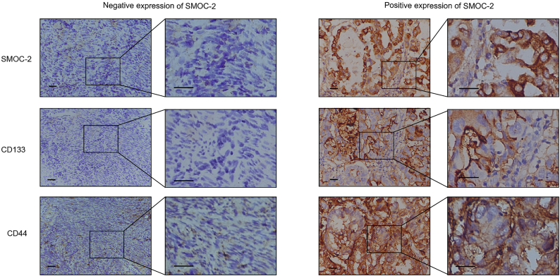
Findings: We found the expression of SPARC-related modular calcium binding 2 (SMOC-2), a member of SPARC family, was higher in endometrial CSCs than that in non-CSCs. SMOC-2 was also more highly expressed in spheres than in monolayer cultures. The silencing of SMOC-2 suppressed cell sphere ability; reduced the expression of the stemness-associated genes SOX2, OCT4 and NANOG; and enhanced chemosensitivity in endometrial cancer cells. By co-culture IP assay, we demonstrated that SMOC-2 directly interacted with WNT receptors (Fzd6 and LRP6), enhanced ligand-receptor interaction with canonical WNT ligands (Wnt3a and Wnt10b), and finally, activated the WNT/β-catenin pathway in endometrial cancer. SMOC-2 expression was closely correlated with CSC markers CD133 and CD44 expression in endometrial cancer tissue.

Interpretation: Taken together, we conclude that SMOC-2 might be a novel endometrial cancer stem cell signature gene and therapeutic target for endometrial cancer. FUND: National Natural Science Foundation of China, Scientific and Technological Innovation Act Program of Shanghai Science and Technology Commission, Scientific and Technological Innovation Act Program of Fengxian Science and Technology Commission, Natural Science Foundation of Shanghai.

Keywords: Cancer stem cells; Chemoresistance; Endometrial carcinoma; SMOC-2; WNT/β-catenin pathway.


Figures

Fig. 1
Fig. 1
SMOC-2 is a signature gene of endometrial cancer stem cells. (a) Flow cytometry analysis of CD133+/CD44+ and CD133/CD44 cells sorted from AN3CA and Ishikawa. (b) The expression of SMOC2 in CD133+/CD44+ and CD133/CD44 cells from AN3CA and Ishikawa was detected by western blotting and normalized by tubulin expression. (c) Relative mRNA expression levels of SMOC2 in AN3CA/ CD133+/CD44+ and Ishikawa/ CD133+/CD44+ cells compared with AN3CA/ CD133/CD44 and Ishikawa/ CD133/CD44 cells. (mean ± SD, **P < 0.01, ***P < 0.001. Experiments were statistically analyzed using two-tailed Student's t-test). (d) Relative mRNA expression levels of SMOC2 in spherical cultures of AN3CA and Ishikawa cells compared with those in monolayer cultures. (mean ± SD, *P < 0.05, ***P < 0.001. Experiments were statistically analyzed using two-tailed Student's t-test). (e) Silencing efficacy of SMOC-2 in AN3CA and Ishikawa cells was detected by real-time PCR. (f) Silencing efficacy of SMOC-2 in AN3CA and Ishikawa cells was detected by western blotting. (g) Sphere assay was performed with siSMOC2–1-, siSMOC2–2- or siCT-transfected AN3CA and Ishikawa cells and passage cells. Scale bars, 50 μm. Quantification of sphere number was shown. (mean ± SD, *P < 0.05, **P < 0.01, ***P < 0.001. Experiments were statistically analyzed using two-tailed Student's t-test). (h) The expression of SOX2, OCT4, and NANOG in SMOC-2-silenced cells AN3CA/Ishikawa were detected by western blotting. (i) Overexpression efficacy of SMOC-2 in HEC-1A and ECC-1 cells was determined by western blotting. The expression of SOX2, OCT4, and NANOG in SMOC-2-overexpressioning cells HEC-1A/ECC-1 were detected by western blotting.
Fig. 2
Fig. 2
SMOC-2 promotes endometrial cancer cell growth in vitro and in vivo. (a) The cell proliferation of siCT and siSMOC-2-1, siSMOC-2-2 groups in AN3CA/ Ishikawa cells was determined by CCK8 assay at low or high density. (mean ± SD, **P < 0.01, ***P < 0.001. Experiments were statistically analyzed using two-tailed Student's t-test). (b) The cell proliferation of the vector and overexpressing SMOC-2 groups in HEC-1A/ECC-1 cells was determined by CCK8 assay in low or high density. (mean ± SD, ***P < 0.001. Experiments were statistically analyzed using two-tailed Student's t-test). (c and d) Morphologic characteristics of tumors from mice inoculated with Lenti-vector and Lenti-shSMOC-2/AN3CA cells. Tumor weights and volumes of Lenti-vector and Lenti-shSMOC-2 groups were shown, n = 5. (mean ± SD, *P < 0.05. Experiments were statistically analyzed using two-tailed Student's t-test).
Fig. 3
Fig. 3
Silencing SMOC-2 enhanced paclitaxel and cisplatin sensitivity. (a) siCT, siSMOC-2-1, and siSMOC-2-2 groups of AN3CA and Ishikawa cells were treated with a series of concentrations paclitaxel/cisplatin to obtain half maximal inhibitory concentration (IC50). (b) Sphere assay was performed with vector, SMOC-2 DNA transfected HEC-1A and ECC-1 cells and SMOC-2 overexpressioning cells treated with paclitaxel or cisplatin. Scale bars, 50 μm. Quantification of sphere number was shown. (mean ± SD, *P < 0.05, **P < 0.01, ***P < 0.001. Experiments were statistically analyzed using two-tailed Student's t-test). (c) Morphologic characteristics of tumors from mice inoculated with sh-vector/Saline, sh-vector/Cisplatin, sh-SMOC-2/Saline and sh-SMOC-2/Cisplatin cells. (d) Tumor volumes of 4 groups from c. n = 5. (mean ± SD, ***P < 0.001. Experiments were statistically analyzed using two-tailed Student's t-test). (e) TUNEL assay was detected in 4 groups from c. Scale bars, 50 μm. (mean ± SD, ***P < 0.001. Experiments were statistically analyzed using two-tailed Student's t-test). (f) PCNA staining was detected in 4 groups from c. Scale bars, 50 μm. (mean ± SD, ***P < 0.001. Experiments were statistically analyzed using two-tailed Student's t-test).
Fig. 4
Fig. 4
SMOC-2 activates the WNT/β-catenin pathway. (a) Luciferase reporter gene assay of AN3CA and HEC-1A cells transfected with siSMOC-2-1, siSMOC-2-2, or siCT. The results shown are the mean ± SD of the relative firefly/Renilla ratio. (mean ± SD, **P < 0.01. Experiments were statistically analyzed using two-tailed Student's t-test). (b) Luciferase reporter gene assay of AN3CA and HEC-1A cells transfected with SMOC-2 plasmid or vector. The results shown are the mean ± SD of the relative firefly/Renilla ratio. (mean ± SD, **P < 0.01. Experiments were statistically analyzed using two-tailed Student's t-test). (c) Silencing SMOC2 induced a reduction of Wnt/β-catenin target gene (MYC and CCND1) mRNA expression. (mean ± SD, *P < 0.05, **P < 0.01. Experiments were statistically analyzed using two-tailed Student's t-test). (d) Representative images of the distribution of β-catenin in HEC-1A and ECC-1 cells transfected with vector or SMOC-2 plasmid by immunofluorescence staining. Scale bars: 20 μm. (e) The expression of β-catenin from nuclear and whole cell lysates of SMOC-2-overexpression cell lines (HEC-1A and ECC-1) and vector cells were detected by western blotting. (f) Correlation between SMOC-2 expression and number of nuclear β-catenin+ cells was determined based on the IHC staining (R = 0.4206, P < 0.0001. HPF, high power field. Spearman correlation analysis was used). (g) Cell survivor rate was determined by CCK8 in HEC-1A and ECC-1 cells treated with vector, SMOC-2 or SMOC-2 + 5 μM XAV-939 and 50 nM paclitaxel or 1 μM cisplatin (mean ± SD, *P < 0.05, **P < 0.01, ***P < 0.001. Experiments were statistically analyzed using two-tailed Student's t-test).
Fig. 5
Fig. 5
SMOC-2 enhances ligand-receptor interaction of WNT/β-catenin pathway. (a) Co-IP assay between SMOC-2 and canonical WNT components (ligand: Wnt3a or Wnt10b; receptor: Fzd6 or LRP6). AN3CA cells were transfected with SMOC-2-Flag or a control vector. The input on the right panel shows the levels of transfected Flag-SMOC-2 and endogenous WNT components (Wnt3a, Wnt10b, Fzd6 and LRP6) in Flag-tagged SMOC-2 or vector control. (b) SMOC-2 enhanced the binding between canonical WNT protein (Wnt3a or Wnt10b) and Fzd6. HA-tagged Fzd6-expressing cells were co-cultured with Myc-tagged WNT ligand-expressing (Wnt3a or Wnt10b) cells and Flag-tagged SMOC-2 expressing cells both separately and in combination. The HA-tagged Fzd6 was immunoprecipitated in this experiment. (c and d) Densitometric analysis showed the relative amounts of precipitated WNT ligand (Wnt3a or Wnt10b) interacted with Fzd6 or LRP6 affected by SMOC-2. Values are normalized to intensities without SMOC-2 as 1. (e) SMOC-2 enhanced the binding between the canonical WNT protein (Wnt3a or Wnt10b) and LRP6. Similarly, HA-tagged LRP6-expressing cells were co-cultured with WNT ligands-expressing cells and Flag-tagged SMOC-2-expressing cells both separately and in combination. (f) The interactions of SMOC-2 and LRP6/Fzd6 were detected by in situ proximity ligation assay (red dots, n = 3, mean ± SD, *P < 0.05, **P < 0.01, ***P < 0.001. Experiments were statistically analyzed using two-tailed Student's t-test). Scale bars: 20 μm. (g) Vector, SMOC-2, SMOC-2 + siLRP6 and SMOC-2 + siFzd6 groups of HEC-1A and ECC-1 cells were treated with a series of concentrations paclitaxel/cisplatin to obtain half maximal inhibitory concentration (IC50). (For interpretation of the references to colour in this figure legend, the reader is referred to the web version of this article.)
Fig. 6
Fig. 6
SMOC-2 and CSCs markers are co-expressed in endometrial cancer. 151 endometrial cancer tissues were stained with SMOC-2, CD44 and CD133 antibodies using serial sections. The left images represented a typical case of low expression of SMOC-2, and CD44 and CD133. The right images represented a typical case with high expression of SMOC-2, CD44 and CD133. Scale bars, 100 μm.

Similar articles

Cited by

References

    1. Torre L.A., Bray F., Siegel R.L., Ferlay J., Lortet-Tieulent J., Jemal A. Global cancer statistics, 2012. CA Cancer J Clin. 2015;65(2):87–108. - PubMed
    1. Siegel R.L., Miller K.D., Jemal A. Cancer statistics, 2017. CA Cancer J Clin. 2017 - PubMed
    1. Cancer Genome Atlas Research N., Kandoth C., Schultz N., Cherniack A.D., Akbani R., Liu Y. Integrated genomic characterization of endometrial carcinoma. Nature. 2013;497(7447):67–73. - PMC - PubMed
    1. Church D.N., Stelloo E., Nout R.A., Valtcheva N., Depreeuw J., ter Haar N. Prognostic significance of POLE proofreading mutations in endometrial cancer. J Natl Cancer Inst. 2015;107(1):402. - PMC - PubMed
    1. Basil J.B., Goodfellow P.J., Rader J.S., Mutch D.G., Herzog T.J. Clinical significance of microsatellite instability in endometrial carcinoma. Cancer. 2000;89(8):1758–1764. - PubMed

MeSH terms