. 2019 Apr;174(2):413-422.
doi: 10.1007/s10549-018-05103-w. Epub 2018 Dec 29.

Adipose stem cell crosstalk with chemo-residual breast cancer cells: implications for tumor recurrence

Affiliations

Adipose stem cell crosstalk with chemo-residual breast cancer cells: implications for tumor recurrence

Matthew A Lyes et al. Breast Cancer Res Treat. 2019 Apr.

Abstract

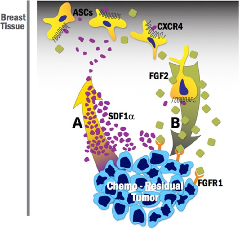
Purpose: Most triple-negative breast cancer (TNBC) patients exhibit an incomplete response to neoadjuvant chemotherapy, resulting in chemo-residual tumor cells that drive tumor recurrence and patient mortality. Accordingly, strategies for eliminating chemo-residual tumor cells are urgently needed. Although stromal cells contribute to tumor cell invasion, to date, their ability to influence chemo-residual tumor cell behavior has not been examined. Our study is the first to investigate cross-talk between adipose-derived stem cells (ASCs) and chemo-residual TNBC cells. We examine if ASCs promote chemo-residual tumor cell proliferation, having implications for tumor recurrence.

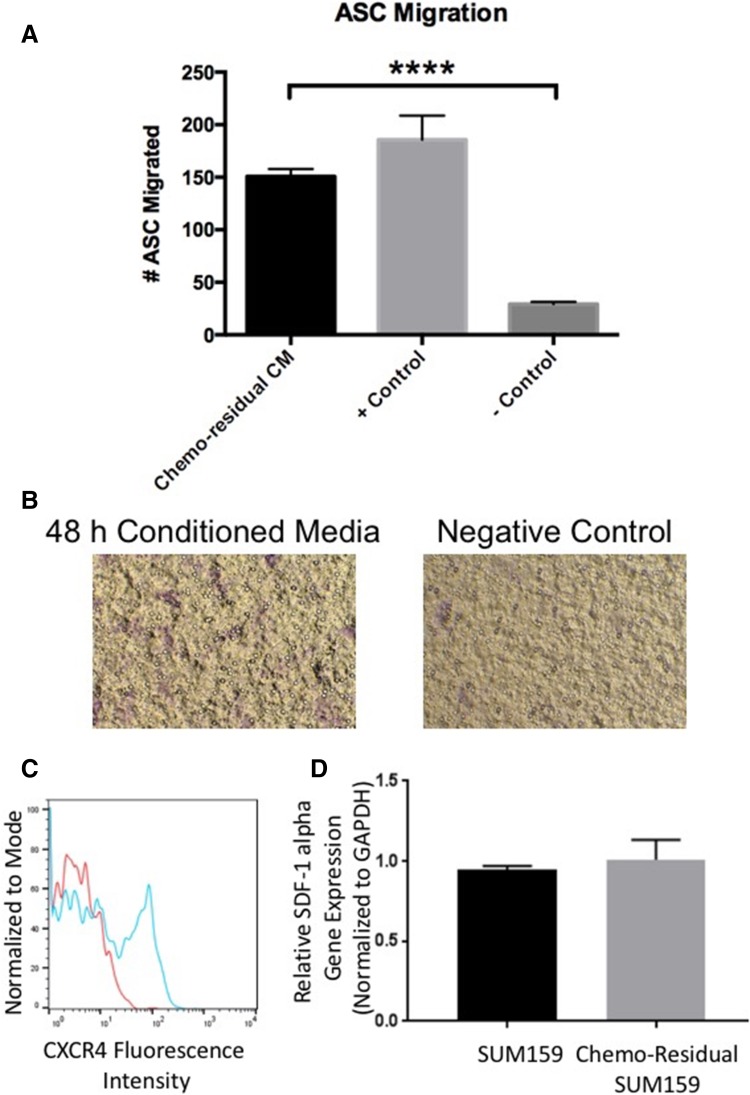
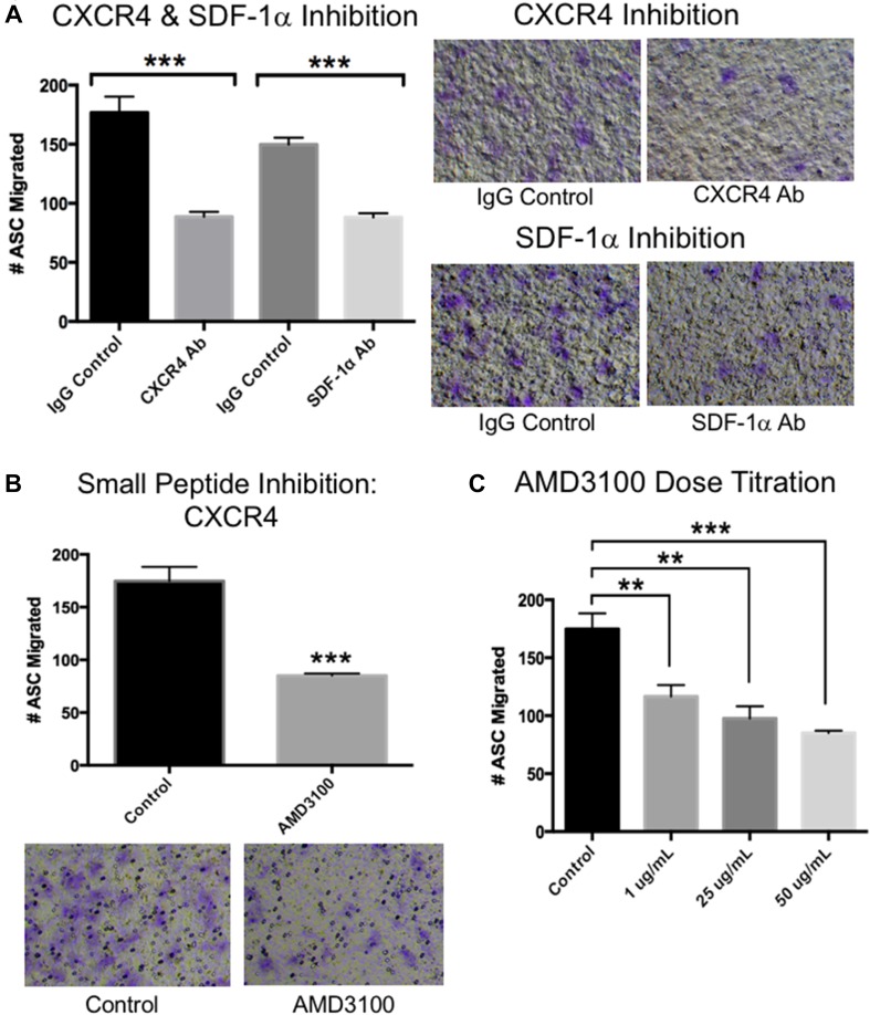
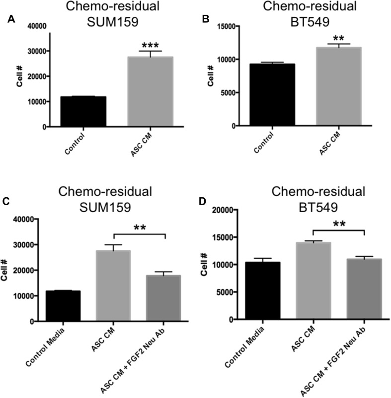
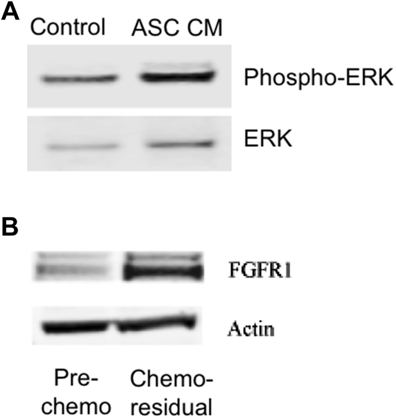
Methods: ASC migration toward chemo-residual TNBC cells was tested in a transwell migration assay. Importance of the SDF-1α/CXCR4 axis was determined using neutralizing antibodies and a small molecule inhibitor. The ability of ASCs to drive tumor cell proliferation was analyzed by culturing tumor cells ± ASC conditioned media (CM) and determining cell counts. Downstream signaling pathways activated in chemo-residual tumor cells following their exposure to ASC CM were studied by immunoblotting. Importance of FGF2 in promoting proliferation was assessed using an FGF2-neutralizing antibody.

Results: ASCs migrated toward chemo-residual TNBC cells in a CXCR4/SDF-1α-dependent manner. Moreover, ASC CM increased chemo-residual tumor cell proliferation and activity of extracellular signal-regulated kinase (ERK). An FGF2-neutralizing antibody inhibited ASC-induced chemo-residual tumor cell proliferation.

Conclusions: ASCs migrate toward chemo-residual TNBC cells via SDF-1α/CXCR4 signaling, and drive chemo-residual tumor cell proliferation in a paracrine manner by secreting FGF2 and activating ERK. This paracrine signaling can potentially be targeted to prevent tumor recurrence.

Keywords: Adipose-derived stem cells (ASCs); Fibroblast growth factor 2 (FGF2); Migration; Proliferation; Recurrence; Triple-negative breast cancer (TNBC).


Conflict of interest statement

Conflict of interest

No authors on this manuscript declare a conflict of interest.

Ethical approval

This article does not contain any studies with human participants or animals performed by any of the authors.

Figures

Fig. 1
Fig. 1
Adipose stem cells (ASCs) Migrate toward Conditioned Media (CM) from Chemo-Residual TNBC Cells. A CM was prepared by growing chemo-residual SUM159 TNBC cells in serum-free media for 48 h. The ability of primary human ASCs (Zenbio) to migrate toward this CM (Chemo-residual CM), FBS (+ Control) or serum-free media (− Control) was evaluated in a 15 h transwell assay. Total number of migrated cells from 5 representative fields (100 × magnification) was determined for each well, and mean cell number from triplicate wells (± SD) was calculated. Significance was determined by two tailed t test (****p < 0.0001). Similar results were obtained in at least three independent trials. B Representative fields (100 ×) showing migration of ASCs towards chemo-residual TNBC CM in a 15 h transwell assay. C CXCR4 expression was assessed by incubating ASCs with CXCR4 antibody (blue line) or control IgG (red line), followed by FITC-conjugated secondary antibody. CXCR4 cell surface expression was determined by flow cytometry. D RNA was isolated from untreated and chemo-residual SUM159 cells and subjected to SDF-1 alpha and GAPDH real-time PCR. SDF-1 alpha gene expression relative to GAPDH is presented for both cell lines
Fig. 2
Fig. 2
CXCR4 and SDF-1α Neutralizing Antibodies Block ASC Migration toward chemo-residual TNBC cell Conditioned Media. A Human ASC migration toward chemo-residual TNBC cell CM (prepared as described in Fig. 1) was measured in the presence of a CXCR4-neutralizing Ab (EMDMillipore, 10 µg/mL), an SDF1α -neutralizing antibody (R&D Systems, 10 µg/mL), or control IgG (R&D Systems, 10 µg/mL). Chemotaxis was measured as in Fig. 1. The right panels show representative fields of migrated ASCs in the presence of the indicated antibodies. Significance was measured with a two tailed t test (***p < 0.0005). B The ability of a CXCR4 small molecule inhibitor (AMD3100; EMD Millipore, 50 µg/mL) to block ASC migration toward chemo-residual TNBC cell CM was tested in a 15 h transwell assay. The bottom panels are representative fields of migrated ASCs ± AMD3100. Significance was measured with a two tailed t test (***p < 0.0005). C The ability of ASCs to migrate toward chemo-residual TNBC cell CM was tested in a 15 h transwell assay in the presence of the indicated concentrations of CXCR4 small molecule inhibitor (AMD3100). Significance was measured using a two tailed t test (**p < 0.005; ***p < 0.0005)
Fig. 3
Fig. 3
ASC CM Increases Proliferation of Chemo-residual TNBC Cells in an FGF2-dependent manner. A and B ASC CM was prepared from human ASCs (ZenBio) grown in reduced serum media for 72 h. This ASC CM (ASC CM), or control reduced serum media (Control) was added to chemo-residual SUM159 (A) or chemo-residual BT549 (B) cells. The number of chemo-residual TNBC cells was determined after 24 h by trypan blue staining. Results are reported as mean cell number (± SEM) from triplicate wells. Similar results were obtained in 3 trials. Significance was measured using a two tailed t test, ***p < 0.0005. (C and D) Chemo-residual SUM159 cells (C) or chemo-residual BT549 cells (D) were incubated with control media, ASC CM + control IgG, or ASC CM + FGF2-neutralizing antibody (EMD Millipore, 10 µg/mL) for 24 h. Cell numbers were determined as in A. Significance was measured with a two tailed t test (**p < 0.005). This effect was independently observed in three trials. Of note, incubation of chemo-naïve SUM159 cells with ASC CM did not induce cell proliferation (data not shown)
Fig. 4
Fig. 4
Chemo-residual TNBC signaling. A Cytosolic extracts were obtained from chemo-residual SUM159 tumor cells pre-treated ± ASC CM for 24 h. Equivalent amounts were immunoblotted with ERK and phospho-ERK antibodies, followed by the appropriate Alexa Fluor secondary antibody. Protein bands were detected by LI-COR Odyssey Fluorescent imaging. Protein bands were quantified using Image J (NIH) and the ratio of phospho-ERK/ERK for each sample was determined. ASC conditioned media induced a two-fold increase in the phospho-ERK/ERK ratio in chemo-residual cells. B Cytosolic extracts were obtained from untreated SUM159 cells and from chemo-residual SUM159 cells. Equivalent amounts were immunoblotted with FGFR1 or Actin antibody, followed by secondary antibody. Protein bands were detected as in A
Fig. 5
Fig. 5
ASC cross-talk with chemo-residual TNBC cells- implications for tumor recurrence. A Chemo-residual TNBC cells secrete the chemokine SDF1α, resulting in recruitment of ASCs, which express the SDF1α chemokine receptor CXCR4, to the tumor site. B ASCs secrete FGF2, which binds to an FGF2 receptor on chemo-residual tumor cells, initiating signaling that drives tumor cell proliferation. Considering that chemo-residual tumor cells can remain dormant in patients for months to years, this ASC/chemo-residual tumor cell cross-talk likely contributes to tumor recurrence in patients post-chemotherapy treatment

Similar articles

Cited by

References

    1. Anders CK, Carey LA. Biology, metastatic patterns, and treatment of patients with triple-negative breast cancer. Clin Breast Cancer. 2009;9(suppl 2):S73–S81. doi: 10.3816/CBC.2009.s.008. - DOI - PMC - PubMed
    1. Hudis CA, Gianni L. Triple negative breast cancer: an unmet medical need. Oncologist. 2011;16(Suppl 1):1–11. doi: 10.1634/theoncologist.2011-S1-01. - DOI - PubMed
    1. Rouzier R, Perou CM, Symmans WF, Ibrahim N, Cristofanilli M, Anderson K, Hess KR, Stec J, Ayers M, Wagner P, Morandi P, Fan C, Rabiul I, Ross JS, Hortobagyi GN, Pusztai L. Breast cancer molecular subtypes respond differently to preoperative chemotherapy. Clinical Cancer Res. 2005;11(16):5678–5685. doi: 10.1158/1078-0432.CCR-04-2421. - DOI - PubMed
    1. Liedtke C, Mazouni C, Hess KR, André F, Tordai A, Mejia JA, Symmans WF, Gonzalez-Angulo AM, Hennessy B, Green M, Cristofanilli M, Hortobagyi GN, Pusztai L. Response to neoadjuvant therapy and long-term survival in patients with triple-negative breast cancer. J Clin Oncol. 2008;26(8):1275–1281. doi: 10.1200/JCO.2007.14.4147. - DOI - PubMed
    1. Li S, Kennedy M, Payne S, Kennedy K, Seewaldt VL, Pizzo SV, Bachelder RE. Model of tumor dormancy/recurrence after short-term chemotherapy. PLoS ONE. 2014;9(5):e98021. doi: 10.1371/journal.pone.0098021. - DOI - PMC - PubMed

MeSH terms

LinkOut - more resources