

Pregnancy-Associated Cardiac Hypertrophy in Corin-Deficient Mice: Observations in a Transgenic Model of Preeclampsia
- PMID: 30595185
- PMCID: PMC6314216
- DOI: 10.1016/j.cjca.2018.11.001
Pregnancy-Associated Cardiac Hypertrophy in Corin-Deficient Mice: Observations in a Transgenic Model of Preeclampsia
Abstract
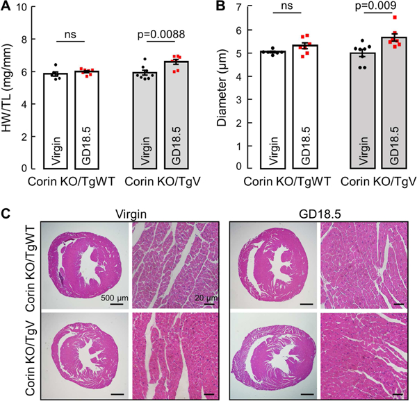
Background: Preeclampsia increases the risk of heart disease. Defects in the protease corin, including the variant T555I/Q568P found in approximately 12% of blacks, have been associated with preeclampsia and cardiac hypertrophy. The objective of this study was to investigate the role of corin and the T555I/Q568P variant in preeclampsia-associated cardiac alterations using genetically modified mouse models.
Methods: Virgin wild-type (WT) and corin knockout mice with or without a cardiac WT corin or T555I/Q568P variant transgene were mated at 3 or 6 months of age. Age- and genotype-matched virgin mice were used as controls. Cardiac morphology and function were assessed at gestational day 18.5 or 28 days postpartum by histologic and echocardiographic analyses.
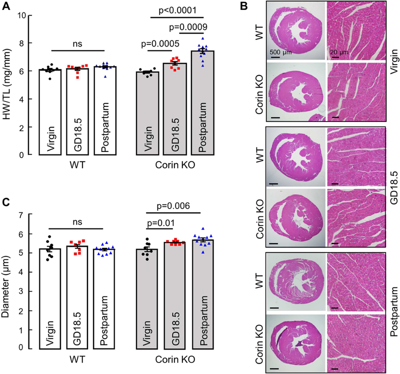
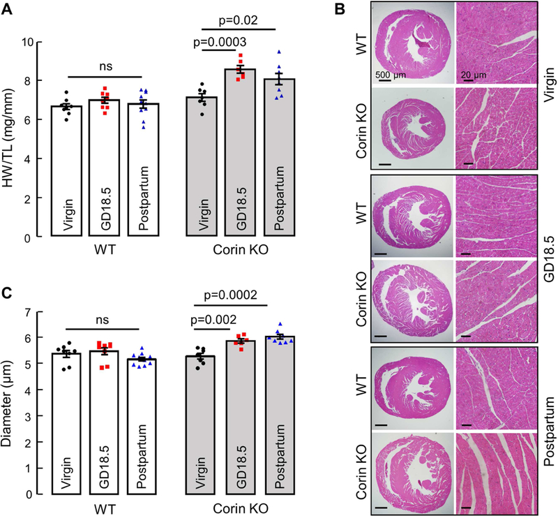
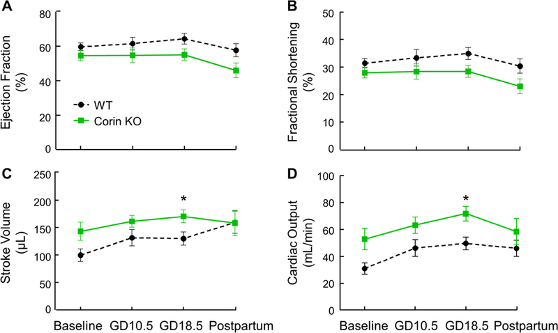
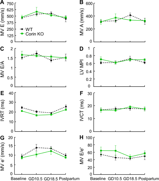
Results: Pregnant corin knockout mice at gestational day 18.5 developed cardiac hypertrophy. Such a pregnancy-associated phenotype was not found in WT or corin knockout mice with a cardiac WT corin transgene. Pregnant corin knockout mice with a cardiac T555I/Q568P variant transgene developed cardiac hypertrophy similar to that in pregnant corin knockout mice. The cardiac hypertrophy persisted postpartum in corin knockout mice and was worse if the mice were mated at 6 instead of 3 months of age. There was no hypertrophy-associated decrease in cardiac function in pregnant corin knockout mice.
Conclusions: In mice, corin deficiency causes cardiac hypertrophy during pregnancy. Replacement of cardiac WT corin, but not the T555I/Q568P variant found in blacks, rescues this phenotype, indicating a local antihypertrophic function of corin in the heart. Corin deficiency may represent an underlying mechanism in preeclampsia-associated cardiomyopathies.
Copyright © 2018 Canadian Cardiovascular Society. Published by Elsevier Inc. All rights reserved.
Conflict of interest statement
Disclosures
The authors have no conflicts of interest to disclose.
Figures
Comment in
-
At the Core of Preeclampsia Genetics: Key Insights into the Neurohormonal Contribution to Hypertensive Diseases of Pregnancy and Their Complications.Can J Cardiol. 2019 Jan;35(1):19-22. doi: 10.1016/j.cjca.2018.11.026. Epub 2018 Dec 3. Can J Cardiol. 2019. PMID: 30595178 No abstract available.
Similar articles
-
Salt-sensitive hypertension and cardiac hypertrophy in transgenic mice expressing a corin variant identified in blacks.Hypertension. 2012 Nov;60(5):1352-8. doi: 10.1161/HYPERTENSIONAHA.112.201244. Epub 2012 Sep 17. Hypertension. 2012. PMID: 22987923 Free PMC article.
-
Corin variant associated with hypertension and cardiac hypertrophy exhibits impaired zymogen activation and natriuretic peptide processing activity.Circ Res. 2008 Aug 29;103(5):502-8. doi: 10.1161/CIRCRESAHA.108.177352. Epub 2008 Jul 31. Circ Res. 2008. PMID: 18669922 Free PMC article.
-
Dysfunctional corin i555(p568) allele is associated with impaired brain natriuretic peptide processing and adverse outcomes in blacks with systolic heart failure: results from the Genetic Risk Assessment in Heart Failure substudy.Circ Heart Fail. 2009 Nov;2(6):541-8. doi: 10.1161/CIRCHEARTFAILURE.109.866822. Epub 2009 Sep 28. Circ Heart Fail. 2009. PMID: 19919978 Free PMC article.
-
Role of corin and atrial natriuretic peptide in preeclampsia.Placenta. 2013 Feb;34(2):89-94. doi: 10.1016/j.placenta.2012.11.016. Epub 2012 Dec 2. Placenta. 2013. PMID: 23211473 Free PMC article. Review.
-
Role of corin in the regulation of blood pressure.Curr Opin Nephrol Hypertens. 2017 Mar;26(2):67-73. doi: 10.1097/MNH.0000000000000297. Curr Opin Nephrol Hypertens. 2017. PMID: 27898523 Free PMC article. Review.
Cited by
-
Impact of Adverse Gestational Milieu on Maternal Cardiovascular Health.Endocrinology. 2023 Apr 17;164(6):bqad060. doi: 10.1210/endocr/bqad060. Endocrinology. 2023. PMID: 37042476 Free PMC article. Review.
-
Pregnancy-associated cardiac dysfunction and the regulatory role of microRNAs.Biol Sex Differ. 2020 Apr 6;11(1):14. doi: 10.1186/s13293-020-00292-w. Biol Sex Differ. 2020. PMID: 32252821 Free PMC article. Review.
-
Single-Nucleotide Polymorphisms in the 3' Untranslated Region of CORIN Associated With Cardiovascular Diseases in a Chinese Han Population: A Case-Control Study.Front Cardiovasc Med. 2021 Aug 2;8:625072. doi: 10.3389/fcvm.2021.625072. eCollection 2021. Front Cardiovasc Med. 2021. PMID: 34409072 Free PMC article.
-
Natriuretic peptide pathways in heart failure: further therapeutic possibilities.Cardiovasc Res. 2023 Feb 3;118(18):3416-3433. doi: 10.1093/cvr/cvac125. Cardiovasc Res. 2023. PMID: 36004816 Free PMC article.
-
TFEC contributes to cardiac hypertrophy by inhibiting AMPK/mTOR signaling.Exp Ther Med. 2021 Nov;22(5):1271. doi: 10.3892/etm.2021.10706. Epub 2021 Sep 7. Exp Ther Med. 2021. PMID: 34594408 Free PMC article.
References
-
- Hypertension in pregnancy. Report of the American College of Obstetricians and Gynecologists’ Task Force on Hypertension in Pregnancy. Obstet Gynecol 2013;122:1122–1131. - PubMed
-
- Sibai B, Dekker G, Kupferminc M. Pre-eclampsia. Lancet 2005;365:785–799. - PubMed
-
- Bamfo JE, Kametas NA, Chambers JB, Nicolaides KH. Maternal cardiac function in normotensive and pre-eclamptic intrauterine growth restriction. Ultrasound Obstet Gynecol 2008;32:682–686. - PubMed
-
- Melchiorre K, Sutherland GR, Baltabaeva A, Liberati M, Thilaganathan B. Maternal cardiac dysfunction and remodeling in women with preeclampsia at term. Hypertension 2011; 57:85–93. - PubMed
Publication types
MeSH terms
Substances
Grants and funding
LinkOut - more resources
Full Text Sources
Molecular Biology Databases
Miscellaneous
