. 2019 Jan 10;176(1-2):167-181.e21.
doi: 10.1016/j.cell.2018.10.053. Epub 2018 Dec 27.

The CMG Helicase Bypasses DNA-Protein Cross-Links to Facilitate Their Repair

Affiliations

The CMG Helicase Bypasses DNA-Protein Cross-Links to Facilitate Their Repair

Justin L Sparks et al. Cell. .

Abstract

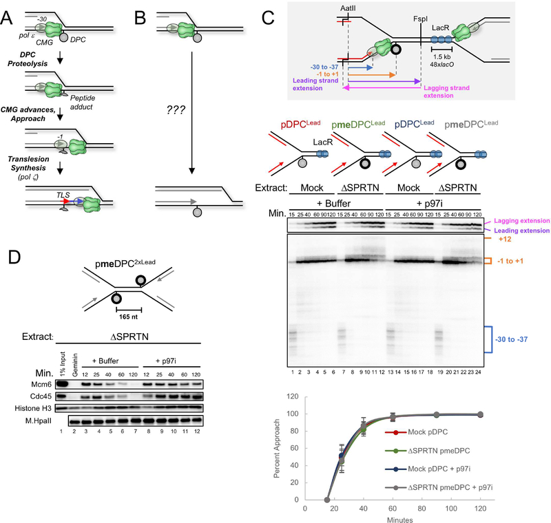
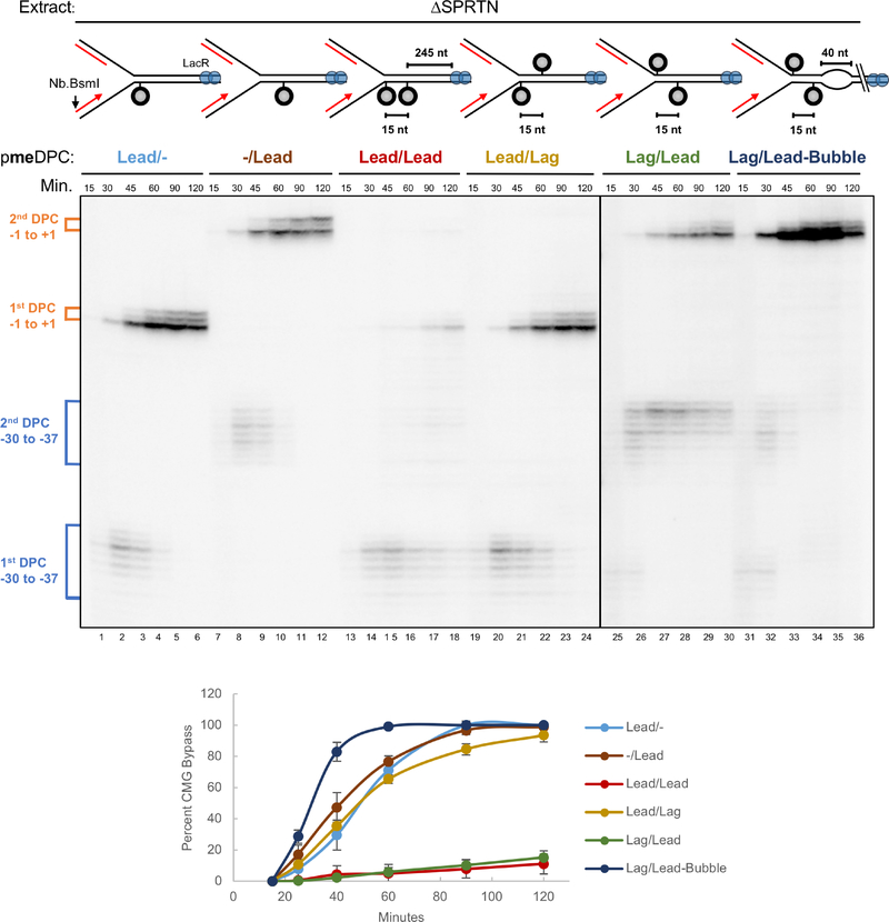
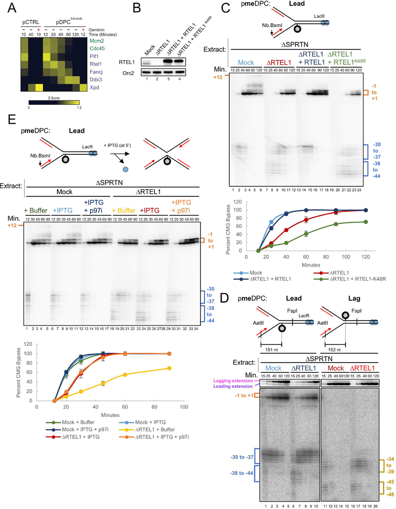
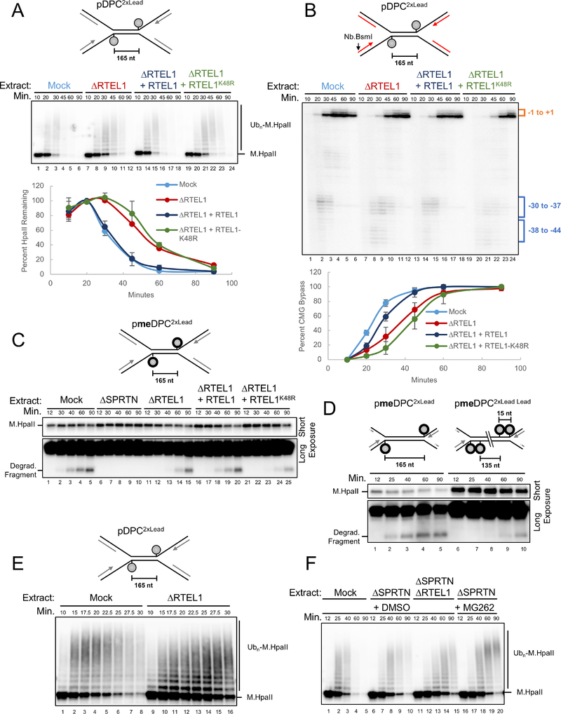
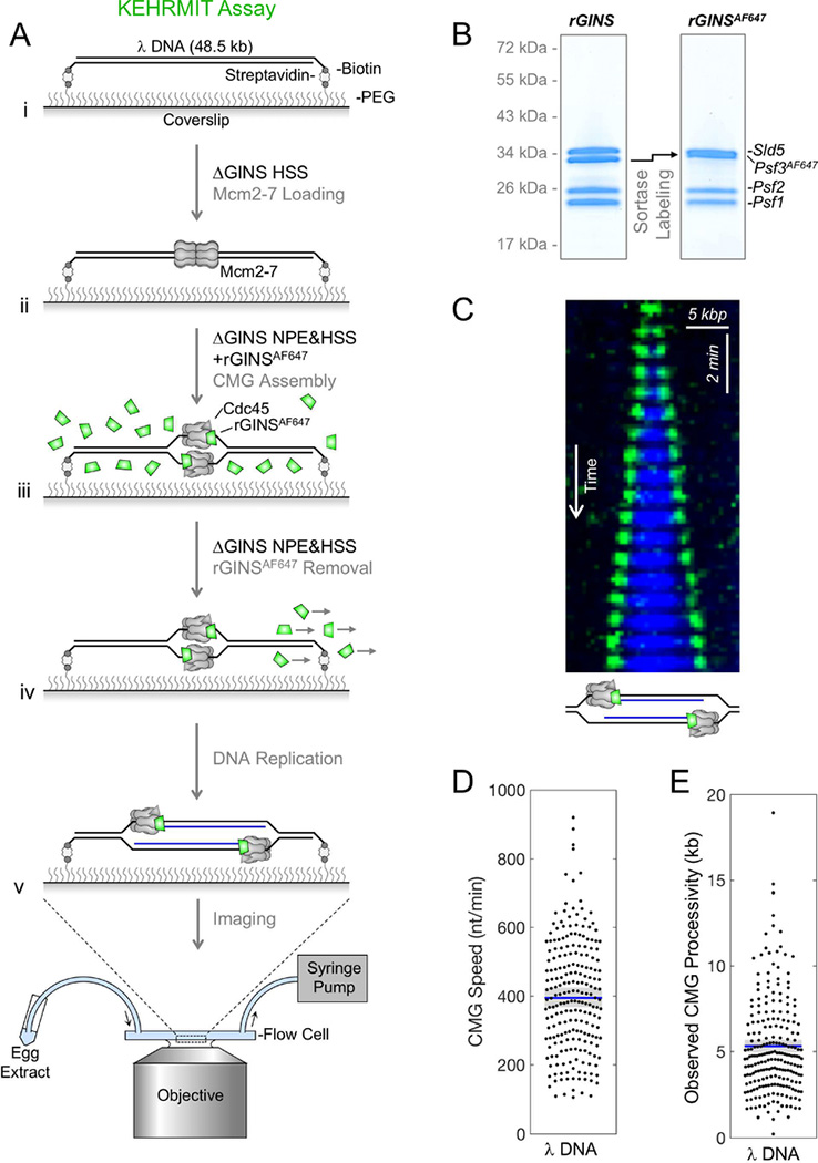
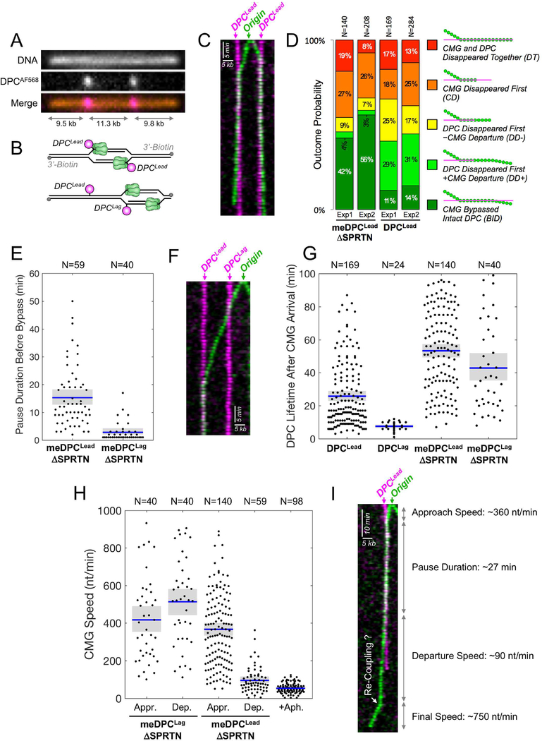
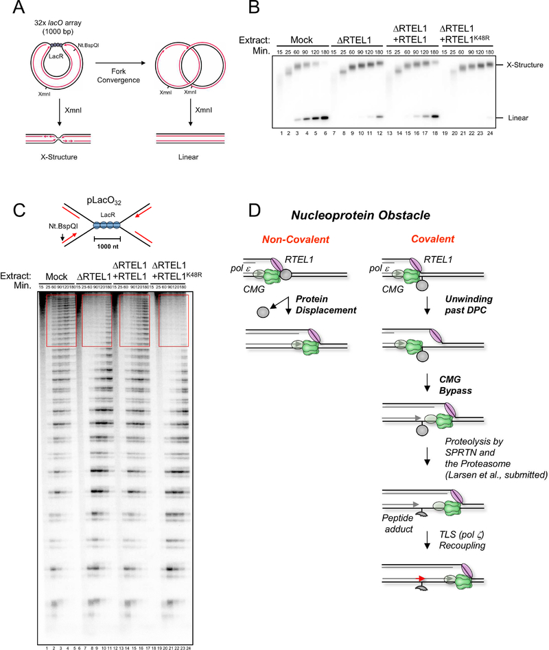
Covalent DNA-protein cross-links (DPCs) impede replication fork progression and threaten genome integrity. Using Xenopus egg extracts, we previously showed that replication fork collision with DPCs causes their proteolysis, followed by translesion DNA synthesis. We show here that when DPC proteolysis is blocked, the replicative DNA helicase CMG (CDC45, MCM2-7, GINS), which travels on the leading strand template, bypasses an intact leading strand DPC. Single-molecule imaging reveals that GINS does not dissociate from CMG during bypass and that CMG slows dramatically after bypass, likely due to uncoupling from the stalled leading strand. The DNA helicase RTEL1 facilitates bypass, apparently by generating single-stranded DNA beyond the DPC. The absence of RTEL1 impairs DPC proteolysis, suggesting that CMG must bypass the DPC to enable proteolysis. Our results suggest a mechanism that prevents inadvertent CMG destruction by DPC proteases, and they reveal CMG's remarkable capacity to overcome obstacles on its translocation strand.

Keywords: CMG; DNA-protein cross-links; KEHRMIT; Xenopus laevis egg extracts; bulky lesion bypass; eukaryotic DNA replication; eukaryotic replicative helicase; helicase uncoupling; replication-coupled DNA repair; single-molecule approach.


Conflict of interest statement

Declaration of Interests

The authors declare no competing interests.

Figures

Figure 1.
Figure 1.. Disappearance of the CMG footprint at DPCLead is unaffected by proteolysis or p97 inhibition.
(A) Previous model of replication-coupled DPC repair (Duxin et al., 2014). (B) Schematic of what happens in the presence of Ub-VS (Duxin et al., 2014). (C) pDPCLead or pmeDPCLead were pre-bound with LacR to prevent one replication fork from reaching the DPC. The plasmids were replicated in mock-depleted or SPRTN-depleted egg extract containing 32P[α]-dATP and supplemented with buffer or the p97 inhibitor NMS-873 (p97i). At different times, DNA was recovered and digested with AatII and FspI, separated on a denaturing polyacrylamide gel, and visualized by autoradiography. Grey inset: Schematic of nascent leading strand products released by AatII and FspI digestion of pmeDPCLead or pDPCLead. The lower autoradiogram shows nascent leading strands generated by the rightward replication fork, and the upper autoradiogram shows both extension products. Blue bracket, CMG footprint (−30 to −37); orange bracket, products stalled at the adducted base (−1 to +1). The percentage of leading strands that approached from the −30 cluster to the −1 cluster was quantified (see methods), and the mean of n=5 experiments is graphed. Error bars represent the standard deviation. See Figure S1E for description of −1 to +12 products in lanes 7–12 and 19–24. (D) pmeDPC2xLead was replicated in SPRTN-depleted egg extracts and supplemented with buffer or p97i. At different times, plasmid-associated proteins were recovered and blotted with the indicated antibodies. Samples were also examined for DPC proteolysis (Figure S1D). A model of CMG unloading from this template is shown in Figure S1G.
Figure 2.
Figure 2.. ssDNA downstream of an intact DPC facilitates CMG bypass.
The indicated plasmids were pre-incubated with LacR, replicated in SPRTN-depleted egg extract, and analyzed as in Figure S1J. Approach was used as a proxy for CMG bypass and quantified as in Figure 1C (see methods). Figure S2 depicts the proposed events on each plasmid. The mean of n=3 experiments is graphed. Error bars represent the standard deviation.
Figure 3.
Figure 3.. RTEL1 is required for efficient CMG bypass.
(A) Recovery of 5’ to 3’ helicases in the mass spectrometry dataset of Larsen et al. (submitted). Relative abundance of each protein in the specified conditions is expressed as a z-score with yellow indicating higher abundance. Where indicated, Geminin was added to block replication initiation. (B) Mock-depleted, RTEL1-depleted, and RTEL1-depleted egg extracts supplemented with wild-type RTEL1 or RTEL1-K48R were blotted with RTEL1 and ORC2 (loading control) antibodies. (C) pmeDPCLead was replicated in the indicated extracts, and supplemented with buffer, wild-type RTEL1, or RTEL1-K48R. Leading strand approach was visualized as in Figure S1J and quantified as in Figure 1C. The mean of n=3 independent experiments is graphed. Error bars represent the standard deviation. (D) pmeDPCLead or pmeDPCLag was replicated in the indicated extracts and analyzed as in Figure 1C. CMG bypass was quantified as in Figure 1C. (E) pmeDPC was replicated in the indicated extracts and supplemented with IPTG (at 5 minutes after replication initiation) and/or p97i, as indicated. Leading strand approach was visualized as in Figure S1J and quantified as in Figure 1C. The mean of n=3 independent experiments is graphed. Error bars represent the standard deviation. The slower CMG bypass observed in RTEL1-depleted extract containing IPTG relative to mock-depleted extract (light blue vs. red graph) is largely accounted for by the fact that CMG progression through the lacO array is delayed by LacR, even in the presence of IPTG, as seen from the slower appearance of resolved, linear species in Figure S3F (compare light blue vs. red graphs).
Figure 4.
Figure 4.. RTEL1 is required for efficient DPC proteolysis.
(A) pDPC2xLead was replicated in the indicated extracts and supplemented with buffer, wild-type RTEL1, or RTEL1-K48R. Plasmid was recovered under stringent conditions, the DNA digested, and the resulting samples blotted for HpaII. Signal from the entire lane was quantified, and peak signal was assigned a value of 100%. The mean of n=3 independent experiments is graphed. Error bars represent standard deviation. (B) Parallel reactions to those in (A) were supplemented with [α−32P]-dATP. Leading strand approach was visualized as in Figure S1J and quantified as in Figure 1C. The mean of n=3 independent experiments is graphed. (C) pmeDPC2XLead was replicated in the indicated extracts. Samples were processed by the pulldown procedure described in (A). Short and long (lower panel) exposures of the same blot are shown. (D) pmeDPC2xLead or pmeDPC2xLead Lead were replicated in non-depleted extract. Plasmid pull downs were performed as in (A) and presented as in (C). (E) pDPC2XLead was replicated in the indicated extracts, and plasmid pull-down was performed as in (A). RTEL1 depletion was verified in Figure S4B. (F) pDPC2XLead was replicated in the indicated egg extracts that also contained DMSO or MG262. Stringent plasmid pull-down was performed as in (A).
Figure 5.
Figure 5.. KEHRMIT – a single molecule assay for CMG dynamics
(A) Schematic of KEHRMIT assay. (B) Coomasie-stained SDS-PAGE gel of recombinant GINS before and after sortase labeling of Psf3 with AF647, which shifts its mobility (arrow). (C) Kymogram of a replication bubble from a KEHRMIT experiment. Green, GINSAF647 signal. Blue, Fen1mKikGR - a fluorescent protein that binds nascent DNA (Loveland et al., 2012). (D-E) Beeswarm plots of CMG speed and processivity (i.e. distance travelled) measured via KEHRMIT (dots represent n=218 individual helicase molecules). Blue line, mean; gray box, 95% Confidence Interval (CI) estimated by bootstrapping.
Figure 6.
Figure 6.. Direct observation of DPC bypass by CMG
(A) Stretched and immobilized DNA-DPC substrate. DNA was stained with sytox orange (top panel). The DPC was labeled on its C-terminus with AF568 (middle panel). Merge, bottom panel. (B) Cartoons depicting how the location of initiation determines whether CMGs encounter DPCLead (top) or DPCLag followed by DPCLead (bottom) (C) Kymogram of a meDPC substrate undergoing replication in SPRTN-depleted extract from an origin that fired between the DPCs. Both CMGs undergo DPC bypass. Images were acquired at 1 frame/min. Green, AF647; pink, AF568. (D) Quantification of five different classes of CMG-DPCLead encounters in n=2 independent biological repeats: (i) BID, Bypass of intact DPC, representing unambiguous bypass events; (ii) DD+, DPC disappeared first, followed by CMG departure from the pause site. When proteolysis was inhibited (meDPC, ΔSPRTN extract), DD+ events likely involve DPC bypass but do not meet the BID criteria due to DPC photobleaching; (iii) DD-, DPC disappeared first, without CMG departure from the pause site, including potential bypass events where CMG photobleached or DNA ruptured soon after the DPC signal vanished; (iv) CD, CMG disappeared first, likely due to photobleaching, obscuring any subsequent bypass events; (v) DT, CMG and DPC disappeared together, including events where the experiment ended or the DNA ruptured before bypass could be detected. Due to rounding errors, probabilities may not add up to 100%. N represents the number of molecules. (E) Beeswarm plot of the time needed to bypass meDPCLead or meDPCLag in SPRTN-depleted extract. Blue lines and gray boxes correspond to the mean and the 95% CI for the mean, respectively. N represents the number of molecules. (F) Same as (C) but showing a kymogram in which an origin fired to the right of both lesions. The leftward-moving helicase (green) first bypassed a meDPCLag in ~1 min, then reached a meDPCLead where it paused. (G) Beeswarm plot of DPC lifetime after CMG arrival at the lesion. Blue lines, gray boxes, and N as in (E). (H) Beeswarm plot of CMG speed during approach to (Appr.) and departure from (Dep.) DPC lesions versus the speed of aphidicolin-uncoupled helicases. Blue lines, gray boxes, and N as in (E). The aphidicolin condition was performed on λ DNA. (I) Kymogram of CMG-meDPCLead encounter (SPRTN-depleted extract) that resulted in DPC bypass and CMG uncoupling, followed by apparent re-coupling (white arrow).
Figure 7.
Figure 7.. RTEL1 is required for CMG bypass of non-covalent nucleoprotein complexes.
(A) Top, structures generated with and without XmnI digestion before and after forks progress through the LacR array. (B) pLacO32 was pre-incubated with LacR and replicated in the indicated egg extracts containing [α−32P]-dATP. DNA was recovered, digested with the single cutter XmnI, resolved by native agarose gel electrophoresis, and visualized by autoradiography. (C) DNA samples from (B) were nicked with Nt. BspQI to release the rightward leading strand (red arrow), separated on a denaturing polyacrylamide gel, and visualized by autoradiography. lacO sites are located ~30 nt downstream of each stalling product (Dewar et al., 2015). (D) Model of replisome bypass of nucleoprotein barriers. When the replisome encounters a non-covalent nucleoprotein complex, RTEL1 and CMG cooperate to unwind the DNA underlying the protein, leading to its displacement and immediate resumption of fork progression. At a covalent DPC, RTEL1 translocates along the undamaged lagging strand template, exposing ssDNA that facilitates CMG bypass. Given the stable interaction of pol ε (grey oval) with CMG (Langston et al., 2014), we envision that it bypasses the DPC with CMG. After CMG bypass, the DPC undergoes proteolysis by SPRTN or the proteasome. Finally, the leading strand is extended past the peptide adduct using translesion synthesis polymerases.

References

    1. Arias EE, and Walter JC (2005). Replication-dependent destruction of Cdt1 limits DNA replication to a single round per cell cycle in Xenopus egg extracts. Genes Dev 19, 114–126. - PMC - PubMed
    1. Bochman ML, and Schwacha A (2008). The Mcm2–7 complex has in vitro helicase activity. Mol Cell 31, 287–293. - PubMed
    1. Byun TS, Pacek M, Yee MC, Walter JC, and Cimprich KA (2005). Functional uncoupling of MCM helicase and DNA polymerase activities activates the ATR-dependent checkpoint. Genes Dev 19, 1040–1052. - PMC - PubMed
    1. Chen I, Dorr BM, and Liu DR (2011). A general strategy for the evolution of bond-forming enzymes using yeast display. Proc Natl Acad Sci U S A 108, 11399–11404. - PMC - PubMed
    1. Dewar JM, Budzowska M, and Walter JC (2015). The mechanism of DNA replication termination in vertebrates. Nature 525, 345–350. - PMC - PubMed

Publication types

LinkOut - more resources