. 2019 Feb 11;48(3):406-419.e5.
doi: 10.1016/j.devcel.2018.11.047. Epub 2018 Dec 27.

Probing the Function of Metazoan Histones with a Systematic Library of H3 and H4 Mutants

Affiliations

Probing the Function of Metazoan Histones with a Systematic Library of H3 and H4 Mutants

Weimin Zhang et al. Dev Cell. .

Abstract

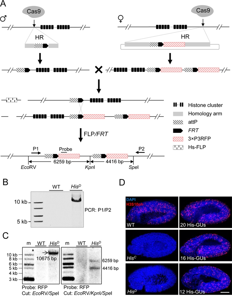
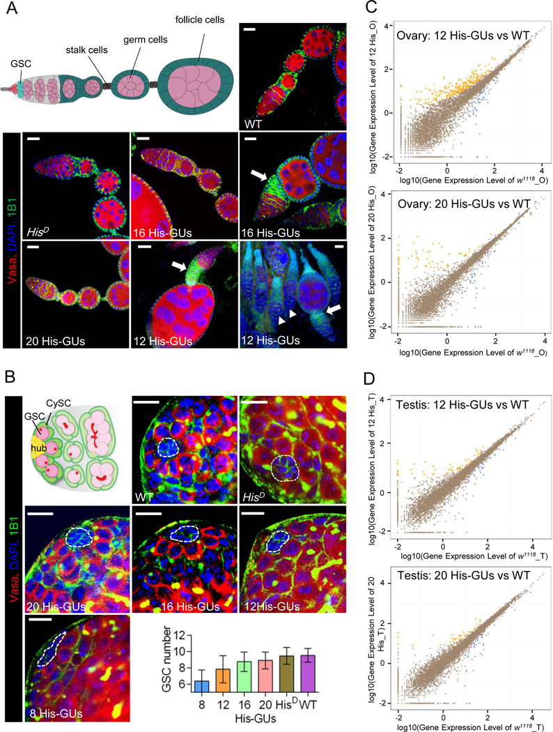
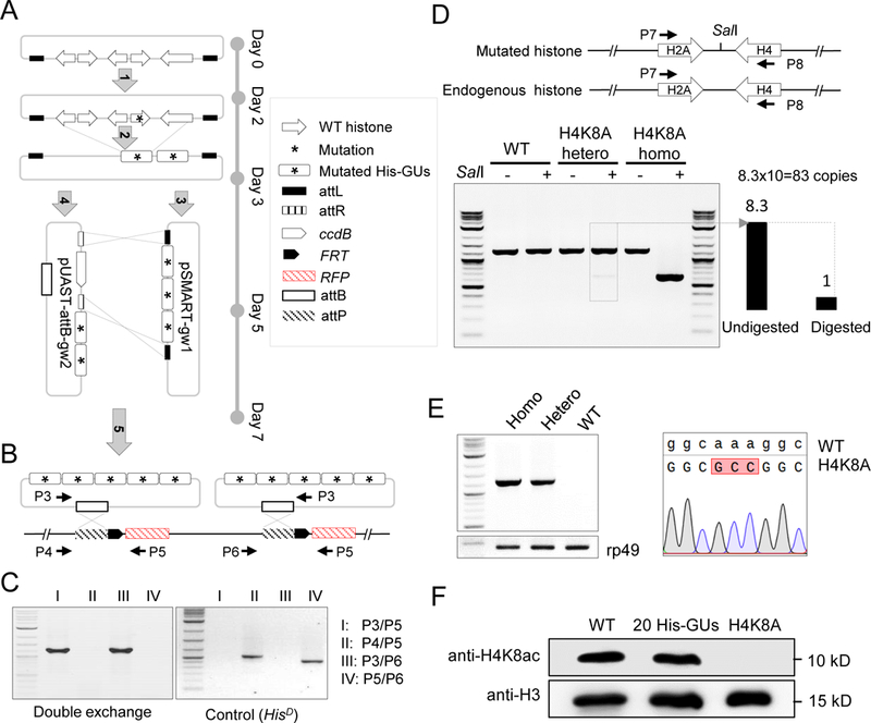
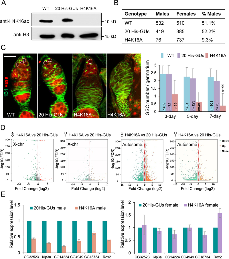
Replication-dependent histone genes often reside in tandemly arrayed gene clusters, hindering systematic loss-of-function analyses. Here, we used CRISPR/Cas9 and the attP/attB double-integration system to alter numbers and sequences of histone genes in their original genomic context in Drosophila melanogaster. As few as 8 copies of the histone gene unit supported embryo development and adult viability, whereas flies with 20 copies were indistinguishable from wild-types. By hierarchical assembly, 40 alanine-substitution mutations (covering all known modified residues in histones H3 and H4) were introduced and characterized. Mutations at multiple residues compromised viability, fertility, and DNA-damage responses. In particular, H4K16 was necessary for expression of male X-linked genes, male viability, and maintenance of ovarian germline stem cells, whereas H3K27 was essential for late embryogenesis. Simplified mosaic analysis showed that H3R26 is required for H3K27 trimethylation. We have developed a powerful strategy and valuable reagents to systematically probe histone functions in D. melanogaster.

Keywords: CRISPR/Cas9; Drosophila; FLP-FRT; H4K16; attB-attP; dosage effects; histone mutant library; mosaic system.


Conflict of interest statement

Competing interests: The authors declare no competing interests.

DECLARATION OF INTERESTS

The authors declare no competing interests.

Figures

Figure 1
Figure 1. Schematic overview of histone cluster deletion and molecular verification.
(A) Strategy for knocking out the entire histone cluster. CRISPR/Cas9-mediated homologous recombination (HR) pathway was used to knock-in attP-FRT cassette on both sides of the histone cluster. On the left side, single-stranded oligonucleotide DNA was used as the HR donor. On the right side, a plasmid with homologous arms was used. Flies with ‘ends-in’ HR events were identified during knocking-in at the right side, creating an attP-FRT duplication (top). The two fly lines were crossed and flippase activity was induced. Through HR between FRT sequences (middle), a histone null mutant was generated (bottom). Primers (P1 and P2) for long-range PCR, probe and restriction-enzyme sites for Southern blotting are indicated. The distances between these restriction sites are labeled. (B) PCR analysis with P1 and P2 primers was used to validate histone cluster deletion (HisD). w1118 wild-type (WT) flies were used as a control. (C) Southern-blot analysis of HisD. Size markers (m). Genomic DNA from either WT (w1118) or HisD was digested with EcoRV/SpeI (Left) or EcoRV/KpnI/SpeI (Right), separated and blotted with the probe indicated in Figure 1A. (D) Confocal images of cycle 15 embryos immunostained with H3S10ph antibody (red) and DAPI (blue). WT: w1118; HisC: homozygous histone deletion (Bloomington Stock Center 8670); HisD: homozygous histone cluster deletion (generated in this study); 20His-GUs, 16 His-GUs, and 12His-GUs indicate homozygous histone cluster deletions with different copy numbers of histone gene units (His-GUs). No H3S10ph signal was detected in HisD and HisC embryos. Scale bar: 100 µm. See also Figure S1.
Figure 2
Figure 2. Low histone dosage affects the fertility of rescued adults.
(A) Results of rescue tests for adult flies with different copy numbers of wild-type (WT) histone gene units (His-GUs; 4, 6, 8, 10, 12, 16, and 20 units) that were reintroduced into histone null mutants (HisD) to rescue the lethal phenotype. Results are means ± SD from triplicate determinations. (B) Western blot analysis of levels of H3, H4, H2A, H2B, and H1 histones in virgin adult flies with 12, 16, or 20 His-GUs, HisD, and WT (w1118). (C, D) Results of fertility tests of male (C) and female (D) adult flies with 12, 16, or 20 His-GUs, HisD, and WT (w1118). The fertility test is done by counting the number of surviving adult progeny produced by male or female flies of the given genotype crossed with w1118. Each point represents a vial of flies. The horizontal bar indicates the mean number of adult progeny produced. Error bars represent standard error of the mean (± SEM). (E, F) Testes (E) and ovaries (F) from flies with different histone copy numbers were classified by their morphology into three categories, as show in the top panels: WT (w1118), moderate defect, and severe defect. Below, the numbers of adults with the given testis (E) and ovary (F) morphology in flies with 12, 16, or 20 His-GUs, HisD, and WT are shown. Total number of dissected testes and ovaries is listed on the right. See also Figure S2.
Figure 3
Figure 3. Low histone dosage affects testis and ovary development.
(A) Low copy numbers of histone gene units (His-GUs) caused severe budding defect in egg-chamber development. Illustration (top left) of a wild-type (WT) Drosophila germarium and egg chamber. Germline stem cells (GSCs; dark red) reside in a germline niche and divide asymmetrically and form cystoblasts, which develop into 16-cell cysts surrounded by follicle cells (green). The cyst cells then bud from the germarium as individual egg chambers. As the egg chambers continue to grow, they move further to the posterior and form a chain of egg chambers connected by stalk cells (blue). Confocal images of ovaries from adult flies carrying different copy numbers of His-GUs stained with anti-Vasa (red), DAPI (blue), anti-1B1 (green). Scale bar, 20 µm. The majority of ovaries in flies with 12 His-GUs had severe budding problems and did not have mid-stage egg chambers (solid arrow head). (B) Low copy numbers of His-GUs caused GSC loss in the testis. Illustration of WT Drosophila testis (top left). Non-dividing hub cells (yellow) are surrounded by GSCs (dark red) and cyst stem cells (CySCs, dark green). GSCs with round fusome (red) divide asymmetrically and produce differentiated spermatogonia (green) with branched fusome. Low copy number (≤12 His-GUs) testes have fewer GSCs (P-value <0.00005). Testes from adult flies with different copy numbers of His-GUs were stained with anti-Vasa (red), DAPI (blue), anti-1B1 (green). Scale bar: 10 µm. The GSC number of testes was counted and results are means ± SEM (bottom right). (C, D) Transcriptome comparison of ovaries (C) and testes (D) in flies with 12 or 20 His-GUs and WT (w1118). The scatter plots (yellow or blue) indicate differentially expressed genes. The RNA-seq experiments were performed with two biological replicates. See also Figure S3.
Figure 4
Figure 4. Systematic mutagenesis of Drosophila melanogaster histone H3 and H4 residues.
(A) Schematic representation of histone-mutagenesis procedures with Gateway assembly system (see details in Methods). ‘Five-step’ assembly was performed to insert five mutated-histone gene units (His-GUs) into the pUAST-attB integration vector. (B) PhiC31-mediated ‘attB/attP’ exchange system for integration of two mutated 5 His-GUs into the genome in the HisD fly. Primer pairs for PCR verification are indicated. (C) PCR verification of double attB/attP recombination. (D) Molecular characterization of H4K8A mutant flies. The top panel is the schematic representation of the synthetic and native histone genes. Primers to amplify part of the histone genes are indicated. The SalI site introduced in the synthetic construct is shown. The bottom panel shows the gel image of amplified PCR fragments with or without SalI digestion. The copy number of His-GU was estimated as the ratio of band intensity of digested and undigested fragments, and is shown at the bottom left. (E) Sequence confirmation of the H4K8A mutant. The left panel shows the PCR result using primers specifically to amplify the mutated DNA. The rp49 gene was used as a loading control. The sequencing trace is shown on the right. (F) Analysis of H4K8ac (acetylated H4K8) in different fly lines by western blot. See also Figure S4.
Figure 5
Figure 5. H4K16 was required for ovarian germline stem cell (GSC) maintenance and male viability.
(A) Western blot of adult fly extract from wild type (WT; w1118), 20 His-GUs (20 copies of wild-type histone gene units), and H4K16A genotypes. (B) The number of male and female progeny from wild type, 20 His-GUs and H4K16A parents. (C) Confocal images of ovaries from w1118, 20 His-GUs and H4K16A flies. GSCs (dotted line) stained with 1B1 (green) and Vasa (red). Histogram shows the number of GSCs per germarium at days 3, 5, and 7 adulthood. The number of germaria examined per genotype is shown on each column. Values are means ± SEM. Scale bar: 20 µm. (D) Transcriptome comparisons of third larvae salary glands between H4K16A mutants and 20His-GUs. (E) RT-PCR verification of effects of H4K16A mutation. Six-linked genes (CG32523, Klp3a, CG14224, CG4949, CG18734, and Rox2) were chosen as the targets. Values are means ± SEM of three biological replicates (rp49 was the reference gene for normalization, RNA was extracted from salivary gland). See also Figure S6.
Figure 6
Figure 6. A rapid screen of mosaic analysis identifies histone modification sites involved in polycomb-group repressin.
(A) Pattern of cell division in epidermal cells from flies with 20 His-GUs (20 copies of wild-type histone gene units) and H3K27A mutations. Panels show time-matched individual embryos labeled for Cyclin B (white), H3K27me3 antibody (red), and DAPI (blue). Scale bar: 100 µm. (B) Clonal analysis demonstrates H3K27 is required for polycomb target-gene repression. Wing imaginal disc clones of HisD homozygous cells in animals with transgenic His-GUsWT (left) or His-GUsH3K27A (right) were immunostained with anti-Ubx and anti-H3K27me3. HisD homozygous cells with transgene are marked by the absence of green fluorescent protein (GFP). The yellow box shows the clone cells. Scale bar: 50 µm. (C) H3R26 is essential for efficient polycomb-mediated gene repression in Drosophila. Wing imaginal discs clones of HisD homozygous cells in animals with transgenic His-GUsH3R26A were immunostained with anti-Ubx or anti-Abd-B, anti-H3K27me3 or anti-H3K9me3. HisD homozygous cells with His-GUsH3R26A are marked by the absence of green fluorescent protein (GFP). Both Ubx and Abd-B were mis-expressed in homozygous H3R26A mutant cells (left column) and H3K27me3 was strongly reduced (top right corner). By contrast, H3K9me3 was unaffected in H3R26A clones (lower right corner). (D) H3K27 tri-methylation is decreased in H3R26A Drosophila embryos. Western blot assay on lysate from 20 His-GUs, H3K27A, H3R26A, H3S28A, and H3K9A flies immunoblotted using anti-H3K27me3 antibody. (E) Quantification of Figure 6D western blots. Densitometry of western blot bands was quantified with Image J software. Values are means ± SEM of three biological replicates. See also Figure S7.

Similar articles

Cited by

References

    1. Bischof J, Maeda RK, Hediger M, Karch F, and Basler K (2007). An optimized transgenesis system for Drosophila using germ-line-specific phi C31 integrases. P Natl Acad Sci USA 104, 3312–3317. - PMC - PubMed
    1. Celic I, Masumoto H, Griffith WP, Meluh P, Cotter RJ, Boeke JD, and Verreault A (2006). The sirtuins hst3 and Hst4p preserve genome integrity by controlling histone h3 lysine 56 deacetylation. Curr Biol 16, 1280–1289. - PubMed
    1. Chaiyachati BH, Jani A, Wan YS, Huang HC, Flavell R, and Chi T (2013). BRG1-mediated immune tolerance: facilitation of Treg activation and partial independence of chromatin remodelling. Embo J 32, 395–408. - PMC - PubMed
    1. Cheung WL, Turner FB, Krishnamoorthy T, Wolner B, Ahn SH, Foley M, Dorsey JA, Peterson CL, Berger SL, and Allis CD (2005). Phosphorylation of histone H4 serine 1 during DNA damage requires casein kinase II in S. cerevisiae. Curr Biol 15, 656–660. - PubMed
    1. Coleman RT, and Struhl G (2017). Causal role for inheritance of H3K27me3 in maintaining the OFF state of a Drosophila HOX gene. Science 356, 41–+. - PMC - PubMed

Publication types

LinkOut - more resources