. 2019 Jan 9;25(1):153-165.e5.
doi: 10.1016/j.chom.2018.11.007. Epub 2018 Dec 27.

A Phytophthora Effector Suppresses Trans-Kingdom RNAi to Promote Disease Susceptibility

Affiliations

A Phytophthora Effector Suppresses Trans-Kingdom RNAi to Promote Disease Susceptibility

Yingnan Hou et al. Cell Host Microbe. .

Abstract

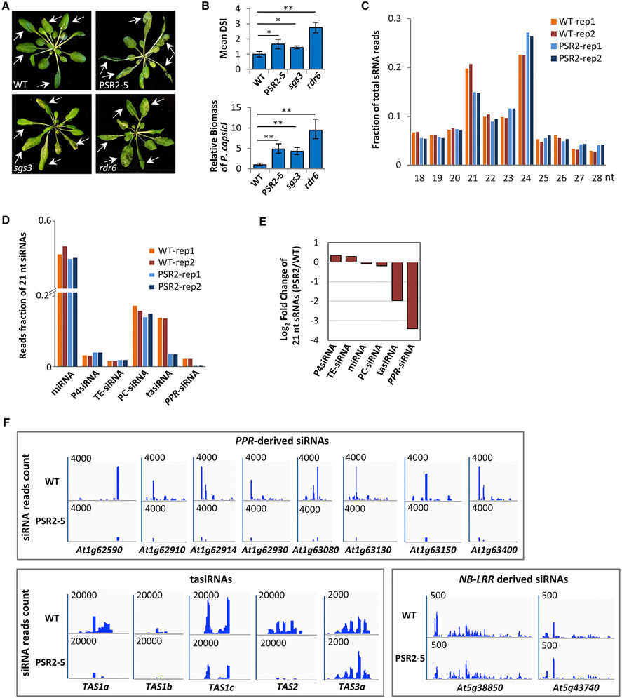
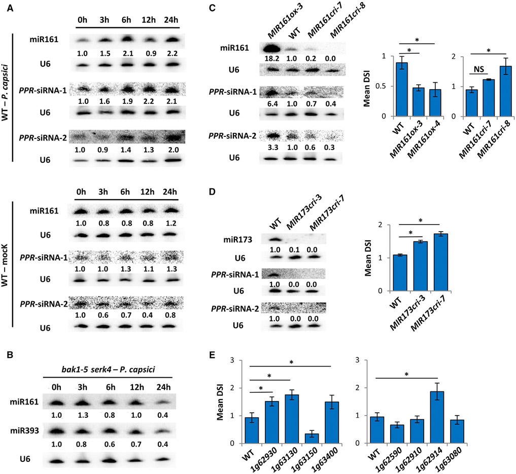
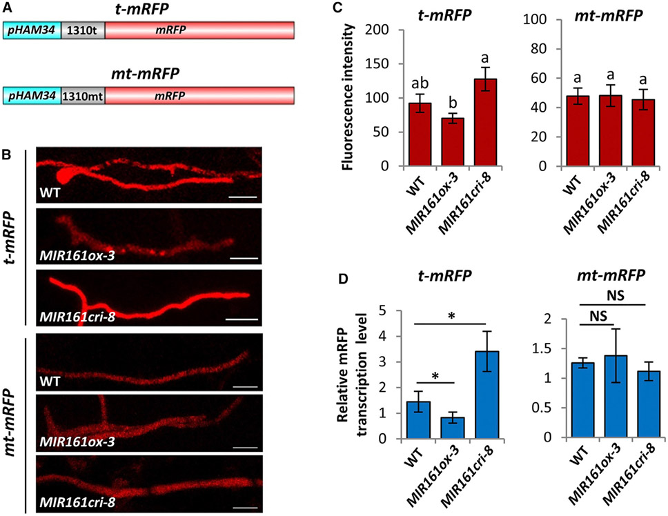
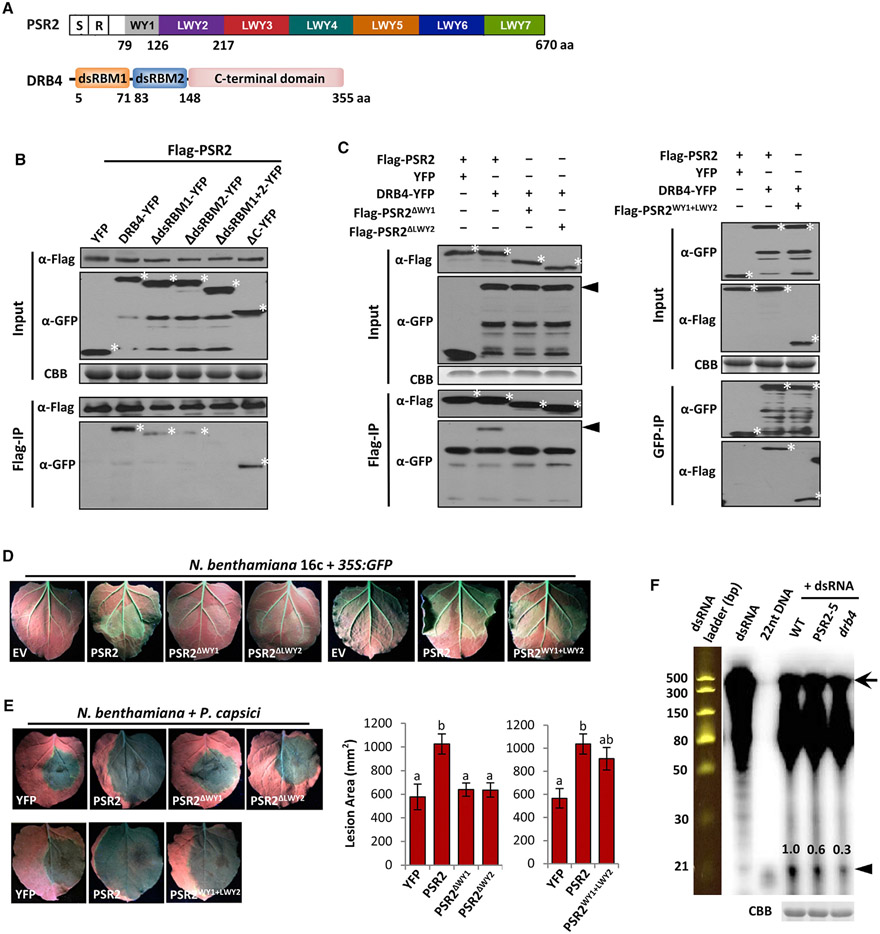
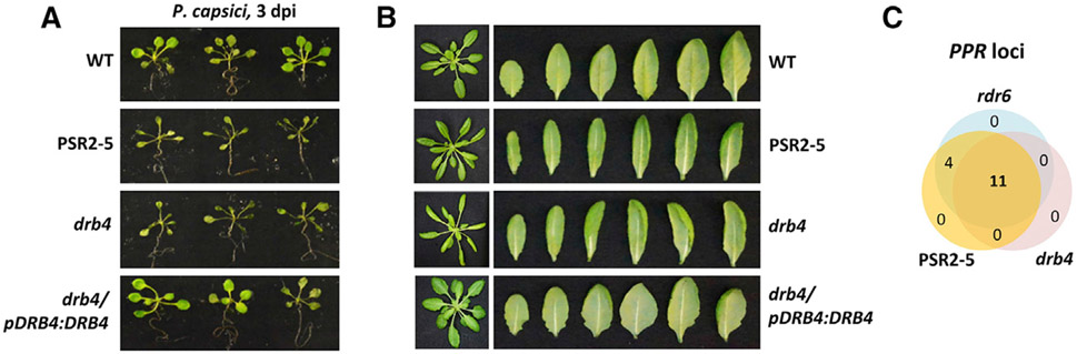
RNA silencing (RNAi) has a well-established role in anti-viral immunity in plants. The destructive eukaryotic pathogen Phytophthora encodes suppressors of RNAi (PSRs), which enhance plant susceptibility. However, the role of small RNAs in defense against eukaryotic pathogens is unclear. Here, we show that Phytophthora infection of Arabidopsis leads to increased production of a diverse pool of secondary small interfering RNAs (siRNAs). Instead of regulating endogenous plant genes, these siRNAs are found in extracellular vesicles and likely silence target genes in Phytophthora during natural infection. Introduction of a plant siRNA in Phytophthora leads to developmental deficiency and abolishes virulence, while Arabidopsis mutants defective in secondary siRNA biogenesis are hypersusceptible. Notably, Phytophthora effector PSR2 specifically inhibits secondary siRNA biogenesis in Arabidopsis and promotes infection. These findings uncover the role of siRNAs as antimicrobial agents against eukaryotic pathogens and highlight a defense/counter-defense arms race centered on trans-kingdom gene silencing between hosts and pathogens.

Keywords: RNA interference; RNA-silencing suppressor; host-induced gene silencing; microRNA; pathogenesis; plant immunity; secondary small RNAs; virulence factor.


Conflict of interest statement

DECLARATION OF INTERESTS

The authors declare no competing interests.

Figures

Figure 1.
Figure 1.. PSR2 Affects the Accumulation of Specific 21-nt siRNAs in Arabidopsis
(A) Mutants defective in secondary siRNA production and a transgenic line expressing PSR2 (PSR2-5) are hypersusceptible to Phytophthora capsici. Arabidopsis plants were inoculated with zoospore suspensions of P. capsici isolate LT263. Photos were taken at 3 days post inoculation (dpi). Arrows indicate inoculated leaves. WT, wild-type Col-0. (B) Disease severity index (DSI) and pathogen biomass in inoculated plants at 3 dpi. Values are mean ± SEM of three biological replicates (n ≥ 20 in each replicate). *p < 0.05, **p < 0.01 (Student’s t test). (C) Size distribution of total sRNAs in WT and PSR2-5 plants. Data from two biological replicates are presented. (D) Reads fraction of 21-nt sRNAs in WT and PSR2-5 plants. Percentage of reads count of miRNAs and siRNAs produced from Pol IV transcripts (P4siRNA), transposable elements (TE), protein-coding transcripts (PC), TAS and PPR transcripts are shown. Data from two biological replicates are presented. (E) Changes in the abundance (in a log2 scale) of 21-nt sRNAs derived from different classes of transcripts in PSR2-5. (F) Secondary siRNAs generated from PPR, TAS, and NB-LRR loci in WT and PSR2-5 plants. The number in each plot indicates the scale of sRNA reads count derived from each locus.
Figure 2.
Figure 2.. Secondary siRNAs Generated from a Cluster of PPR Genes Contribute to Arabidopsis Resistance to P. capsici
(A) Northern blotting showing induced accumulation of miR161 and two representative PPR-siRNAs in WT Arabidopsis during P. capsici infection or mock treatment (water). Numbers represent relative signal intensities. U6 was used as a loading control. Similar results were obtained from two biological replicates. (B) Northern blotting showing unchanged abundance of miR161 and miR393 in bak1 serk4 mutant after P. capsici inoculation. (C) miR161 contributes to plant immunity. The abundance of miR161 and two PPR-siRNAs was determined in WT, MIR161ox, and MIR161cri plants by northern blotting. Disease severity (represented by DSI) was determined at 3 days after inoculation by P. capsici. Values are mean ± SEM of three biological replicates. *p < 0.05 (Student’s t test, n ≥ 20); NS, no statistical difference. (D) MIR173cri mutants are hypersusceptible to P. capsici. The abundance of two PPR-derived siRNAs was evaluated in WT and MIR173cri plants. DSI was determined at 3 dpi. Values are mean ± SEM of three biological replicates. *p < 0.05 (Student’s t test, n ≥ 20). (E) Secondary siRNA-producing PPR genes contribute to Arabidopsis resistance to P. capsici. DSI of eight PPR mutants was determined at 3 dpi. Values are mean ± SEM of three biological replicates. *p < 0.05 (Student’s t test, n ≥ 20). See also Figures S1-S3.
Figure 3.
Figure 3.. A PPR-Derived siRNA Silences a Gene in Phytophthora to Confer Resistance
(A) A flow chart describing the experimental procedure of the functional analysis of PPR-siRNAs. (B) Base pairing of the PPR-derived siR1310 and siR0513 with their predicted target site in Phyca_554980 of P. capsici. (C) qRT-PCR determining the transcript abundances of Phyca_554980 and Phyca_538731 (an off-target control) in P. capsici transformants harboring synthesized siR1310. P. capsici transformed with siRGFP was used as a negative control. Values are mean ± SEM of three biological replicates. (D) Numbers of sporangia produced by WT or transformants of P. capsici harboring siR1310 or siRGFP. Sporangia (indicated by arrows) were numerated from four randomly selected fields of view under a microscope for each strain. Scale bars, 200 μm. Values are mean ± SEM of three biological replicates. One-way ANOVA and post hoc Tukey testing were used for statistical analysis. Different letters label significantly different values (p < 0.05). (E) P. capsici transformants carrying siR1310 lost virulence activity. Mycelial plugs were used to inoculate detached leaves of N. benthamiana. Photos were taken at 3 dpi under UV to better visualize the lesions. Lesion sizes are presented as mean ± SEM of three replicates. One-way ANOVA and post hoc Tukey testing were used for statistical analysis. Different letters label significant different values (p < 0.05). (F) qRT-PCR determining the abundances of siR1310 in leaves or EVs of WT and PSR2-5 Arabidopsis plants with or without P. capsici infection. Values are mean ± SEM of three biological replicates. *p < 0.05, **p < 0.01 (Student’s t test). (G) Transcript abundances of Phyca_554980 determined by qRT-PCR in WT, MIR161ox, PSR2-5, and rdr6 plants inoculated with P. capsici. Values are mean ± SEM of three biological replicates. *p < 0.05, ***p < 0.001 (Student’s t test). See also Figures S4 and S5.
Figure 4.
Figure 4.. PPR-Derived siR1310 Silences a Reporter Gene during Phytophthora Infection
(A) Schematic illustration of the construction of mRFP reporters containing either a target site of siR1310 (t-mRFP) or a mutated target site (mt-mRFP). (B and C) (B) Red fluorescence intensity was monitored during P. capsici infection of WT, MIR161ox, or MIR161cri Arabidopsis plants. Photos were taken at 2 dpi. Scale bars, 20 μm. Values shown in (C) are mean ± SEM and analyzed by one-way ANOVA and post hoc Tukey testing. Different letters label statistically different values (p < 0.05, n ≥ 8). Because t-mRFP and mt-mRFP constructs were independently transformed into P. capsici, their basal mRFP expression levels were different. (D) mRFP transcript levels of P. capsici infecting WT, MIR161ox, and MIR161cri plants were determined by qRT-PCR. Values are mean ± SEM of four replicates. *p < 0.05 (Student’s t test,); NS, no statistical difference. See also Figure S5.
Figure 5.
Figure 5.. PSR2 Associates with DRB4 in Arabidopsis
(A) Schematic representation of the domain structure of PSR2 and DRB4. S, secretion signal; R, RxLR motif. Numbers indicate amino acid positions of the motifs. (B) The two dsRNA-binding domains of DRB4 mediate interaction with PSR2. FLAG-PSR2, DRB4-YFP, and DRB4 truncates were transiently expressed in N. benthamiana. PSR2 was pulled down using anti-FLAG agarose. Enrichment of DRB4 or its truncated mutants in the agarose was detected by western blotting. Asterisk (*) labels corresponding protein band. Protein gel was stained with Coomassie brilliant blue (CBB) as a loading control. (C) WY1 and LWY2 are necessary and sufficient for PSR2 interaction with DRB4. FLAG-tagged PSR2, PSR2ΔWY1, PSR2ΔLWY2, or PSR2WY1+LWY2 were expressed in N. benthamiana with DRB4-YFP. Enrichment of DRB4 in anti-FLAG agarose was detected by western blotting. Asterisk (*) labels corresponding protein band. The arrowhead labels DRB4. (D) WY1 and LWY2 are necessary and sufficient for the transgene-silencing suppression activity of PSR2. Leaves of N. benthamiana 16c plants were co-infiltrated with Agrobacterium carrying 35S:GFP and 35S:PSR2 constructs. Pictures were taken 5 days after Agrobacterium infiltration. EV, empty vector. (E) WY1 and LWY2 are necessary and sufficient for the virulence activities of PSR2. PSR2 or its derivatives were expressed in N. benthamiana leaves, which were subsequently inoculated with P. capsici strain LT263. YFP was used as a control. Lesions were examined at 3 dpi. Values are mean ± SEM of three biological replicates. One-way ANOVA and post hoc Tukey testing were used for statistical analysis. Different letters label significantly different values (p < 0.01). (F) Reduced dsRNA cleavage in PSR2-5 and a drb4 plants. In vitro synthesized dsRNAs (510 bp in length) were labeled with 32P and incubated with protein extracts. Cleavage products were then analyzed by electrophoresis. A 32P-labeled 22-nt DNA and a dsRNA ladder were used as size markers. Arrow and arrowhead label dsRNA precursor and sRNA products respectively. Numbers represent the relative abundances of the sRNA products. Total proteins in the extracts were analyzed on SDS-PAGE and stained with CBB as a loading control. See also Figure S6.
Figure 6.
Figure 6.. A drb4 Mutant Phenocopies PSR2-Expressing Plants
(A) drb4 and PSR2-5 were hypersusceptible to P. capsici. Roots of 14-day-old seedlings were inoculated by zoospore suspensions and photos were taken at 3 dpi. This phenotype was complemented by introducing DRB4-YFP under its native promoter into the drb4 mutant. (B) Five-week-old drb4 and PSR2-5 plants exhibited a similar curly/narrow leaf phenotype. (C) Venn diagram showing PPR loci with reduced secondary siRNA production in rdr6, drb4, and PSR2-5 compared with WT Arabidopsis. See also Figure S6.

Comment in

Similar articles

Cited by

References

    1. Addo-Quaye C, Eshoo TW, Bartel DP, and Axtell MJ (2008). Endogenous siRNA and miRNA targets identified by sequencing of the Arabidopsis degradome. Curr. Biol 18, 758–762. - PMC - PubMed
    1. Adenot X, Elmayan T, Lauressergues D, Boutet S, Bouche N, Gasciolli V, and Vaucheret H (2006). DRB4-dependent TAS3 trans-acting siRNAs control leaf morphology through AGO7. Curr. Biol 16, 927–932. - PubMed
    1. An Q, Ehlers K, Kogel KH, van Bel AJ, and Huckelhoven R (2006a). Multivesicular compartments proliferate in susceptible and resistant MLA12-barley leaves in response to infection by the biotrophic powdery mildew fungus. New Phytol. 172, 563–576. - PubMed
    1. An Q, Huckelhoven R, Kogel KH, and van Bel AJ (2006b). Multivesicular bodies participate in a cell wall-associated defence response in barley leaves attacked by the pathogenic powdery mildew fungus. Cell Microbiol. 8, 1009–1019. - PubMed
    1. Axtell MJ (2013). Classification and comparison of small RNAs from plants. Annu. Rev. Plant Biol 64, 137–159. - PubMed

Publication types

MeSH terms