. 2018 Nov 22:2018:3021863.
doi: 10.1155/2018/3021863. eCollection 2018.

Anti-TNF- α Therapy Suppresses Proinflammatory Activities of Mucosal Neutrophils in Inflammatory Bowel Disease

Affiliations

Anti-TNF- α Therapy Suppresses Proinflammatory Activities of Mucosal Neutrophils in Inflammatory Bowel Disease

Cui Zhang et al. Mediators Inflamm. .

Abstract

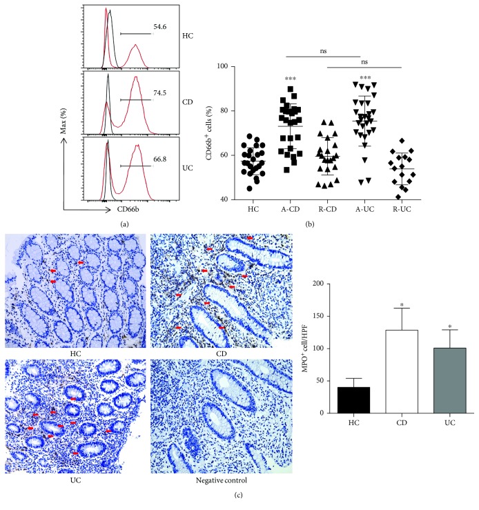
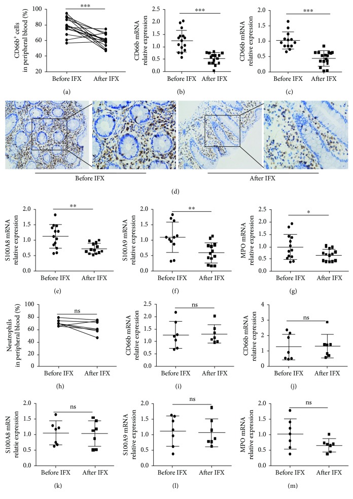
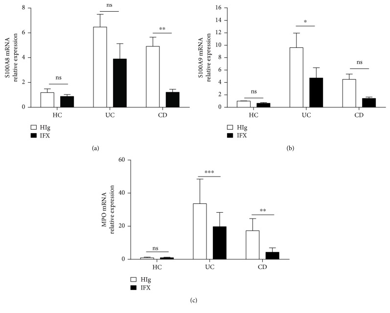
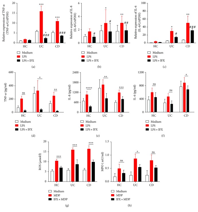
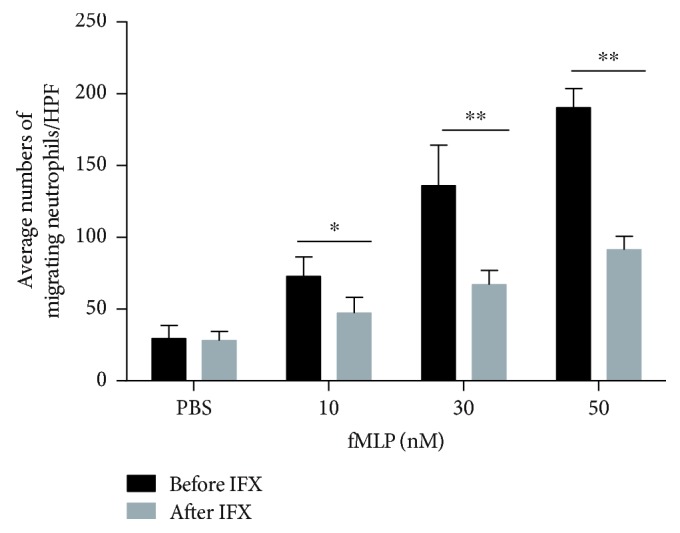
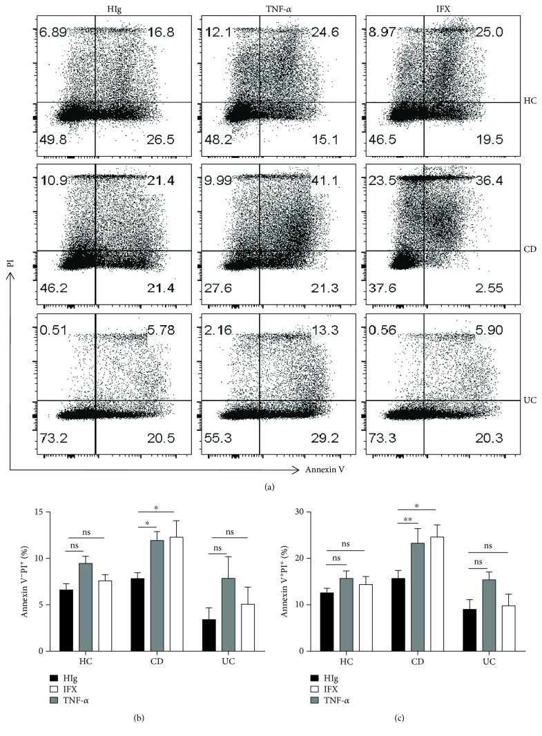
Neutrophils have been found to play an important role in the pathogenesis of inflammatory bowel disease (IBD), and anti-TNF-α mAb (i.e., infliximab) therapy is demonstrated to be effective in the induction of clinical remission and mucosal healing in these patients. However, how anti-TNF-α mAb regulates the functions of neutrophils is still unknown. Herein, we found that anti-TNF-α therapy significantly downregulated infiltration of neutrophils in inflamed mucosa of IBD patients. Importantly, anti-TNF-α mAb could inhibit neutrophils to produce proinflammatory mediators, such as ROS, calprotectin, IL-8, IL-6, and TNF-α. These data indicate that TNF-α plays a critical role in the induction of mucosal inflammatory response, and that blockade of TNF-α modulates intestinal homeostasis through balancing immune responses of neutrophils.


Figures

Figure 1
Figure 1
Neutrophils are more activated in the peripheral blood and intestinal mucosa of active IBD patients. (a, b) Peripheral blood samples were collected from healthy donors (HC, n = 24), patients with active CD (A-CD, n = 26), patients with CD in remission (R-CD, n = 22), patients with active UC (A-UC, n = 28), and patients with UC in remission (R-UC, n = 16). The whole blood excluding RBC was analyzed by flow cytometry. ∗∗∗ P < 0.001 compared with HC. (c) Immunohistochemical staining of MPO in inflamed intestinal mucosa from healthy control (HC, n = 10), patients with A-CD (n = 10), and patients with A-UC (n = 10). The arrows indicate MPO+ cells. Original magnification × 200. P < 0.05 compared with HC.
Figure 2
Figure 2
The activity of neutrophils is inhibited in the peripheral blood, and the infiltration of neutrophils is decreased in the intestinal mucosa of CD patients after anti-TNF-α treatment. Response group: (a) percentages of CD66b+ neutrophils in the peripheral blood of patients with active CD (n = 15) before and after IFX treatment; (b) expression of CD66b mRNA in the peripheral blood neutrophils from patients with active CD (n = 15) before and after IFX treatment by qRT-PCR; (c) expression of CD66b mRNA in intestinal mucosa from patients with active CD (n = 13) before and after IFX treatment by qRT-PCR; (d) immunohistochemical staining of CD66b in the intestinal mucosa of active CD (n = 10) before and after IFX treatment; and (e) expression of S100A8, (f) S100A9, and (g) MPO in the intestinal mucosa of patients with active CD (n = 13) before and after IFX treatment. Failure group: percentage (h) and expression of CD66b (i) of peripheral blood neutrophils from active CD patients (n = 7) before and after IFX. Expression of CD66b (j), S100A8 (k), S100A9 (l), and MPO (m) in intestinal mucosa was detected by qRT-PCR from CD patients in the failure group. P < 0.05, ∗∗ P < 0.01, and ∗∗∗ P < 0.001.
Figure 3
Figure 3
IFX suppresses the expression of neutrophil-derived S100A8, S100A9, and MPO in intestinal mucosa. Fresh colon biopsies were collected from inflamed mucosa of patients with active CD (n = 12) and active UC (n = 12), and cultured ex vivo with IFX or control human IgG (HIg) (both at 50 μg/ml) for 24 h. Tissues were harvested for detection of expression of S100A8 (a), S100A9 (b), and MPO (c) by qRT-PCR and compared with healthy controls (n = 5). P < 0.05 and ∗∗ P < 0.01.
Figure 4
Figure 4
Anti-TNF-α therapy inhibits peripheral neutrophils to produce proinflammatory cytokines, chemokines, ROS, and MPO. Peripheral neutrophils (5 × 106) from healthy donors (n = 10), patients with active CD (n = 10), and active UC (n = 10) were stimulated by LPS (200 ng/ml) and incubated with or without IFX (50 μg/ml) for 3 h. Cells were collected and expressions of TNF-α (a), IL-8 (b), and IL-6 (c) were detected by qRT-PCR. Peripheral neutrophils (2 × 106) from healthy donors (n = 6), patients with active CD (n = 11), and patients with active UC (n = 4) were simulated with LPS (200 ng/ml) and incubated with or without IFX (50 μg/ml) for 3 h. Culture media were replenished and incubated for another 24 h. Supernatants and protein production of TNF-α (d), IL-8 (e), and IL-6 (f) were measured by ELISA. Peripheral neutrophils (1 × 104) isolated from healthy donors (n = 5), patients with active CD (n = 5), and patients with active UC (n = 5) were measured for ROS (h) and MPO (i) with Amplex Red Hydrogen Peroxide Assay Kit. P < 0.05, ∗∗ P < 0.01, and ∗∗∗ P < 0.001 compared with medium control and # P < 0.05, ## P < 0.01, and ### P < 0.001 compared with LPS stimulation.
Figure 5
Figure 5
Anti-TNF-α therapy suppresses neutrophil migration in active CD patients. Peripheral neutrophils (5 × 105) were isolated from patients with active CD (n = 4) and measured with an 8 μm-Transwell plate under attraction with fMLP (10, 30, and 50 nM) for 30 min. The histogram represents the number of migrating neutrophils per high-power field (HPF). P < 0.05 and ∗∗ P < 0.01.
Figure 6
Figure 6
Anti-TNF-α therapy promotes apoptosis of neutrophils in active CD patients. (a–c) Peripheral neutrophils were isolated from healthy donors (n = 8), patients with active CD (n = 10), and patients with active UC (n = 5) and incubated with HIg (50 μg/ml), IFX (50 μg/ml), or TNF-α (20 ng/ml) for 24 h. Cells were collected and detected for apoptosis by flow cytometry. P < 0.05.

References

    1. Ng S. C., Tang W., Ching J. Y., et al. Incidence and phenotype of inflammatory bowel disease based on results from the Asia-Pacific Crohn’s and colitis epidemiology study. Gastroenterology. 2013;145(1):158–165.e2. doi: 10.1053/j.gastro.2013.04.007. - DOI - PubMed
    1. Sun M., He C., Cong Y., Liu Z. Regulatory immune cells in regulation of intestinal inflammatory response to microbiota. Mucosal Immunology. 2015;8(5):969–978. doi: 10.1038/mi.2015.49. - DOI - PMC - PubMed
    1. Kaser A., Zeissig S., Blumberg R. S. Inflammatory bowel disease. Annual Review of Immunology. 2010;1(28):573–621. - PMC - PubMed
    1. Liu Z., Cao A. T., Cong Y. Microbiota regulation of inflammatory bowel disease and colorectal cancer. Seminars in Cancer Biology. 2013;23(6):543–552. doi: 10.1016/j.semcancer.2013.09.002. - DOI - PMC - PubMed
    1. Danese S., Vuitton L., Peyrin-Biroulet L. Biologic agents for IBD: practical insights. Nature Reviews Gastroenterology & Hepatology. 2015;12(9):537–545. doi: 10.1038/nrgastro.2015.135. - DOI - PubMed

MeSH terms

LinkOut - more resources