

Analysis of Cause of Endodontic Failure of C-Shaped Root Canals
- PMID: 30595786
- PMCID: PMC6286757
- DOI: 10.1155/2018/2516832
Analysis of Cause of Endodontic Failure of C-Shaped Root Canals
Abstract
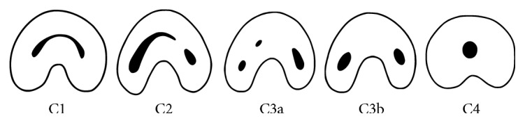
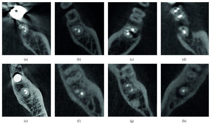
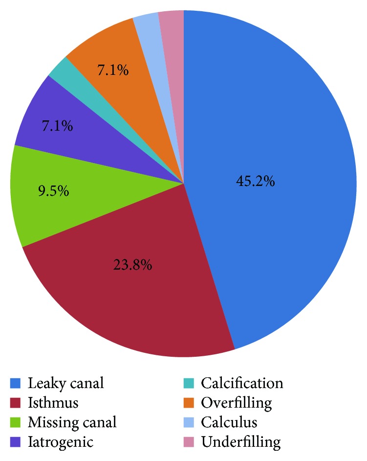
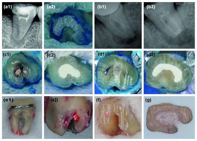
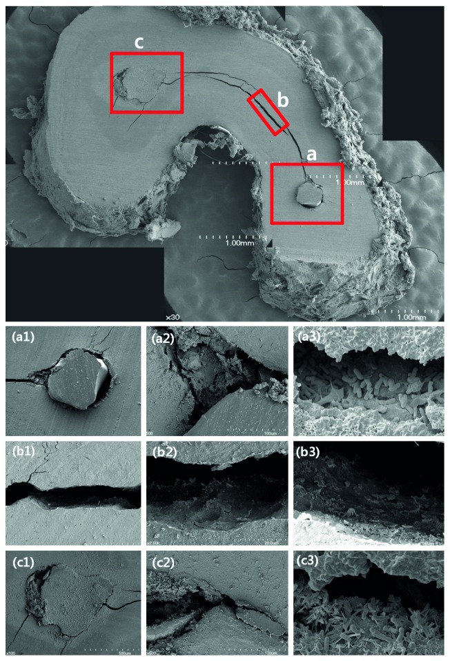
The purpose of this study was to analyze various characteristics and classification of C-shaped root canals and evaluate the causes of endodontic failure of C-shaped root canals by examining the resected root surface with an endodontic microscope and a scanning electron microscope (SEM). Forty-two teeth with C-shaped root canals were included in this study and had undergone intentional replantation surgery. Before surgery, periapical radiography and cone-beam computed tomography were taken. The root canal configuration was analyzed and classified according to Melton's classification at coronal and apical level. After injection of 1 : 100,000 epinephrine with 2% lidocaine, the tooth was carefully extracted. After the root-end resection, the resected root surface was examined using an operating microscope and SEM. Mandibular second molars were most frequently involved teeth (90.4%). The most frequently observed root canal configurations were C1 at the coronal level (45.2%) and C3 at the apical 3 mm level (45.2%). The most common cause of failure for a C-shaped root canal treatment was a leaky canal (45.2%), followed by an isthmus (23.8%), missing canal, overfilling, and iatrogenic problems. In conclusion, C-shaped root canals were most frequently found in mandibular second molars. The most common cause of failure was a leaky canal and isthmus.
Figures
Similar articles
-
A biometric study of C-shaped root canal systems in mandibular second molars using cone-beam computed tomography.Int Endod J. 2012 Sep;45(9):807-14. doi: 10.1111/j.1365-2591.2012.02037.x. Epub 2012 Mar 20. Int Endod J. 2012. PMID: 22432971
-
Analysis of the root canal configuration and C-shaped canal frequency of mandibular second molars: a cone beam computed tomography study.Folia Morphol (Warsz). 2018;77(4):752-757. doi: 10.5603/FM.a2018.0040. Epub 2018 May 26. Folia Morphol (Warsz). 2018. PMID: 29802711 Clinical Trial.
-
C-shaped canals-prevalence and root canal configuration by cone beam computed tomography evaluation in first and second mandibular molars-a cross-sectional study.Clin Oral Investig. 2017 Jul;21(6):2039-2044. doi: 10.1007/s00784-016-1993-y. Epub 2016 Nov 14. Clin Oral Investig. 2017. PMID: 27844150
-
C-shaped root canal in a maxillary first molar: a case report.Int Endod J. 2006 Feb;39(2):162-6. doi: 10.1111/j.1365-2591.2006.01069.x. Int Endod J. 2006. PMID: 16454798 Review.
-
Evaluation of root canal morphology of human primary molars by using CBCT and comprehensive review of the literature.Acta Odontol Scand. 2016;74(4):250-8. doi: 10.3109/00016357.2015.1104721. Epub 2015 Nov 2. Acta Odontol Scand. 2016. PMID: 26523502 Review.
Cited by
-
Robotic and Microrobotic Tools for Dental Therapy.J Healthc Eng. 2022 Feb 18;2022:3265462. doi: 10.1155/2022/3265462. eCollection 2022. J Healthc Eng. 2022. PMID: 35222881 Free PMC article. Review.
-
Evaluation of various obturation techniques with bioceramic sealers in 3D-printed C-shaped canals.BMC Oral Health. 2024 May 12;24(1):554. doi: 10.1186/s12903-024-04334-2. BMC Oral Health. 2024. PMID: 38735924 Free PMC article.
-
Deep-learning for predicting C-shaped canals in mandibular second molars on panoramic radiographs.Dentomaxillofac Radiol. 2021 Jul 1;50(5):20200513. doi: 10.1259/dmfr.20200513. Epub 2021 Jan 6. Dentomaxillofac Radiol. 2021. PMID: 33405976 Free PMC article.
-
Endodontic Retreatment of a Mandibular Second Molar With a C-shaped Root Canal Configuration: A Case Report.Cureus. 2024 Jan 23;16(1):e52812. doi: 10.7759/cureus.52812. eCollection 2024 Jan. Cureus. 2024. PMID: 38389597 Free PMC article.
-
Root canal disinfection and maintenance of the remnant tooth tissues by using grape seed and cranberry extracts.Odontology. 2023 Jul;111(3):541-553. doi: 10.1007/s10266-022-00766-w. Epub 2022 Dec 10. Odontology. 2023. PMID: 36495398 Review.
References
MeSH terms
Substances
LinkOut - more resources
Full Text Sources
Miscellaneous
