

CD44 Immunoexpression in the Progression of Actinic Keratosis and Cutaneous Squamouscarcinoma
- PMID: 30595883
- PMCID: PMC6284843
- DOI: 10.12865/CHSJ.43.03.10
CD44 Immunoexpression in the Progression of Actinic Keratosis and Cutaneous Squamouscarcinoma
Abstract
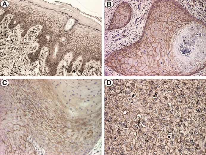
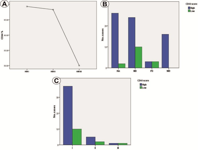
CD44 seems to confer the needed conditions for malignant neoplasms to grow and progress. The present study aims to investigate the role of CD44 expression in pre-invasive and invasive squamous lesions of the skin. We investigated 89 cases of preinvasive and invasive cutaneous lesions, of which 28 corresponded to actinic keratosis (KIN- keratinocyte intraepithelial neoplasia) with varying degrees of severity and 61 cases of squamous cell carcinoma with variable degrees of differentiation. The statistical analysis of CD44 immunoexpression indicated significantly higher values for KIN I and II compared to KIN III, as well as for KIN lesions in comparison with squamous cell carcinomas. Similar results were observed in well differentiated carcinomas compared to moderate and poorly differentiated lesions. These observations suggest that CD44 expression plays a role in the progression of cutaneous squamous neoplasia.
Keywords: CD44; actinic keratosis; squamous cell carcinomas.
Figures
Similar articles
-
P21 Immunoexpression in Actinic Keratosis and Cutaneous Carcinomas.Curr Health Sci J. 2017 Jul-Sep;43(3):226-230. doi: 10.12865/CHSJ.43.03.07. Epub 2017 Sep 28. Curr Health Sci J. 2017. PMID: 30595880 Free PMC article.
-
Cyclin A and beta-catenin expression in actinic keratosis, Bowen's disease and invasive squamous cell carcinoma of the skin.Br J Dermatol. 2005 Dec;153(6):1166-75. doi: 10.1111/j.1365-2133.2005.06898.x. Br J Dermatol. 2005. PMID: 16307653
-
The development of actinic keratosis into invasive squamous cell carcinoma: evidence and evolving classification schemes.Clin Dermatol. 2004 May-Jun;22(3):189-96. doi: 10.1016/j.clindermatol.2003.12.006. Clin Dermatol. 2004. PMID: 15262304 Review.
-
Expression Profile of Fibroblast Growth Factor Receptors, Keratinocyte Differentiation Markers, and Epithelial Mesenchymal Transition-Related Genes in Actinic Keratosis: A Possible Predictive Factor for Malignant Progression?Biology (Basel). 2021 Apr 15;10(4):331. doi: 10.3390/biology10040331. Biology (Basel). 2021. PMID: 33920760 Free PMC article.
-
[Actinic keratosis, Bowen's disease, keratoacanthoma and squamous cell carcinoma of the skin].Pathologe. 2015 Feb;36(1):16-29. doi: 10.1007/s00292-014-2063-3. Pathologe. 2015. PMID: 25663185 Review. German.
Cited by
-
Adhesion Molecules in Non-melanoma Skin Cancers: A Comprehensive Review.In Vivo. 2021 May-Jun;35(3):1327-1336. doi: 10.21873/invivo.12385. Epub 2021 Apr 28. In Vivo. 2021. PMID: 33910810 Free PMC article. Review.
-
Retrospective Study of a Patient with Multiple de Novo Skin Tumor Formations.Curr Health Sci J. 2023 Jul-Sep;49(3):434-437. doi: 10.12865/CHSJ.49.03.18. Epub 2023 Sep 30. Curr Health Sci J. 2023. PMID: 38314212 Free PMC article.
References
-
- Grimme HU, Termeer CC, Bennett KL, Weiss JM, Schöpf E, Aruffo A, Simon JC. Colocalization of basic fibroblast growth factor and CD44 isoforms containing the variably spliced exon v3 (CD44v3) in normal skin and in epidermal skin cancers. Br J Dermatol. 1999;141(5):824–832. - PubMed
-
- Dietrich A, Tanczos E, Vanscheidt W, Schöpf E, Simon JC. Detection of CD44 splice variants in formalin-fixed, paraffin-embedded specimens of human skin cancer. J Cutan Pathol. 1997;24(1):37–42. - PubMed
-
- Bourguignon LY, Singleton PA, Diedrich F. Hyaluronan-CD44 interaction with Rac1-dependent protein kinase N-gamma promotes phospholipase Cgamma1 activation, Ca(2+) signaling, and cortactin-cytoskeleton function leading to keratinocyte adhesion and differentiation. J Biol Chem. 2004;279(28):29654–29669. - PubMed
-
- Bourguignon LY, Ramez M, Gilad E, Gilad E, Singleton PA, Man MQ, Crumrine DA, Elias PM, Feingold KR. Hyaluronan-CD44 interaction stimulates keratinocyte differentiation, lamellar body formation/secretion, and permeability barrier homeostasis. J Invest Dermatol. 2006;126(6):1356–1365. - PubMed
Publication types
LinkOut - more resources
Full Text Sources
Miscellaneous