Review
. 2018 Jul 27:12:82-89.
doi: 10.1016/j.pacs.2018.07.004. eCollection 2018 Dec.

Photoacoustic tomography for human musculoskeletal imaging and inflammatory arthritis detection

Affiliations
Review

Photoacoustic tomography for human musculoskeletal imaging and inflammatory arthritis detection

Janggun Jo et al. Photoacoustics. .

Abstract

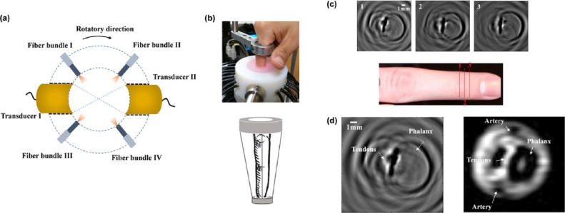
With the capability of assessing high resolution optical contrast in soft tissues, photoacoustic imaging (PAI) can offer valuable structural and functional information of human joints, and hold potential for diagnosis and treatment monitoring of inflammatory arthritis. Recent studies have demonstrated that PAI can map 2D and 3D morphology of the cartilage, synovium, vascularity, and bone tissue in human peripheral joints. Initial trials with patients affected by inflammatory arthritis have also suggested that PAI can detect the hemodynamic properties in articular tissues as well as their changes due to active inflammation. This review focuses on the recent progress in technical development of PAI for human musculoskeletal imaging and inflammation detection. PAI can provide non-invasive and non-ionizing serial measurements for monitoring of therapeutic interventions with the potential for higher sensitivity than existing imaging modalities such as ultrasound. However, further investigation is needed to validate the value of PAI in rheumatology clinical settings.

Keywords: Human joint; Inflammatory arthritis; Photoacoustic tomography.


Figures

Fig. 1
Fig. 1
(a) Schematic of a home-fabricated tomographic PAI system for imaging of human joints ex vivo. (b) A cross-sectional image along a PIP joint. (c) A cross-sectional image along a DIP joint. (d)–(e) Corresponding anatomical photographs from the same joints confirming the imaging results in (b) and (c), respectively. AP: aponeurosis, PH: phalanx, SK: skin, SU: subcutaneous tissue, TE: tendon, VP: volar plate. Adapted with permission from Ref. [30].
Fig. 2
Fig. 2
(a) Schematic of a PAI system for joint imaging utilizing virtual detector concept in image reconstruction and the cylindrical scanning in data collection. (b) Photograph showing the PAI-joint interface for DIP joint imaging. (c) Cross-sectional PA images along three slices in a DIP joint of a female middle finger. (d) Comparison between the PA image and the corresponding MRI image. Adapted with permission from Ref. [32].
Fig. 3
Fig. 3
(a) Schematic of a home-built PAI setup involving 32 transducers and 6 optical fibers. (b) Photograph of the system. (c)-(j) PA cross-sectional images along different slices in a healthy index finger of a volunteer. The images are taken at the positions shown in the longitudinal US image (l) and are concentrated at the PIP joint and the DIP joint. (c#), (e#), (g#), and (i#) Enlarged images from the boxes in (c), (e), (g), and (i), respectively. (k) Cross-sectional and (l) longitudinal US images of the finger. Adapted with permission from Ref. [33].
Fig. 4
Fig. 4
(a) Schematic of a PA and US dual modality imaging system for human peripheral joints built on a commercial US unit and a linear array probe. (b) Photograph of the experimental setup for coronal middle plane from the volar side. (c) Photograph of the experimental setup for sagittal middle plane. (d) PA and US images of coronal plane (volar) of a male PIP joint. (e) PA and US images of sagittal plane of a male PIP joint. (f) PA and US images of coronal plane (volar) of a female PIP joint. (g) PA and US images of sagittal plane of a female PIP joint. TE: tendon, JO: joint, PE: periosteum, BO: bone. Adapted with permission from Ref. [34].
Fig. 5
Fig. 5
(a) Schematic of PA and US dual modality real-time imaging system for human peripheral joints built on a research US platform and a linear array probe. (b)–(e) PA and US imaging of a human proximal interphalangeal joint. (b) PA image using traditional back-projection method. (c) PA image using optimized back-projection method. (d) PA image after envelop detection of the image in (c). (e) Grey-scale US image. TE: tendon, JO: joint, PE: periosteum, BO: bone, and IN: inner structure of tendon. Adapted with permission from Ref. [35].
Fig. 6
Fig. 6
(a) Photograph of a PAI system applied to the imaging of finger joints from normal volunteers and OA patients. (b)–(e) Recovered absorption coefficient images for four normal joints. (f)–(g) Recovered absorption coefficient images for two OA joints. (h) Average absorption coefficient of cartilage (red) and fluid (blue) for the healthy (H1-H4) and the OA (OA1-OA2) joints. Adapted with permission from Ref. [36].
Fig. 7
Fig. 7
(a) Photograph of the PIP joint 3 of a patient affected by early rheumatoid arthritis. (b) Cross-sectional PA image at location indicated in (a). A magnified view of indicated region shows small thread shaped and point shaped blood vessels at large depth (4–6 mm), which is thought to be associated with arthritis. Adapted with permission from Ref. [37].
Fig. 8
Fig. 8
(a) The pseudo-color PA image superimposed on the gray scale US image of a right hand 2nd MCP joint affected by arthritis, demonstrating the presence of active vascularity in the joint. Yellow arrow: bone; Green arrow: tendon; Red arrow: skin. (b) US Doppler image acquired by a commercial US unit confirming the active synovitis in the result in (a). (c) The pseudo-color PA image superimposed on the gray scale US image of a right hand 2nd MCP joint of a healthy control subject. No active vascularity was noticed in the joint. (d) US Doppler image confirming the normal finding in (c). (e)–(f) Statistical studies comparing single-wavelength PA imaging results from the 16 arthritic joints to those from the 16 healthy controls. (e) The quantified density of the pseudo-color pixels in the joint area (p < 0.001). (f) The averaged intensity of the pseudo-color pixels in the joint area (p < 0.001). Adapted with permission from Ref. [38].

Similar articles

Cited by

References

    1. Barbour K.E., Helmick C.G., Boring M., Brady T.J. Vital signs: prevalence of doctor-diagnosed arthritis and arthritis-attributable activity limitation – United States, 2013–2015. MMWR Morb. Mortal. Wkly. Rep. 2017;66(9):246–253. - PMC - PubMed
    1. Hootman J.M., Helmick C.G., Barbour K.E., Theis K.A., Boring M.A. Updated projected prevalence of self-reported doctor-diagnosed arthritis and arthritis-attributable activity limitation among US adults, 2015-2040 . 2016;68(7):1582–1587. - PMC - PubMed
    1. McNeil J.M., Binette J. Cdc, prevalence of disabilities and associated health conditions among adults – United States, 1999 (reprinted from MMWR, vol 50, pg 120–125, 2001) JAMA-J. Am. Med. Assoc. 2001;285(12):1571–1572.
    1. Yelin E., Weinstein S., King T. The burden of musculoskeletal diseases in the United States. Semin. Arthritis Rheum. 2016;46(3):259–260. - PubMed
    1. Garnero P., Rousseau J.-C., Delmas P.D. Molecular basis and clinical use of biochemical markers of bone, cartilage, and synovium in joint diseases. Arthritis Rheum. 2000;43(5):953–968. - PubMed

LinkOut - more resources