. 2018 Nov 25:2018:2942679.
doi: 10.1155/2018/2942679. eCollection 2018.

HLA-A2-Restricted Epitopes Identified from MTA1 Could Elicit Antigen-Specific Cytotoxic T Lymphocyte Response

Affiliations

HLA-A2-Restricted Epitopes Identified from MTA1 Could Elicit Antigen-Specific Cytotoxic T Lymphocyte Response

Yahong Wu et al. J Immunol Res. .

Abstract

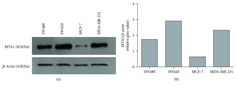
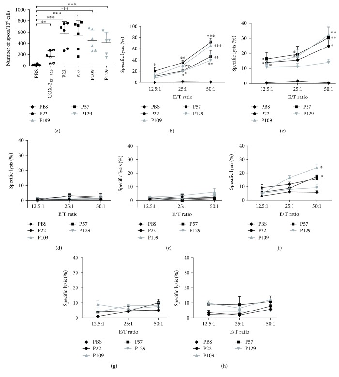
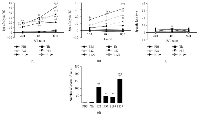
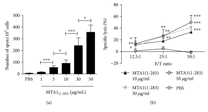
Overexpression of metastasis-associated protein 1 (MTA1) has been observed in many human malignancies and is significantly related to tumor invasion and metastasis, therapeutic resistance to radiation and chemotherapy, making MTA1 an ideal candidate tumor antigen. We identified several human leukocyte antigen- (HLA-) A2-restricted epitopes in MTA1 and evaluated their binding ability to HLA-A0201 molecules. Subsequently, a recombinant fragment encompassing the dominant epitopes, MTA1(1-283), was expressed, and the abilities of the selected epitopes of MTA1 and the MTA1(1-283) fragment to induce cytotoxic T lymphocytes (CTLs) were examined. Our results indicated that the epitopes and MTA1(1-283) fragment elicited HLA-A2-restricted and antigen-specific CTL responses both in vitro and in vivo. The new epitopes identified here may help promote the development of new therapeutic vaccines for HLA-A2+ patients expressing MTA1.


Figures

Figure 1
Figure 1
Protein expression of MTA1 by Western Blot analysis. The protein expression of MTA1 (81 kDa) was evaluated in four human cancer cell lines. β-Actin (42 kDa) was used as loading control, and rabbit polyclonal anti-MTA1 antibody (cat: bs-1412R, dilutions: 1 : 1500, Bioss Antibodies, China) or rabbit polyclonal anti-β-actin antibody (cat: bs-0061R, dilutions: 1 : 50,000, Bioss Antibodies, China) was used in this assay. (a) The bands representing MTA1 and β-actin in each cancer cell line. (b) Gray analysis of the MTA1 protein band intensities (relative to the β-actin band intensity) was carried out by Image J.
Figure 2
Figure 2
IFN-γ release and lyse target cell ability of peptide-induced CTLs. PBMCs from six HLA-A02+ healthy donors were collected and induced by the indicated peptides (10 μg/ml) three times in RPMI 1640 medium supplemented with 3 μg/ml β2-M (once at each stimulation), interleukin-2 (50 U/ml, on day 3 and 1 day after each stimulation), and 10% FCS for 21 days. CTLs induced by COX-2321–329 were used as positive controls. On day 21, the CTLs were collected, and the IFN-γ secretion and cytotoxic activity of the CTLs were measured. (a) CTL secretion of IFN-γ was detected by the ELISPOT assay (n = 6). T2 cells loaded with/without peptide (50 μg/ml) for 4 h were used as the stimulating cells. LDH cytotoxicity assays were performed (n = 3) using the following target cell lines: (b) SW620 (HLA-A2+, MTA1+), (c) MDA-MB-231 (HLA-A2+, MTA1+), (d) SW620 incubated with an anti-human HLA-A2 antibody (HLA-A2, MTA1+), (e) HT-29 (HLA-A2, MTA1+), (f) MCF-7 (HLA-A2+, MTA1+), (g) HUVEC (HLA-A2+, MTA1low), and (h) Het-1A (HLA-A2+, MTA1low). CTLs induced by PBS were used as the negative control. Data represent means ± standard deviation (SD), P < 0.05, ∗∗ P < 0.01, and ∗∗∗ P < 0.001 vs. the PBS group.
Figure 3
Figure 3
Specific lysis of target cells and secretion of IFN-γ by induced CTLs in immunizing HLA-A2.1/Kb transgenic mice immunized with the indicated peptides. Peptide (100 μg) and the IAb-restricted HBV-core128 T helper epitope (140 μg) emulsified in IFA were used to immunize HLA-A2.1/Kb transgenic mice on days 0, 5, and 10. PBS only or T helper peptide emulsified in IFA was used as the negative control. On day 11, we isolated splenocytes from the mice and restimulated the cells with 10 μg/ml peptide and 10 U/ml rmIL-2 to repriming these splenocytes in vitro for additional 6 days. An LDH assay was performed to assess the lysis activity of the induced CTLs toward (a) SW620 cells (HLA-A2+, MTA1+), (b) T2 cells pulsed with each peptide, and (c) SW620 cells incubated with an anti-human HLA-A2 antibody (HLA-A2, MTA1+). IFN-γ secretion was detected by the ELISPOT assay. (d) T2 cells incubated with each peptide and an irrelevant peptide (50 μg/ml) for 4 h were used as the stimulating cells. Each sample was measured in triplicate. Data represent means ± SD (n = 5). P < 0.05, ∗∗ P < 0.05, and ∗∗∗ P < 0.001 vs. the T helper epitope group.
Figure 4
Figure 4
Specific T cells induced by MTA1(1–283) secrete IFN-γ and lyse target cells. PBMCs from healthy HLA-A02+ donors were isolated and stimulated with protein MTA1(1–283) (at final concentrations of 0, 1, 5, 10, 30, and 50 μg/ml) in RPMI 1640 medium supplemented with 50 U/ml interleukin-2 and 10% FCS once a week for 21 days. On day 21, the induced T cells were collected; (a) IFN-γ secretion was assessed by the ELISPOT assay; P < 0.05 and ∗∗∗ P < 0.001 between two groups. (b) Cytotoxic activity was assessed by the LDH assay using SW620 cells (HLA-A2+, MTA1+) as the target cells. T cells induced by PBS were used as negative controls. P < 0.05, ∗∗ P < 0.01, and ∗∗∗ P < 0.001 vs. the control group. Data represent means ± SD (n = 4).
Figure 5
Figure 5
Specific lysis of various cell lines and IFN-γ secretion by induced T cells in HLA-A2.1/Kb transgenic mice immunized with MTA1(1–283). MTA1(1–283) (100 μg/ml) or the same volume of PBS (negative control) emulsified in IFA was used to immunize HLA-A2.1/Kb transgenic mice on days 0 and 7. On day 14, the mice were sacrificed, and their splenocytes were collected and restimulated for another 6 days with the peptide pool (10 μg/ml) and 50 U/ml rmIL-2 in vitro. (a) Serum was collected from the immunized mice on day 14, and the serum IFN-γ concentration was measured by ELISA. (b) The percentage of IFN-γ secreting peptide-specific CD8+ T cells induced by MTA1(1–283) and PBS was analyzed by an intracellular staining assay. (c) The percentages of IFN-γ+CD4+ T cells in the MTA1(1–283) and PBS groups were determined. (d) Specific lysis of target cells SW620 cells (HLA-A2+, MTA1+) was assessed by the LDH cytotoxicity assay. P < 0.05 and ∗∗∗ P < 0.001. P < 0.05 vs. the control group. Data represent means ± SD (n = 5).

Similar articles

Cited by

References

    1. Waitz R., Solomon S. B., Petre E. N., et al. Potent induction of tumor immunity by combining tumor cryoablation with anti-CTLA-4 therapy. Cancer Research. 2012;72(2):430–439. doi: 10.1158/0008-5472.CAN-11-1782. - DOI - PMC - PubMed
    1. Ramamurthy C., Godwin J. L., Borghaei H. Immune checkpoint inhibitor therapy: what line of therapy and how to choose? Current Treatment Options in Oncology. 2017;18(6):p. 33. doi: 10.1007/s11864-017-0476-y. - DOI - PubMed
    1. Gros A., Parkhurst M. R., Tran E., et al. Prospective identification of neoantigen-specific lymphocytes in the peripheral blood of melanoma patients. Nature Medicine. 2016;22(4):433–438. doi: 10.1038/nm.4051. - DOI - PMC - PubMed
    1. Coulie P. G., van den Eynde B. J., van der Bruggen P., Boon T. Tumour antigens recognized by T lymphocytes: at the core of cancer immunotherapy. Nature Reviews Cancer. 2014;14(2):135–146. doi: 10.1038/nrc3670. - DOI - PubMed
    1. Toh Y., Pencil S. D., Nicolson G. L. A novel candidate metastasis-associated gene, mta 1, differentially expressed in highly metastatic mammary adenocarcinoma cell lines. cDNA cloning, expression, and protein analyses. Journal of Biological Chemistry. 1994;269(37):22958–22963. - PubMed

MeSH terms