

Review
doi: 10.1016/j.ekir.2018.10.009.
eCollection 2019 Jan.
Pink Urine Syndrome: A Combination of Insulin Resistance and Propofol
Affiliations
- PMID: 30596166
- PMCID: PMC6308841
- DOI: 10.1016/j.ekir.2018.10.009
Item in Clipboard
Review
Pink Urine Syndrome: A Combination of Insulin Resistance and Propofol
Kidney Int Rep.
.
Abstract
Pink urine syndrome is mostly seen in patients treated with propofol anesthesia. The pink color is attributed to the presence of large concentrations of uric acid (and pigment), which is excreted in large amounts when propofol is given. We describe a case of propofol-induced pink urine syndrome and perform a comprehensive, evidence-based review. We discuss prior case studies already published in the literature as we speculate on the pathophysiology and how it translates to a clinically relevant entity.
Keywords: acid-base equilibrium; biological; insulin resistance; oxidative stress; pigments; propofol; uric acid.
Figures
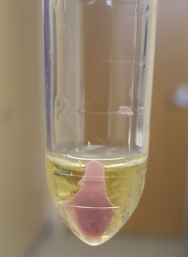
Pink urine sediment after centrifugation in a patient who received propofol.
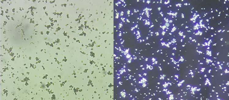
Amorphous crystals. Urine sediment examination demonstrating amorphous crystals. The left is ×40 light microscopy and the right is the same image but polarized.
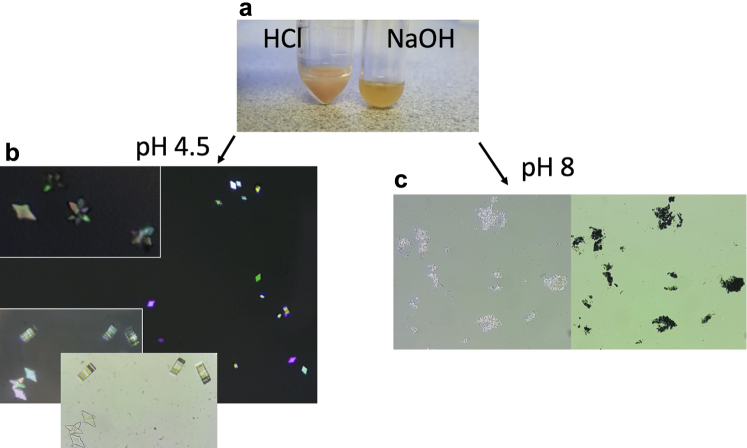
The effect of pH on uric acid crystallization. One drop of 1 N HCl solution (1 normal = 36.5 g hydrochloric acid/1 liter) was mixed with 2 ml of patient’s urine resulting in a reduction of pH, pinkish hue (a, left test tube) on gross inspection, and polychromatic birefringence uric acid crystallization with light microscopy (b). In a similar fashion, 1 drop of 1 N NaOH solution (1 N = 40 g sodium hydroxide/1 liter) was mixed with 2 ml of urine to serve as a negative-treatment response control. On NaOH addition, urine color became a dirty yellow (a, right test tube), urine pH increased, and amorphous crystals were present when analyzed with light microscopy (c).
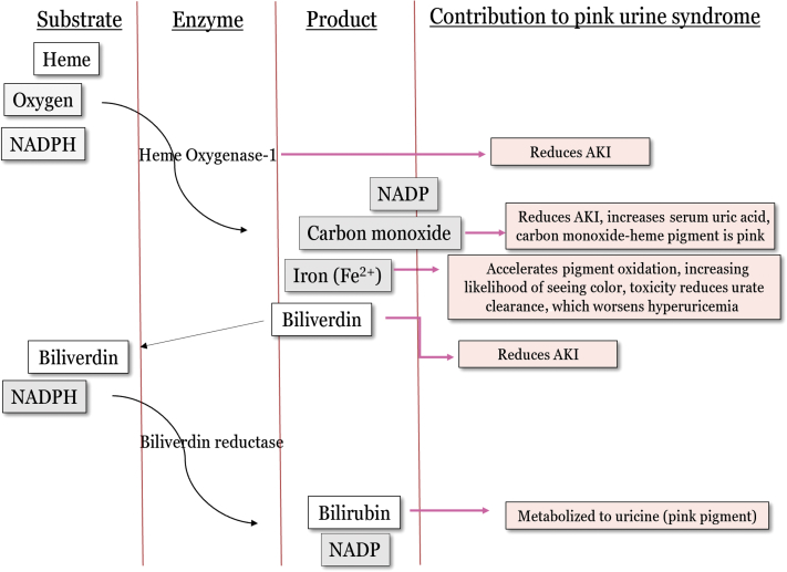
The heme oxygenase-1 pathway. This enzymatic pathway is activated by oxidative stress and is chronically active during a state of insulin resistance. This pathway becomes overwhelmed and unable to become active enough to completely prevent the increasing amount of oxidative damage. Propofol can further activate this enzyme, which reduces oxidative damage and protects the cell. AKI, acute kidney injury; NADPH, nicotinamide adenine dinucleotide phosphate hydrogen.
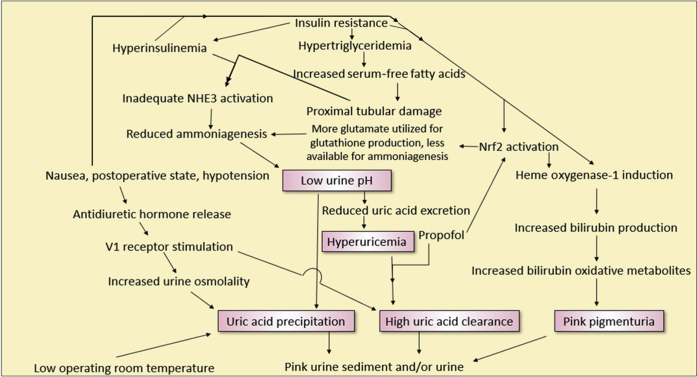
Flow diagram demonstrating the complex interactions involved in pink urine syndrome. NHE3, renal tubular sodium-hydrogen exchanger; Nrf2, nuclear factor-erythroid 2 related factor 2.
References
-
- Diskin C.J. de Ketham revisited: a modern-day urine wheel. Med J Aust. 2008;189:658–659. - PubMed
-
- Masuda A., Hirota K., Satone T., Ito Y. Pink urine during propofol anesthesia. Anesth Analg. 1996;83:666–667. - PubMed
-
- Nates J., Avidan A., Gozal Y., Gertel M. Appearance of white urine during propofol anesthesia. Anesth Analg. 1995;81:210. - PubMed
Publication types
LinkOut - more resources
Full Text Sources
Molecular Biology Databases
