

Use of image-guided bone scalpel for resection of spine tumors: technical note
- PMID: 30596203
- PMCID: PMC6286896
- DOI: 10.21037/acr.2018.11.02
Use of image-guided bone scalpel for resection of spine tumors: technical note
Abstract
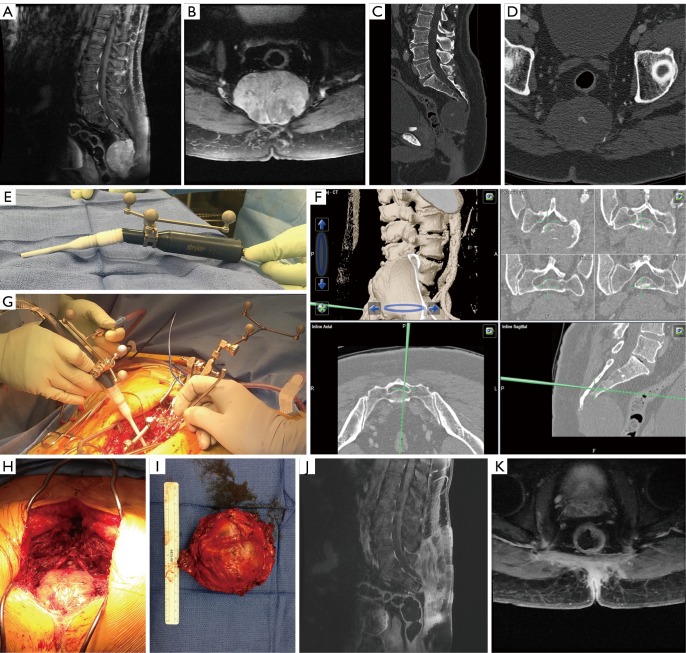
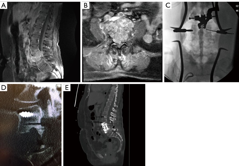
In the literature, the use of navigation for spine tumor surgery has largely centered on implant placement. We describe the cases of two patients with spinal tumors on whom we utilized our resection technique of registering an ultrasonic bone scalpel (UBS) to a navigation system. In both cases, we achieved a satisfactory tumor resection with negative margins and excellent neurologic outcomes. We feel that using the navigation-registered UBS is a valuable tool to increase the operator's ability to achieve desired resections while minimizing the neurologic deficits and operative morbidity associated with these challenging surgical cases.
Keywords: Image-guided navigation; primary spine tumor; sacral chordoma; ultrasonic bone scalpel (UBS).
Conflict of interest statement
Conflicts of Interest: The authors have no conflicts of interest to declare.
Figures
Similar articles
-
Ultrasonic bone scalpel in spine surgery.J Orthop. 2023 May 10;41:1-7. doi: 10.1016/j.jor.2023.05.002. eCollection 2023 Jul. J Orthop. 2023. PMID: 37216021 Free PMC article. Review.
-
Can Navigation-assisted Surgery Help Achieve Negative Margins in Resection of Pelvic and Sacral Tumors?Clin Orthop Relat Res. 2018 Mar;476(3):499-508. doi: 10.1007/s11999.0000000000000064. Clin Orthop Relat Res. 2018. PMID: 29529631 Free PMC article.
-
Maximizing Sacral Chordoma Resection by Precise 3-Dimensional Tumor Modeling in the Operating Room Using Intraoperative Computed Tomography Registration with Preoperative Magnetic Resonance Imaging Fusion and Intraoperative Neuronavigation: A Case Series.World Neurosurg. 2019 May;125:e1125-e1131. doi: 10.1016/j.wneu.2019.01.257. Epub 2019 Feb 18. World Neurosurg. 2019. PMID: 30790740
-
Can Navigation Improve the Ability to Achieve Tumor-free Margins in Pelvic and Sacral Primary Bone Sarcoma Resections? A Historically Controlled Study.Clin Orthop Relat Res. 2019 Jul;477(7):1548-1559. doi: 10.1097/CORR.0000000000000766. Clin Orthop Relat Res. 2019. PMID: 31107331 Free PMC article. Clinical Trial.
-
Image-guided resection of aggressive sacral tumors.Neurosurg Focus. 2017 Jan;42(1):E15. doi: 10.3171/2016.6.FOCUS16125. Neurosurg Focus. 2017. PMID: 28041320 Review.
Cited by
-
Advances in Virtual Cutting Guide and Stereotactic Navigation for Complex Tumor Resections of the Sacrum and Pelvis: Case Series with Short-Term Follow-Up.Bioengineering (Basel). 2023 Nov 22;10(12):1342. doi: 10.3390/bioengineering10121342. Bioengineering (Basel). 2023. PMID: 38135933 Free PMC article.
-
Ultrasonic bone scalpel in spine surgery.J Orthop. 2023 May 10;41:1-7. doi: 10.1016/j.jor.2023.05.002. eCollection 2023 Jul. J Orthop. 2023. PMID: 37216021 Free PMC article. Review.
References
-
- Barnett GH, Kormos DW, Steiner CP, et al. Use of a frameless, armless stereotactic wand for brain tumor localization with two-dimensional and three-dimensional neuroimaging. Neurosurgery 1993;33:674-8. - PubMed
-
- Enneking WF, Spanier SS, Goodman MA. A system for the surgical staging of musculoskeletal sarcoma. Clin Orthop Relat Res 1980;(153):106-20. - PubMed
Publication types
LinkOut - more resources
Full Text Sources