. 2019 Apr;33(4):4866-4882.
doi: 10.1096/fj.201801382R. Epub 2018 Dec 31.

Failure to reabsorb the primary cilium induces cellular senescence

Affiliations

Failure to reabsorb the primary cilium induces cellular senescence

Elizabeth P Jeffries et al. FASEB J. 2019 Apr.

Abstract

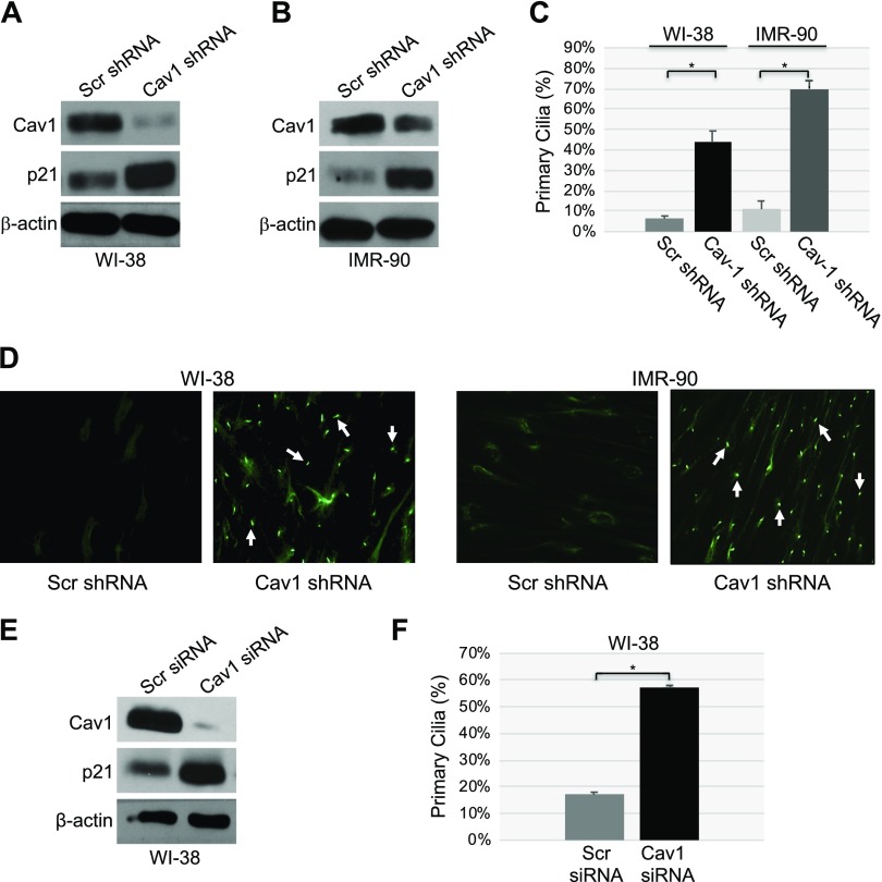
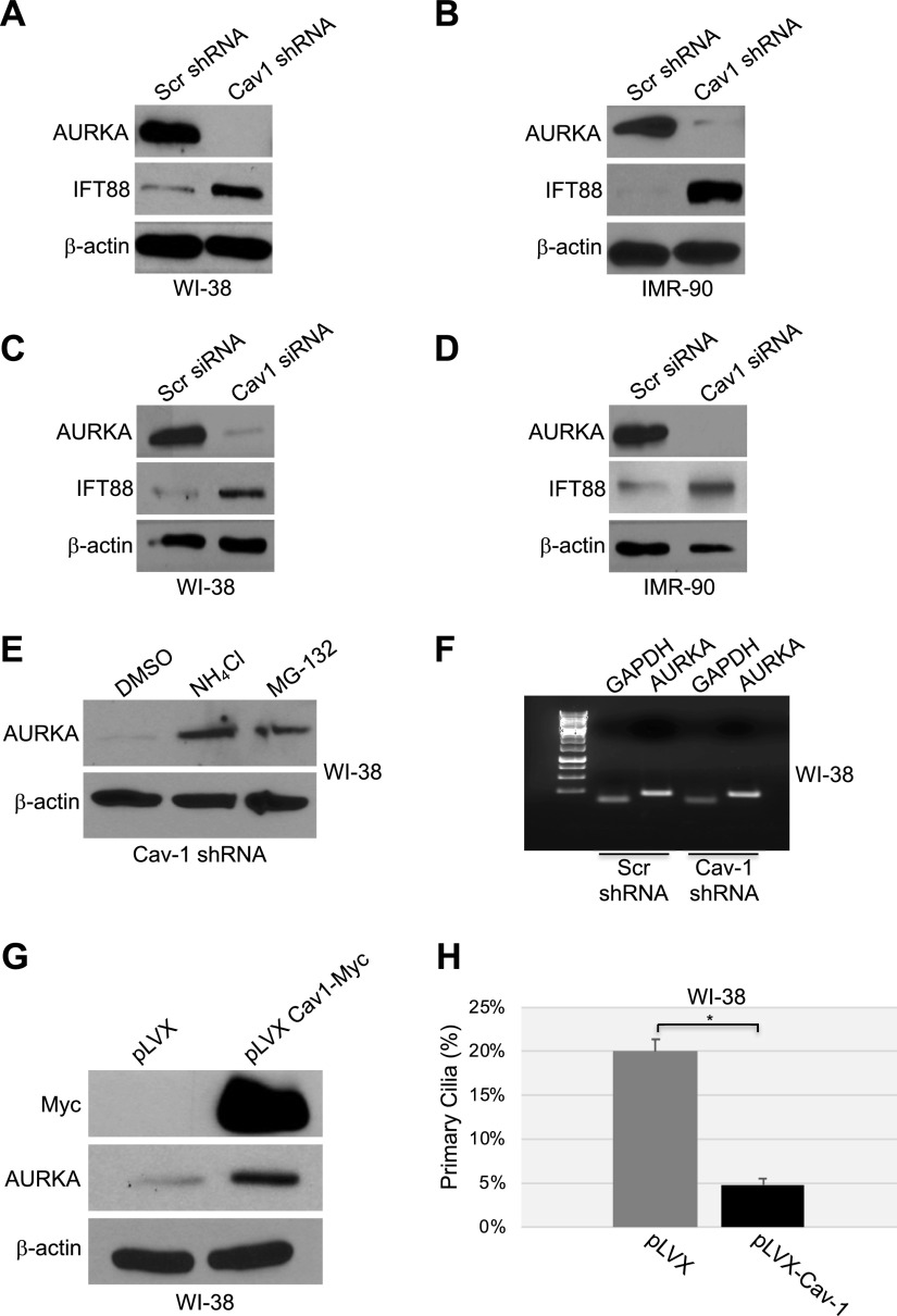
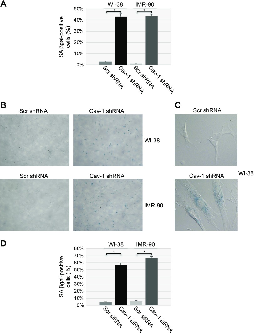
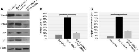
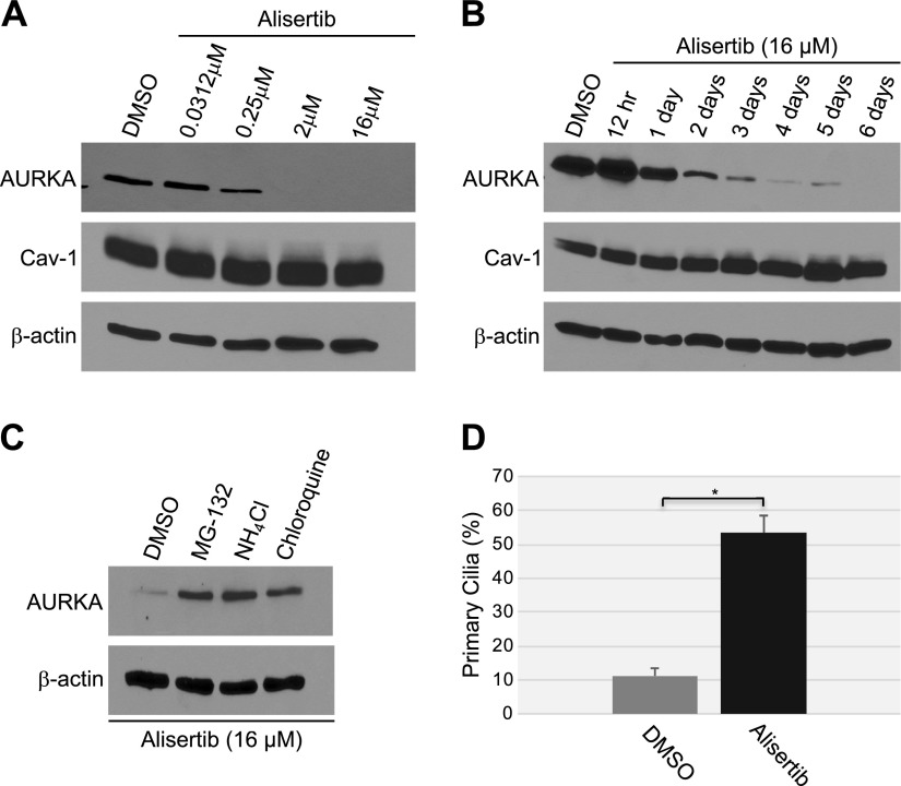
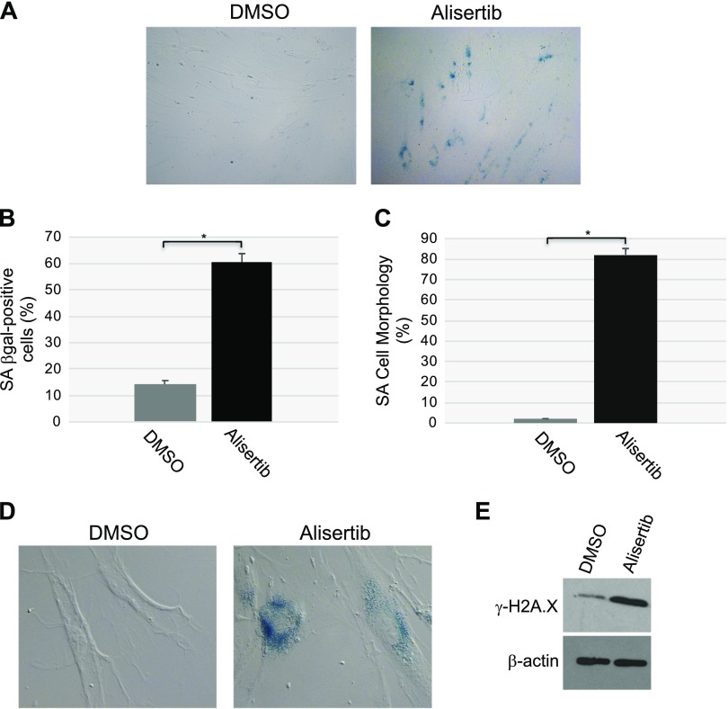
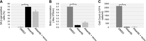
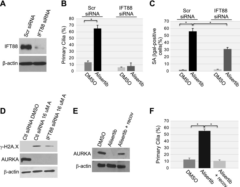
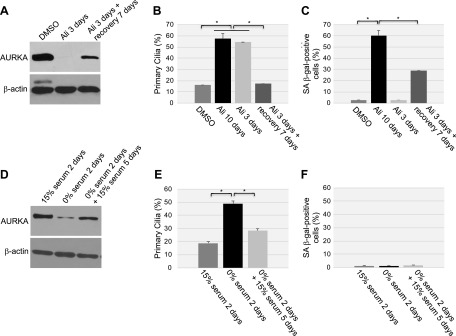
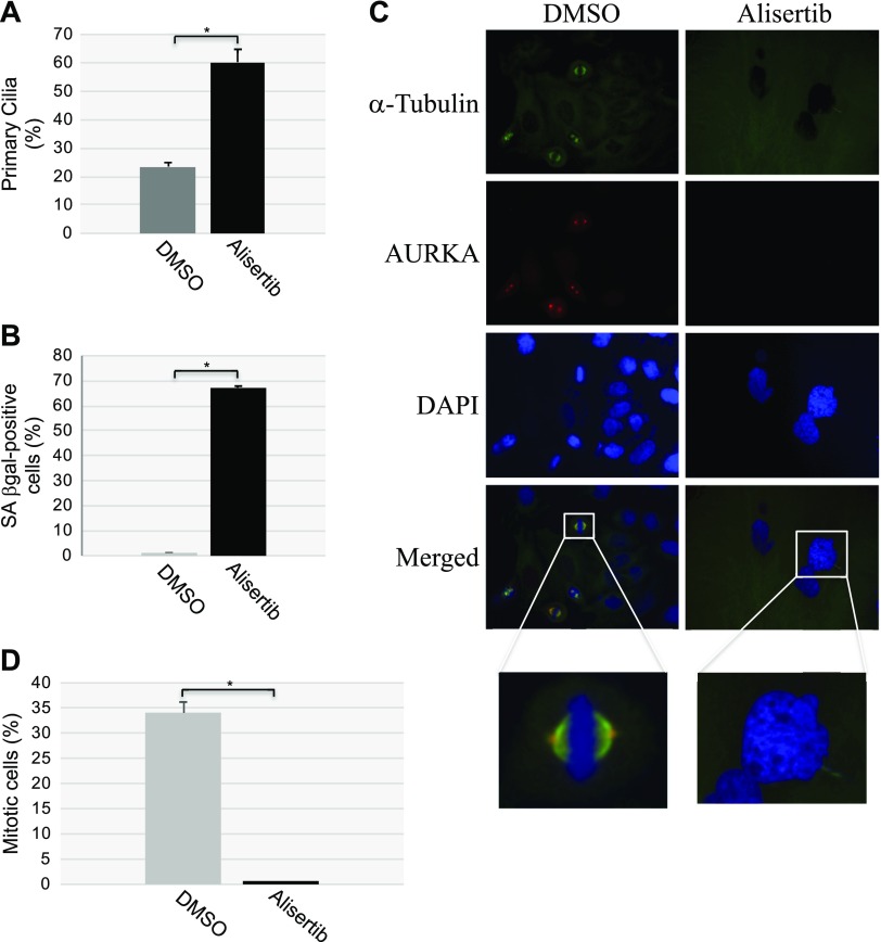
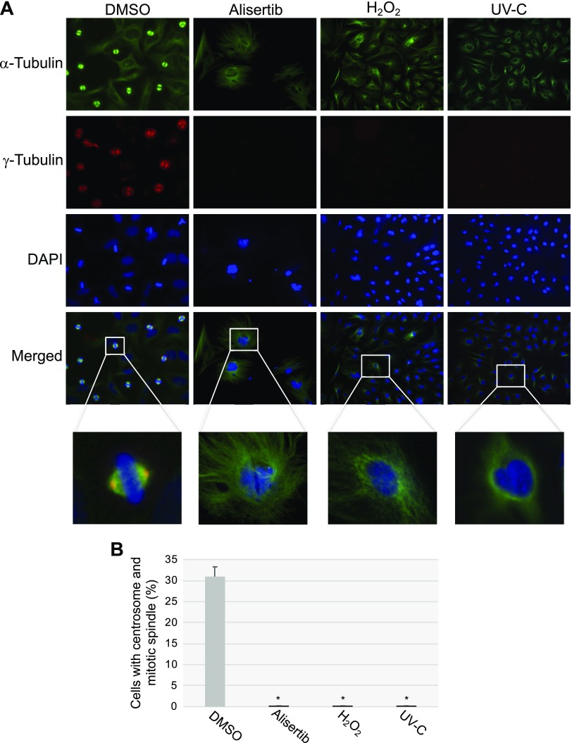
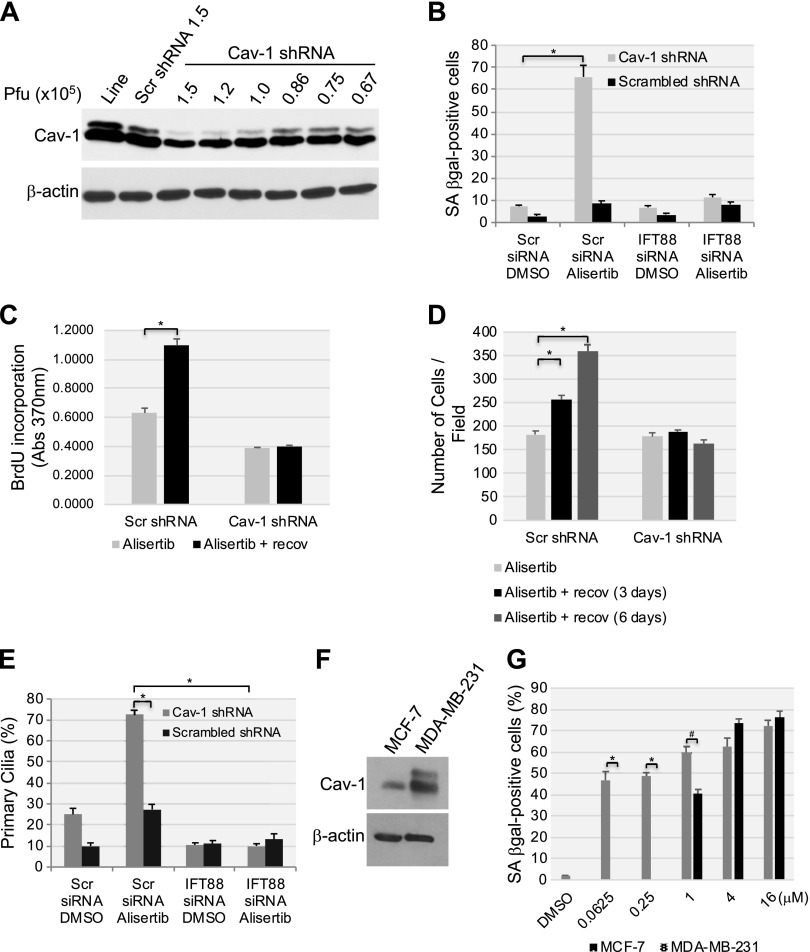
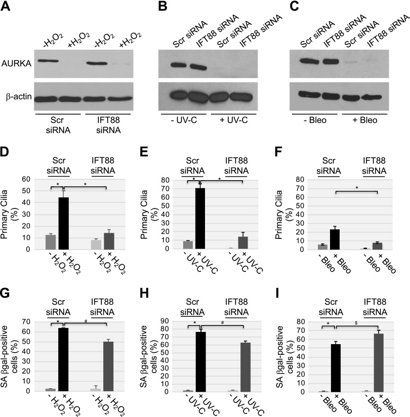
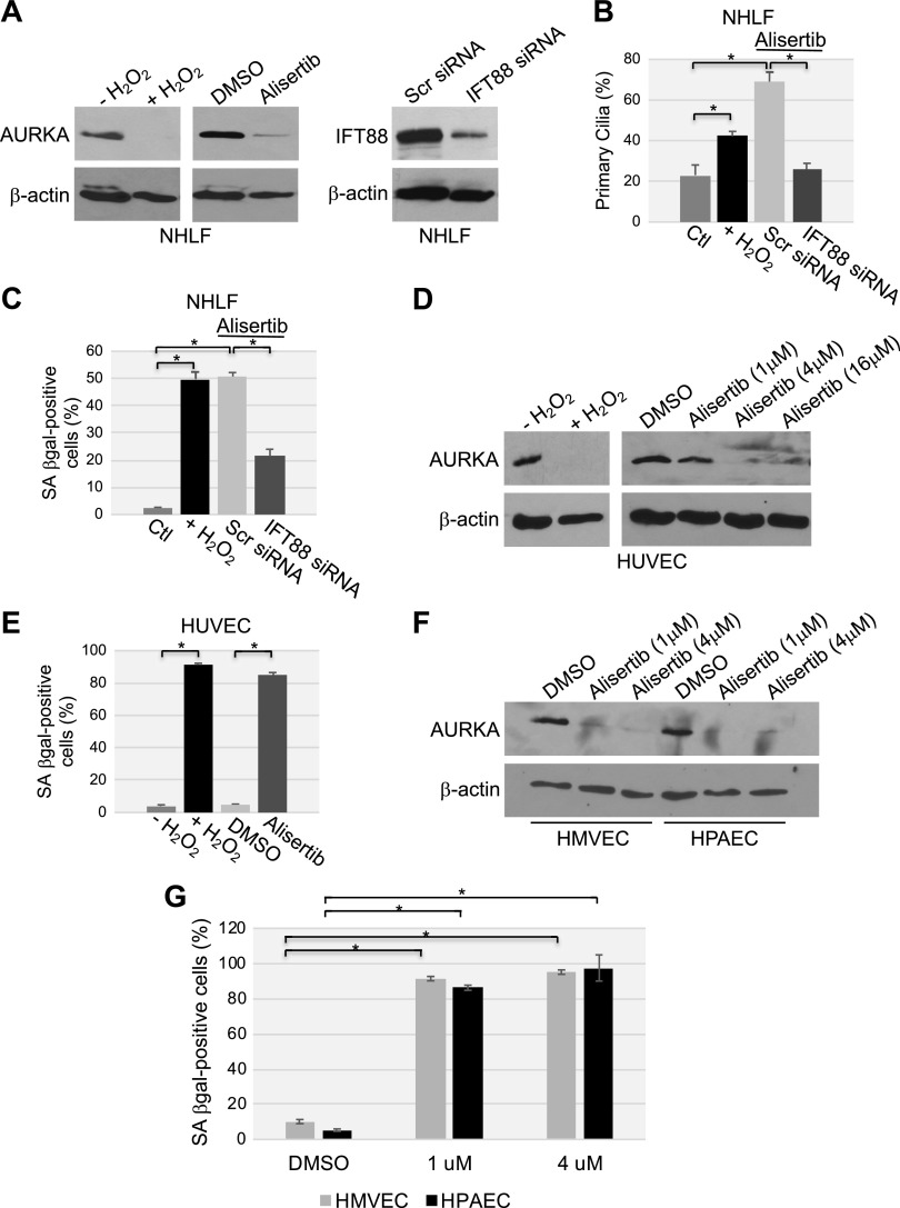
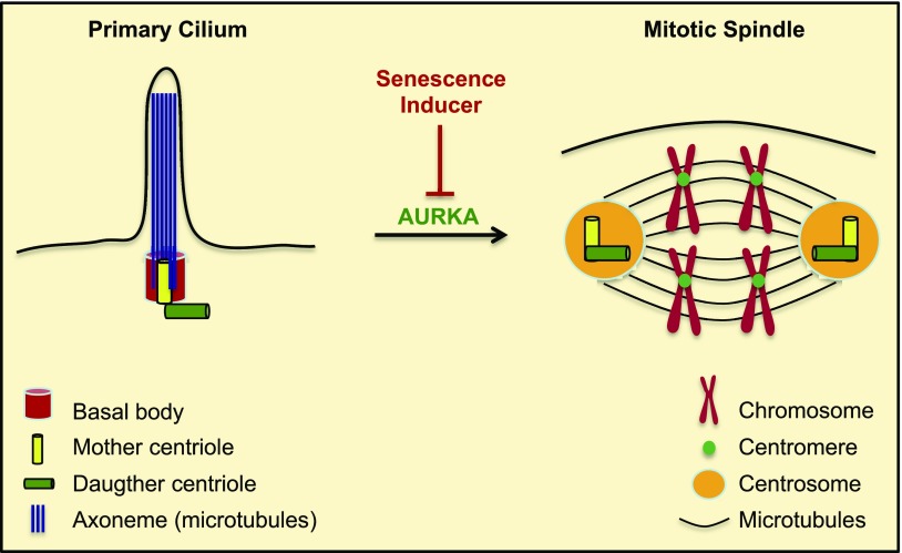
Aurora kinase A (AURKA) is necessary for proper primary cilium disassembly before mitosis. We found that depletion of caveolin-1 expression promotes primary cilia formation through the proteasomal-dependent degradation of aurora kinase A and induces premature senescence in human fibroblasts. Down-regulation of intraflagellar transport-88, a protein essential for ciliogenesis, inhibits premature senescence induced by the depletion of caveolin-1. In support of these findings, we showed that alisertib, a pharmacological inhibitor of AURKA, causes primary cilia formation and cellular senescence by irreversibly arresting cell growth. Suppression of primary cilia formation limits cellular senescence induced by alisertib. The primary cilium must be disassembled to free its centriole to form the centrosome, a necessary structure for mitotic spindle assembly and cell division. We showed that the use of the centriole to form primary cilia blocks centrosome formation and mitotic spindle assembly and prevents the completion of mitosis in cells in which cellular senescence is caused by the inhibition of AURKA. We also found that AURKA is down-regulated and primary cilia formation is enhanced when cellular senescence is promoted by other senescence-inducing stimuli, such as oxidative stress and UV light. Thus, we propose that impaired AURKA function induces premature senescence by preventing reabsorption of the primary cilium, which inhibits centrosome and mitotic spindle formation and consequently prevents the completion of mitosis. Our study causally links the inability of the cell to disassemble the primary cilium, a microtubule-based cellular organelle, to the development of premature senescence, a functionally and pathologically relevant cellular state.-Jeffries, E. P., Di Filippo, M., Galbiati, F. Failure to reabsorb the primary cilium induces cellular senescence.

Keywords: alisertib; aurora kinase A; caveolae; caveolin-1; mitotic spindle.


Conflict of interest statement

This work was supported by Grant R01-CA205165 from the U.S. National Institutes of Health (NIH) National Cancer Institute (to F.G.), and Grant R01-HL124747 from the NIH National Heart, Lung, and Blood Institute (to F.G.). The authors declare no conflicts of interest.

Figures

Figure 1
Figure 1
Down-regulation of caveolin (Cav)-1 promotes ciliogenesis. A, B) WI-38 (A) and IMR-90 (B) human diploid fibroblasts were infected with a lentivirus carrying caveolin-1 shRNA. Infection with a lentivirus carrying scrambled (Scr) shRNA was used as the control. Cells were cultured for 10 d, and cell lysates were collected for immunoblot analysis with antibody probes specific for caveolin-1 and p21. Immunoblot with anti-β-actin IgGs was performed to show equal loading. C, D) Caveolin-1 expression was down-regulated by shRNA in WI-38 and IMR-90 fibroblasts, as described in A and B. After 10 d, primary cilia formation was quantified by immunofluorescence staining with an antibody probe specific for acetylated α-tubulin. Quantification of ciliogenesis (C) and representative images (D) are shown. E, F) Caveolin-1 expression was down-regulated in WI-38 human diploid fibroblasts by caveolin-1 siRNA. Transfection of WI-38 cells with scrambled siRNA was performed as the control. After 10 d, caveolin-1, p21 and β-actin protein expression was determined by immunoblot analysis (E), whereas primary cilia formation was quantified by immunofluorescence staining with anti-acetylated α-tubulin IgGs (F). Values are means ± sem. *P < 0.001 (Student’s t test).
Figure 2
Figure 2
AD) Down-regulation of caveolin-1 expression promotes degradation of AURKA. WI-38 (A, C) and IMR-90 (B, D) fibroblasts were infected with a lentivirus carrying caveolin (Cav)-1 shRNA (A, B) or transfected with caveolin-1 siRNA (C, D). Infection with a lentivirus carrying scrambled (Scr) shRNA (A, B) and transfection with scrambled siRNA (C, D) were used as controls. Cells were collected after 10 d, and cell lysates were subjected to immunoblot analysis using antibody probes specific for AURKA and IFT88. Immunoblotting of anti-β-actin IgGs was performed to show equal loading. E, F) WI-38 human diploid fibroblasts were infected with caveolin-1 shRNA and cultured for 10 d in the presence or absence of ammonium chloride (NH4Cl) or the proteasome inhibitor MG-132. Infection with scrambled shRNA was performed as the control. E) Cells were collected, and the AURKA expression level was determined by immunoblot analysis with anti-AURKA IgGs. Immunoblot of anti-β-actin IgGs was performed as the control. F) Cells were collected, and AURKA mRNA level was determined by RT-PCR with specific primers. RT-PCR with primers specific for GAPDH was performed as an internal control. G, H) WI-38 fibroblasts were infected with a lentiviral vector expressing myc-tagged caveolin-1. Infection with the empty vector pLVX was performed as the control. G) After 6 d, cells were collected, and the AURKA protein level was determined by immunoblot analysis with an antibody probe specific for AURKA. Immunoblot with anti-c-myc IgGs was performed to determine exogenous caveolin-1 expression. Immunoblot of anti-β-actin IgGs was performed to show equal loading. H) Primary cilia formation was quantified by immunofluorescence analysis with anti-acetylated α-tubulin IgGs 6 d after infection. Values represent means ± sem. *P < 0.001 (Student’s t test).
Figure 3
Figure 3
Cellular senescence is promoted by the down-regulation of caveolin-1 expression. AC) Caveolin (Cav)-1 expression was down-regulated in WI-38 and IMR-90 fibroblasts by shRNA. Cells expressing scrambled (Scr) shRNA were used as the control. Ten days after the infection, cellular senescence was quantified by SA-β-gal staining. Quantification (A), representative low-magnification images (B) and representative high-magnification images (C) are shown. D) WI-38 and IMR-90 cells were transfected with caveolin-1 siRNA. Transfection with scrambled (Scr) siRNA was used as the control. After 10 d, cellular senescence was quantified by SA-β-gal staining. Values represent means ± sem. *P < 0.001 (Student’s t test).
Figure 4
Figure 4
Cellular senescence induced by the down-regulation of caveolin-1 is dependent on primary cilia formation. WI-38 human diploid fibroblasts were transfected with caveolin (Cav)-1 siRNA, together with either IFT88 siRNA or scrambled (Scr) siRNA. Transfection of scrambled siRNA alone was used as the control. Cells were collected for analysis 10 d after transfection. A) Cell lysates were subjected to immunoblot analysis with antibody probes specific for caveolin-1, IFT88, p16, and p21. Immunoblot with anti-β-actin IgGs was performed as the control. B) Primary cilia formation was determined by immunofluorescence analysis with anti-acetylated α-tubulin. Quantification of the staining is shown. C) Cells were subjected to SA-β-gal staining; quantification of the staining is shown. Values represent means ± sem. *P < 0.001 (Student’s t test).
Figure 5
Figure 5
Alisertib induces degradation of AURKA and ciliogenesis. A) WI-38 human diploid fibroblasts were treated with different concentrations of alisertib (0.0312, 0.25, 2 and 16 μM) for 6 d. B) WI-38 fibroblasts were treated with 16 μM alisertib for different times (12 h and 1, 2, 3, 4, 5 and 6 d). Treatment with DMSO was used as the control. A, B) Cells were collected, and the expression levels of AURKA and caveolin (Cav)-1 were determined by immunoblot analysis with anti-AURKA and anti-caveolin-1 IgGs. Immunoblot with anti-β-actin IgGs was performed to show equal loading. C) WI-38 fibroblasts were treated with 16 μM alisertib for 6 d in the presence of MG-132 (0.6 μΜ), ammonium chloride (NH4Cl; 10 mM), or chloroquine (50 μΜ). Treatment with DMSO served as the control. Cells were collected, and cell lysates were subjected to immunoblot analysis with antibody probes specific for AURKA and β-actin (to show equal loading). D) WI-38 cells were treated with 16 μM alisertib for 6 d. Cells were then subjected to immunofluorescence analysis using anti-acetylated α-tubulin IgGs. Quantification of the staining is shown. Values represent means ± sem. *P < 0.001 (Student’s t test).
Figure 6
Figure 6
Treatment with alisertib promotes accumulation of cells displaying SA-β-gal activity, senescent cell morphology, and elevation of phosphorylated H2A.X. A, B) WI-38 human diploid fibroblasts were treated with 16 μM alisertib for 10 d. DMSO-treated cells were used as the control. Cells were then subjected to SA-β-gal activity staining. Representative low-magnification images (A) and quantification of the staining (B) are shown;. C, D) WI-38 human diploid fibroblasts were treated with 16 μM alisertib for 10 d. Treatment with DMSO was used as control. Cells showing senescence-associated cell morphology were identified. Quantification of the percentage of cells showing senescence-associated cell morphology (C) and representative high-magnification images (D) are shown. D) SA-β-gal activity staining is also shown. E) WI-38 human diploid fibroblasts were treated with 16 μM alisertib for 10 d. DMSO-treated cells were used as the control. Cells were collected, and cell lysates were subjected to immunoblot analysis with an antibody probe specific for phosphorylated histone H2A.X (γ-H2A.X). Immunoblot analysis with anti-β-actin IgGs was performed to show equal loading. Values represent means ± sem. *P < 0.001 (Student’s t test).
Figure 7
Figure 7
Alisertib causes irreversible growth arrest in human fibroblasts. WI-38 fibroblasts were treated with either DMSO or alisertib (16 μM) for 10 d. Cells were then washed and recovered in alisertib-free medium for an additional 5 d. A) Cells were stained for SA-β-gal activity. B) A BrdU-incorporation assay was performed. C) The number of cells in the cell culture dishes was counted. Values represent means ± sem. *P < 0.001, student’s t test.
Figure 8
Figure 8
The inability to reabsorb the primary cilium contributes to cellular senescence induced by alisertib. A) WI-38 human diploid fibroblasts were transfected with either scrambled (Scr) or IFT88 siRNA. Cells were collected, and cell lysates were subjected to immunoblot analysis using anti-IFT88 IgGs. Immunoblot with anti-β-actin IgGs was performed to show equal loading. BD) WI-38 fibroblasts were transfected with IFT88 siRNA. Transfection with scrambled (Scr) siRNA was used as the control. After 48 h, cells were treated with 16 μM alisertib and cultured for 10 d. Treatment with DMSO was used as the control. B) Primary cilia formation was quantified by immunofluorescence staining with anti-acetylated α-tubulin. C) Cells were subjected to SA-β-gal activity staining. D) The expression levels of phosphorylated histone H2A.X (γ-H2A.X), AURKA, and β-actin were determined by immunoblot analysis using antibody-specific probes. E, F) WI-38 fibroblasts were treated with DMSO or alisertib (16 μM) for 10 d. Cells were then washed and recovered in alisertib-free medium for an additional 5 d. E) Cell lysates were subjected to immunoblot analysis with anti-AURKA IgGs. Immunoblot with anti-β-actin IgGs was performed to show equal loading. F) Primary cilia formation was quantified by immunofluorescence staining with anti-acetylated α-tubulin. Values in B, C and F represent means ± sem. *P < 0.001 (Student’s t test).
Figure 9
Figure 9
Transient down-regulation of AURKA temporarily prevents the disassembly of the primary cilium but does not promote cellular senescence. AC) WI-38 fibroblasts were continuously treated with DMSO or alisertib (16 μM) for 3 or 10 d. Cells were also treated with alisertib (16 μM) for 3 d, washed, and cultured for an additional 7 d in the absence of alisertib. A) Cell lysates were subjected to immunoblot analysis with an antibody probe specific for AURKA. Immunoblot analysis using anti-β-actin IgGs was performed to show equal loading. B) Primary cilia formation was quantified by immunofluorescence staining with an antibody probe specific for acetylated α-tubulin. C) Cellular senescence was quantified after cells were subjected to SA-β-gal activity staining. DF) WI-38 cells were cultured in either serum-containing or serum-free medium for 2 d. Serum-starved cells were then cultured for an additional 5 d in the presence of serum. D) AURKA protein expression was determined by immunoblot analysis using anti-AURKA IgGs. Immunoblot analysis with anti-β-actin IgGs was performed as control. E) Primary cilia formation was quantified by immunofluorescence staining with an antibody probe specific for acetylated α-tubulin. F) Cells were subjected to senescence-associated (SA)-β-galactosidase activity staining. Values represent means ± sem. *P < 0.001 (Student’s t test).
Figure 10
Figure 10
Alisertib inhibits mitotic spindle formation. HeLa cells were treated for 10 d with alisertib (16 μM). Cells were also treated with DMSO as the control. A, B) Primary cilia formation was quantified by immunofluorescence staining with anti-acetylated α-tubulin (A). Cells were subjected to SA-β-gal activity staining (B). C, D) Cells were synchronized in mitosis by double thymidine block. Immunofluorescence staining was then performed with antibody probes specific for α-tubulin (green), AURKA (red), and DAPI (blue). Representative images (C) and quantification of cells displaying a mitotic spindle (D) are shown. Values represent means ± sem. *P < 0.001 (Student’s t test).
Figure 11
Figure 11
Senescence-inducing stimuli inhibit centrosome and mitotic spindle formation. HeLa cells were treated with alisertib (16 μM) for 10 d and H2O2 (450 μM) for 2 h and recovered in complete medium for 7 d, or subjected to UV-C radiation (10 J/m2) and cultured in complete medium for 7 d. Cells were also treated with DMSO as a control. Cells were synchronized in mitosis by double thymidine block. Immunofluorescence staining was then performed using antibody probes specific for α-tubulin (green), γ-tubulin (red), and DAPI (blue). Representative images (A) and quantification of cells displaying centrosome formation and a mitotic spindle (B) are shown. Values represent means ± sem. *P < 0.001 (Student’s t test).
Figure 12
Figure 12
Low endogenous caveolin-1 expression sensitizes human fibroblasts to alisertib-induced primary cilia formation and senescence. A) WI-38 human diploid fibroblasts were infected with a lentivirus-carrying caveolin (Cav)-1 shRNA in the indicated plaque-forming units. Infection with scrambled (Scr) shRNA and uninfected cells were used as controls. Caveolin-1 expression was determined by immunoblot analysis using anti-caveolin-1 IgGs. Immunoblot with anti-β-actin IgGs was performed as the internal control. B) WI-38 human fibroblasts were transfected with either scrambled or IFT88 siRNA. After 48 h, cells were infected with caveolin-1 shRNA at 0.86 × 10⁵ pfu. Infection with scrambled shRNA (0.86 × 10⁵ pfu) was performed as the control. Cells were treated with DMSO or 16 μM alisertib and cultured for 6 d. Cells were then subjected to SA-β-gal staining. C, D) WI-38 cells were transfected/infected and treated for 6 d as described in B. Cell proliferation was then quantified by BrdU incorporation assay (C) and by cell counting (D) at the end of the 6-d treatment or after the cells were washed and recovered in alisertib-free medium for an additional 6 d. E) WI-38 human diploid fibroblasts were transfected/infected and treated for 6 d, as described in B. Primary cilia formation was then quantified by immunofluorescence staining with an antibody probe specific for acetylated α-tubulin. F) Caveolin-1 expression in MCF-7 and MDA-MB-231 breast cancer cells was assessed by immunoblot analysis with a caveolin-1-specific antibody probe. Immunoblot analysis with anti-β-actin IgGs was performed to show equal loading. G) MCF-7 and MDA-MB-231 breast cancer cells were treated with different concentrations of alisertib (0.0625, 0.25, 1, 4 and 16 μM) for 6 d. Treatment with DMSO was the control. Cells were then subjected to SA-β-gal staining. Values represent means ± sem. *P < 0.001, #P < 0.005 (Student’s t test).
Figure 13
Figure 13
Oxidative stress and UV-C radiation induce senescence that is partially dependent on the failure to disassemble the primary cilium. AC) WI-38 human diploid fibroblasts were transfected with either scrambled (Scr) or IFT88 siRNA. A) Cells were treated with sublethal doses (450 μM) of H2O2 for 2 h, washed, and recovered in complete medium for 10 d. B) WI-38 fibroblasts were treated with UV-C radiation (10 J/m2) and cultured in complete medium for 10 d. C) WI-38 cells were treated with bleomycin (10 μg/ml) for 24 h. Cells were washed and cultured in complete medium for 10 d. Cell lysates were then subjected to immunoblot analysis with antibody probes specific for AURKA and β-actin to show equal loading. DF) WI-38 fibroblasts were transfected and treated as described in AC. Cells were then subjected to immunofluorescence staining with anti-acetylated α-tubulin IgGs to detect primary cilia. Quantification of cells carrying a primary cilium is shown. GI) WI-38 fibroblasts were transfected and treated as described in A–C. Cells were then subjected to SA-β-galactosidase activity staining. Values represent means ± sem. *P < 0.001, #P < 0.005, $P < 0.05 (Student’s t test).
Figure 14
Figure 14
Oxidative stress and alisertib promote down-regulation of AURKA, ciliogenesis, and premature senescence in primary human fibroblasts. AC) NHLFs were transfected with either scrambled (Scr) or IFT88 siRNA. Cells were then treated with either alisertib (16 μM) for 10 d or with sublethal doses (450 μM) of H2O2 for 2 h, washed, and recovered in complete medium for 10 d. A) cell lysates were subjected to immunoblot analysis with antibody probes specific for either AURKA or IFT88; immunoblot with anti-β-actin IgGs was performed to show equal loading. B) Cells were subjected to immunofluorescence staining with anti-acetylated α-tubulin IgGs to detect primary cilia. Quantification of cells carrying a primary cilium is shown. C) Cells were subjected to SA-β-gal staining. D, E) HUVECs were treated with either sublethal doses of H2O2 (450 μM) for 2 h and recovered in complete medium for 7 d or alisertib (1, 4 and 16 μM) for 10 d. D) AURKA protein expression was determined by immunoblot analysis with an AURKA-specific antibody probe; immunoblot with anti-β-actin IgGs served as the control. E) Cells were subjected to SA-β-gal activity staining. F, G) HPAECs and human lung microvascular endothelial cells were treated with alisertib (1 and 4 μM) for 10 d. F) Cell lysates were subjected to immunoblot analysis, with antibody probes specific for AURKA; immunoblot with anti-β-actin IgGs was performed as the control. G) Cells were subjected to SA-β-gal activity staining. Values represent means ± sem. *P < 0.001 (Student’s t test).
Figure 15
Figure 15
Summary of the role of primary cilia in the development of cellular senescence. When the cell enters the cell cycle, the primary cilium is disassembled, a process that is regulated by AURKA. The mother and daughter centrioles duplicate and form the centrosomes, which organize the spindle fibers to form the mitotic spindle. The mitotic spindle allows proper chromosomal segregation to form 2 daughter cells during mitosis. Inhibition/degradation of AURKA by senescence inducers prevents primary cilium disassembly, forcing the cell to use the centrioles to maintain the primary cilium. As a result, the centrioles are not available to form the centrosomes and generation of a properly organized mitotic spindle is inhibited. This leads to an irreversible growth arrest/senescent status, because restoration of AURKA expression and absorption of the primary cilium to free the centrioles are not sufficient for cell cycle reentry.

Similar articles

Cited by

References

    1. Sorokin S. (1962) Centrioles and the formation of rudimentary cilia by fibroblasts and smooth muscle cells. J. Cell Biol. 15, 363–377 - PMC - PubMed
    1. Seeley E. S., Nachury M. V. (2010) The perennial organelle: assembly and disassembly of the primary cilium. J. Cell Sci. 123, 511–518 - PMC - PubMed
    1. Ishikawa H., Marshall W. F. (2011) Ciliogenesis: building the cell’s antenna. Nat. Rev. Mol. Cell Biol. 12, 222–234 - PubMed
    1. Pazour G. J., Dickert B. L., Vucica Y., Seeley E. S., Rosenbaum J. L., Witman G. B., Cole D. G. (2000) Chlamydomonas IFT88 and its mouse homologue, polycystic kidney disease gene tg737, are required for assembly of cilia and flagella. J. Cell Biol. 151, 709–718 - PMC - PubMed
    1. Korobeynikov V., Deneka A. Y., Golemis E. A. (2017) Mechanisms for nonmitotic activation of Aurora-A at cilia. Biochem. Soc. Trans. 45, 37–49 - PMC - PubMed

Publication types

LinkOut - more resources