

Diagnostic value of tumor-fascia relationship in superficial soft tissue masses on magnetic resonance imaging
- PMID: 30596710
- PMCID: PMC6312209
- DOI: 10.1371/journal.pone.0209642
Diagnostic value of tumor-fascia relationship in superficial soft tissue masses on magnetic resonance imaging
Abstract
Purpose: Many surgeons participate in the management of superficial soft tissue masses, and a preoperative incorrect diagnosis frequently results in dismal oncological outcomes. The aim of this study was to identify distinguishing magnetic resonance imaging features between malignant and non-malignant lesions.
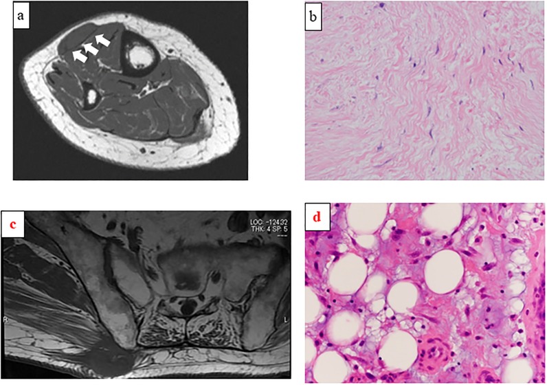
Methods: The clinicopathological data for 219 patients (men 114; women 105) with superficial soft tissue masses treated from January 2007 to December 2016 in our institution were retrospectively analyzed. The median age at the first visit was 55.6 years (range 1-90 years). MRI findings of tumor size, margin, lobulation, intratumoral hemorrhage, peritumoral edema, and tumor-fascia relationship were compared with the final histological diagnosis and tumor grade.
Results: Univariate analysis revealed significant relationships between histologically malignant lesions and tumor size ≥5 cm (p = 0.035), positive peritumoral edema (p = 0.031), and tumor-fascia relationship (p<0.001), but not margin (p = 0.107), lobulation (p = 0.071), and intratumoral hemorrhage (p = 0.17). In addition, using multivariate analysis, the tumor-fascia relationship (p<0.001) and tumor size were significant factors. A significant correlation between tumor-fascia relationship and malignancy (p<0.001) was observed; such a relationship was, however, not observed for tumor grade (p = 0.43).
Conclusions: Tumors measuring ≥5 cm and the tumor-fascia relationship on magnetic resonance imaging are highly indicative of malignancy. When superficial soft tissue masses cross the superficial fascia and form obtuse angles with the fascia, sarcoma should be considered. The tumor-fascia relationship can offer surgeons useful information regarding the status of superficial soft tissue masses.
Conflict of interest statement
The authors have declared that no competing interests exist.
Figures

Similar articles
-
Grading of subcutaneous soft tissue tumors by means of their relationship with the superficial fascia on MR imaging.Skeletal Radiol. 1998 Dec;27(12):657-63. doi: 10.1007/s002560050455. Skeletal Radiol. 1998. PMID: 9921926
-
The incidence and diagnostic relevance of chemical shift artefact in the magnetic resonance imaging characterisation of superficial soft tissue masses.Br J Radiol. 2020 Apr;93(1108):20190828. doi: 10.1259/bjr.20190828. Epub 2019 Dec 19. Br J Radiol. 2020. PMID: 31834812 Free PMC article.
-
MRI of superficial soft tissue masses: analysis of features useful in distinguishing between benign and malignant lesions.Skeletal Radiol. 2012 Dec;41(12):1517-24. doi: 10.1007/s00256-012-1385-6. Epub 2012 Apr 12. Skeletal Radiol. 2012. PMID: 22491777
-
Superficial soft-tissue masses: analysis, diagnosis, and differential considerations.Radiographics. 2007 Mar-Apr;27(2):509-23. doi: 10.1148/rg.272065082. Radiographics. 2007. PMID: 17374866 Review.
-
Magnetic resonance imaging of musculoskeletal tumors: skeletal and soft tissue masses.Curr Probl Diagn Radiol. 1999 Mar-Apr;28(2):29-62. doi: 10.1016/s0363-0188(99)90009-9. Curr Probl Diagn Radiol. 1999. PMID: 10088064 Review.
Cited by
-
Small indeterminate superficial soft tissue masses: relationship between depth and histological grade.Br J Radiol. 2020 Jun;93(1110):20191037. doi: 10.1259/bjr.20191037. Epub 2020 Mar 6. Br J Radiol. 2020. PMID: 32108489 Free PMC article.
-
Case report: Giant atypical granular cell tumor of the median nerve.Front Neurol. 2023 Sep 29;14:1221912. doi: 10.3389/fneur.2023.1221912. eCollection 2023. Front Neurol. 2023. PMID: 37840916 Free PMC article.
-
Tumor-skin invasion is a reliable risk factor for poor prognosis in superficial soft tissue sarcomas.PLoS One. 2022 Sep 2;17(9):e0274077. doi: 10.1371/journal.pone.0274077. eCollection 2022. PLoS One. 2022. PMID: 36054224 Free PMC article.
-
Giant Granular Cell Tumor of the Left Thigh, a Rare Case Report and Literature Review.Orthop Res Rev. 2025 Jan 7;17:1-7. doi: 10.2147/ORR.S499488. eCollection 2025. Orthop Res Rev. 2025. PMID: 39801771 Free PMC article.
References
-
- ESMO /European Sarcoma Network Working Group. Soft tissue and visceral sarcomas: ESMO Clinical Practice Guidelines for diagnosis, treatment and follow-up. See comment in PubMed Commons belowAnn Oncol. 2014; (suppl_3):iii102–12. - PubMed
MeSH terms
LinkOut - more resources
Full Text Sources
Medical
