

Comment
doi: 10.1093/brain/awy314.
Multimodal imaging in familial FTLD: phenoconversion and planning for the future
Affiliations
- PMID: 30596906
- PMCID: PMC6308307
- DOI: 10.1093/brain/awy314
Item in Clipboard
Comment
Multimodal imaging in familial FTLD: phenoconversion and planning for the future
Brain.
.
Abstract
This scientific commentary refers to ‘Longitudinal multimodal MRI as prognostic and diagnostic biomarker in presymptomatic familial frontotemporal dementia’, by Jiskoot et al. (doi:
Figures
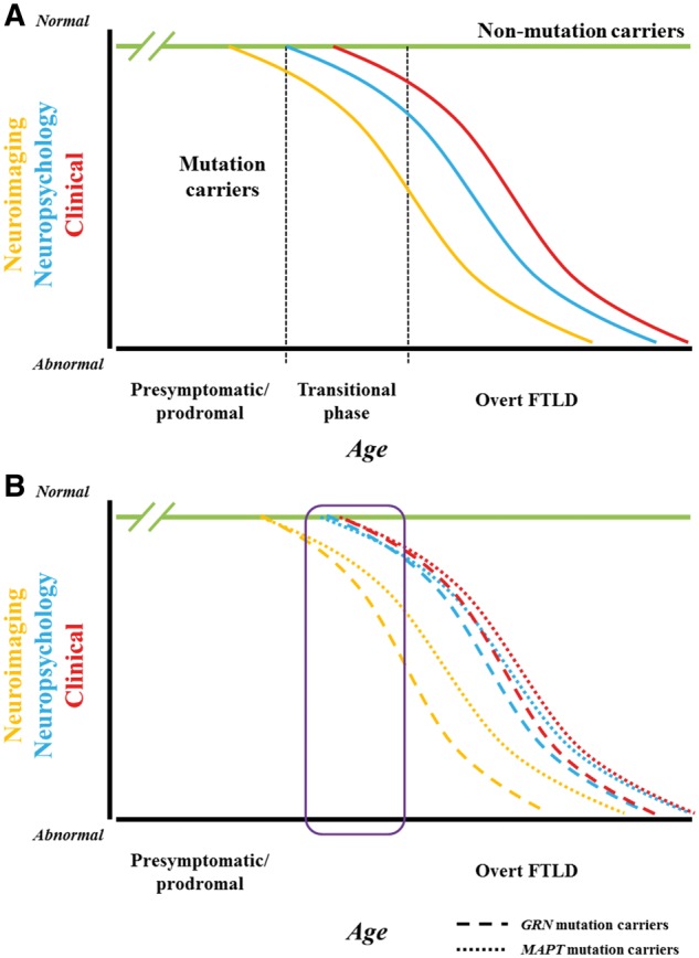
Implications of research findings on the hypothesized framework of evolving familial FTLD. (A) Hypothesized framework of evolving familial FTLD. Compared to non-mutation carrier relatives in familial FTLD kindreds (green line), one would expect that among mutation carriers, molecular alterations would lead to changes in grey and/or white matter indices on brain MRI (orange curve), and these MRI changes would precede the onset of neuropsychological abnormalities (blue curve) and overt clinical features of symptomatic FTLD (red curve). Presumably the curves would be sinusoidal—similar to other neurodegenerative disorders such as Alzheimer’s disease—with a slow initial slope, then an acceleration phase, then a deceleration phase, followed by a terminal gradual change. Inherent in this framework would be a transitional phase (represented by the column bounded by hashed vertical lines) with variable degrees of neuropsychological abnormalities (particularly in social cognition, executive functioning and language functioning) plus subtle clinical changes (such as mild apathy, disinhibition, altered food preferences, reduced empathy, declines in executive functioning and receptive or expressive language functioning) prior to the development of an overt FTLD phenotype such as behavioural variant frontotemporal dementia or primary progressive aphasia. (B) Implications of Jiskoot et al. data on the familial FTLD framework. The data in this manuscript relate to eight individuals—five MAPT mutation carriers who all converted to behavioural variant frontotemporal dementia and three GRN mutation carriers who all converted to non-fluent variant primary progressive aphasia—who were assessed over a 2–4 year timespan (roughly encompassed by the purple rectangle). The data suggest that indices of grey and white matter integrity changed prior to the onset of overt FTLD, with subtle neuropsychological changes around the time of conversion. Plus, the slopes of the imaging and neuropsychological/clinical changes in the GRN mutation carriers appeared to be steeper than those in the MAPT mutation carriers. With only three timepoints in six of the cases and two time points in the other two, it is difficult to determine if an initial slow slope evolved to a more rapid acceleration phase, as implied by these curves. Furthermore, while the period of assessment likely included the transitional phase between normal neurological functioning and overt FTLD for at least some of these cases, it is challenging to characterize this further based on the available data. Regardless, the longitudinal data in this paper provide intriguing preliminary support for the hypothetical familial FTLD model described above.
Comment on
-
Longitudinal multimodal MRI as prognostic and diagnostic biomarker in presymptomatic familial frontotemporal dementia.Brain. 2019 Jan 1;142(1):193-208. doi: 10.1093/brain/awy288. Brain. 2019. PMID: 30508042 Free PMC article.
References
-
- Rosen HJ, Gorno-Tempini ML, Goldman WP, Perry RJ, Schuff N, Weiner M et al. Patterns of brain atrophy in frontotemporal dementia and semantic dementia. Neurology 2002a; 58: 198–208. - PubMed
