Randomized Controlled Trial
. 2019 Jan 1;142(1):103-119.
doi: 10.1093/brain/awy313.

Nitroglycerine triggers triptan-responsive cranial allodynia and trigeminal neuronal hypersensitivity

Affiliations
Randomized Controlled Trial

Nitroglycerine triggers triptan-responsive cranial allodynia and trigeminal neuronal hypersensitivity

Simon Akerman et al. Brain. .

Abstract

Cranial allodynia associated with spontaneous migraine is reported as either responsive to triptan treatment or to be predictive of lack of triptan efficacy. These conflicting results suggest that a single mechanism mediating the underlying neurophysiology of migraine symptoms is unlikely. The lack of a translational approach to study cranial allodynia reported in migraine patients is a limitation in dissecting potential mechanisms. Our objective was to study triptan-responsive cranial allodynia in migraine patients, and to develop an approach to studying its neural basis in the laboratory. Using nitroglycerine to trigger migraine attacks, we investigated whether cranial allodynia could be triggered experimentally, observing its response to treatment. Preclinically, we examined the cephalic response properties of central trigeminocervical neurons using extracellular recording techniques, determining changes to ongoing firing and somatosensory cranial-evoked sensitivity, in response to nitroglycerine followed by triptan treatment. Cranial allodynia was triggered alongside migraine-like headache in nearly half of subjects. Those who reported cranial allodynia accompanying their spontaneous migraine attacks were significantly more likely to have symptoms triggered than those that did not. Patients responded to treatment with aspirin or sumatriptan. Preclinically, nitroglycerine caused an increase in ongoing firing and hypersensitivity to intracranial-dural and extracranial-cutaneous (noxious and innocuous) somatosensory stimulation, reflecting signatures of central sensitization potentially mediating throbbing headache and cranial allodynia. These responses were aborted by a triptan. These data suggest that nitroglycerine can be used as an effective and reliable method to trigger cranial allodynia in subjects during evoked migraine, and the symptom is responsive to abortive triptan treatments. Preclinically, nitroglycerine activates the underlying neural mechanism of cephalic migraine symptoms, central sensitization, also predicting the clinical outcome to triptans. This supports a biological rationale that several mechanisms can mediate the underlying neurophysiology of migraine symptoms, with nitrergic-induced changes reflecting one that is relevant to spontaneous migraine in many migraineurs, whose symptoms of cranial allodynia are responsive to triptan treatment. This approach translates directly to responses in animals and is therefore a relevant platform to study migraine pathophysiology, and for use in migraine drug discovery.


Figures

Figure 1
Figure 1
Original tracings characterizing subtype of dural-evoked trigeminocervical neuronal fibres. Example tracings illustrate (A) early reproducible neuronal discharges classified as receiving only Aδ-fibre input (<20 ms latency; ‘fast’ neuronal responses), and (B) reproducible neuronal spikes with extended discharges beyond 20 ms latency, which were classified as receiving both Aδ and C-fibre inputs (<30 ms latency; ‘fast’ neuronal responses), and also with later unitary discharges that were also classified as receiving C-fibre input (‘slow’ neuronal responses).
Figure 2
Figure 2
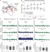
High dose nitroglycerine causes delayed activation of central trigeminocervical neurons. (A) Experimental set-up for electrophysiological recording of neurons in the trigeminocervical complex (TCC), which respond to electrical stimulation of the trigeminal innervation of the dural meninges, with a cutaneous facial receptive field (shaded area) characterization in the ophthalmic dermatome. (B) Time course changes in ongoing spontaneous trigeminocervical neuronal firing [action potentials per second (Hz)] in response to nitroglycerine (NTG, 1 and 10 mg/kg, s.c.) and vasoactive intestinal peptide (VIP, 150 µg/kg, s.c.). The data have been normalized to represent the percentage change from baseline, and are expressed as mean ± SEM. (CE) Representative peri-stimulus time histograms with a single animal for each group demonstrating ongoing spontaneous trigeminocervical neuronal firing before (C) nitroglycerine (1 mg/kg), (D) nitroglycerine (10 mg/kg) and (E) VIP administration, and at 90 min and 3 h post-infusion. The numbers indicate the mean firing [spikes per second (Hz)] over the displayed time period, green neuronal firing indicates baseline and no change in responses, blue neuronal firing indicates a significant increase in neuronal firing. Beneath each of these is a box-and-whisker plot representing median and interquartile range (IQR), with 5th and 95th percentile, and individual points beyond this, of all trigeminocervical complex neurons studied per group, sampled at 1 h intervals after drug administration. Nitroglycerine (10 mg/kg, n = 14) caused a delayed and significant increase in ongoing spontaneous trigeminal neuronal firing that is significant (*P < 0.05) after 60 min and continues until at least 3 h, whereas nitroglycerine (1 mg/kg, n = 9) and VIP (n = 9) do not cause any changes over 3 h. MMA = middle meningeal artery; TG = trigeminal ganglion.
Figure 3
Figure 3
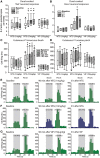
High dose nitroglycerine causes delayed neuronal hypersensitive responses of central trigeminocervical neurons to somatosensory stimulation. Box-and-whisker plots representing median and IQR, and the 5th and 95th percentile, with individual points beyond this, of (A) grouped data of intracranial dural-evoked bursts of trigeminocervical neuronal discharges that include inputs in the Aδ-fibre range (3–20 ms; ‘fast’ neuronal responses), and also those whose latencies were extended and therefore also receive inputs that include both Aδ and C-fibres (3–30 ms; ‘fast’ neuronal responses), and (B) intracranial dural-evoked unitary discharges within the C-fibre latency range (20–80 ms; ‘slow’ neuronal responses), in response to nitroglycerine (1 and 10 mg/kg, s.c.) and VIP (10 µg/kg, s.c.) administration. Nitroglycerine (10 mg/kg) caused a delayed hypersensitivity in neuronal responses to electrical stimulation of the trigeminal innervation of the dural meninges, for ‘fast’ (n = 14) and ‘slow’ (n = 7) neuronal responses that is significant compared to baseline after 1 and 2 h (*P < 0.05), respectively. Nitroglycerine (1 mg/kg) and VIP did not cause any change in neuronal responses. Box-and-whisker plots of (C) innocuous and (D) noxious cutaneous facial-evoked stimulation following nitroglycerine (1 and 10 mg/kg) and VIP. Representative peristimulus time histograms from a single animal for each group depicting trigeminocervical neuronal firing in response to innocuous brush and noxious pinch of the cutaneous facial receptor field before nitroglycerine (10 mg/kg, E), nitroglycerine (1 mg/kg, F) and VIP (G), and at 90 min and 3 h post-infusion. The numbers in parentheses indicate the mean firing (Hz) in response to stimulation (shaded area), green neuronal firing indicates baseline and no change in responses, blue neuronal firing indicates a significant increase in neuronal firing. Nitroglycerine (10 mg/kg) caused a delayed and significant increase in neuronal responses to innocuous and noxious facial cutaneous stimulation in the ophthalmic dermatome after 45 and 135 min, respectively. Nitroglycerine (1 mg/kg) and VIP did not cause any changes. *P < 0.05 significant compared to baseline.
Figure 4
Figure 4
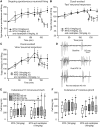
Naratriptan reverses nitroglycerine-evoked activation of central trigeminovascular neurons and hypersensitive responses to somatosensory stimulation. Time course of (A) the mean ongoing spontaneous trigeminal neuronal firing [action potential spikes per second (Hz)], (B) grouped data of intracranial dural-evoked bursts of trigeminocervical neuronal discharges that include inputs in the Aδ-fibre range (3–20 ms; ‘fast’ neuronal responses), and also those whose latencies were extended and therefore also receive inputs that include both Aδ and C-fibres (3–30 ms; ‘fast’ neuronal responses), and (C) intracranial dural-evoked unitary discharges within the C-fibre latency range (20–80 ms; ‘slow’ neuronal responses). All are in response to nitroglycerine (10 mg/kg, s.c.) and after treatment with the migraine abortive, 5-HT1B/1D receptor agonist, naratriptan (10 mg/kg, i.v.). In each panel the data have been normalized to represent a percentage change from baseline and are expressed as mean ± SEM. Naratriptan significantly aborted the increase in ongoing spontaneous neuronal firing, and the hypersensitive neuronal responses to dural-intracranial electrical stimulation for ‘fast’ and ‘slow’ neuronal responses. (D) Original tracings of a single sweep (stimulus) in the same animal for a dural-evoked Aδ-fibre (‘fast’) neuronal burst and unitary C-fibre (‘slow’) discharge at baseline, 2 h after nitroglycerine administration, and 45 min after subsequent administration of the migraine abortive naratriptan. Both the Aδ-fibre neuronal burst and unitary C-fibre discharge become hypersensitive to dural stimulation after nitroglycerine, as evidenced by an increase in the number of action potential spikes at each latency. This is attenuated after treatment with naratriptan. Box-and-whisker plots representing median and IQR, and the 5th and 95th percentile, with individual points beyond this, of time course changes in trigeminocervical neuronal firing, in response to (E) innocuous and (F) noxious cutaneous stimulation of the facial receptive field after nitroglycerine, followed by treatment with naratriptan. The hypersensitivity in neuronal responses to both innocuous and noxious cutaneous facial stimulation 2 h after nitroglycerine is attenuated by naratriptan. *P < 0.05 represents a statistically significant difference compared to baseline (0 min). #P < 0.05 represents a statistically significant difference compared to the time point just prior to administration of naratriptan, which was given post nitroglycerine 135 min. Arrowhead in AC indicates time at which naratriptan was administered. All data are represented as mean ± SEM.

Comment in

References

    1. Afridi SK, Kaube H, Goadsby PJ. Glyceryl trinitrate triggers premonitory symptoms in migraineurs. Pain 2004; 110: 675–80. - PubMed
    1. Afridi SK, Matharu MS, Lee L, Kaube H, Friston KJ, Frackowiak RS et al. A PET study exploring the laterality of brainstem activation in migraine using glyceryl trinitrate. Brain 2005; 128 (Pt 4): 932–9. - PubMed
    1. Akerman S, Goadsby PJ. Neuronal PAC1 receptors mediate delayed activation and sensitization of trigeminocervical neurons: Relevance to migraine. Sci Transl Med 2015; 7: 308ra157. - PubMed
    1. Akerman S, Holland PR, Goadsby PJ. Diencephalic and brainstem mechanisms in migraine. Nat Rev Neurosci 2011; 12: 570–84. - PubMed
    1. Akerman S, Holland PR, Lasalandra MP, Goadsby PJ. Inhibition of trigeminovascular dural nociceptive afferents by Ca(2+)-activated K(+) (MaxiK/BK(Ca)) channel opening. Pain 2010; 151: 128–36. - PubMed

Publication types

MeSH terms