Case Reports
. 2019 Feb;85(2):284-290.
doi: 10.1002/ana.25407. Epub 2019 Jan 17.

Early onset cerebral amyloid angiopathy following childhood exposure to cadaveric dura

Affiliations
Case Reports

Early onset cerebral amyloid angiopathy following childhood exposure to cadaveric dura

Gargi Banerjee et al. Ann Neurol. 2019 Feb.

Abstract

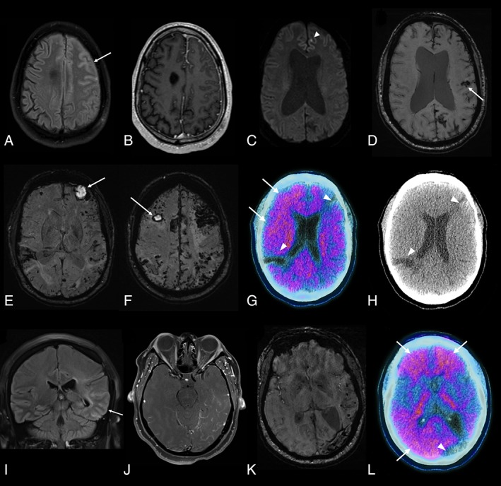
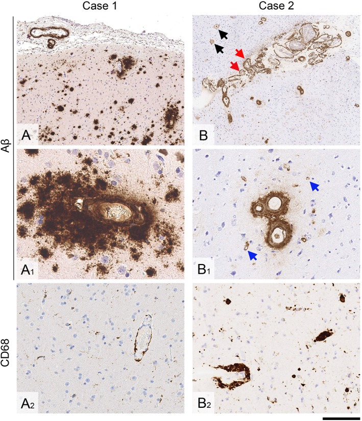
Amyloid-β transmission has been described in patients both with and without iatrogenic Creutzfeldt-Jakob disease; however, there is little information regarding the clinical impact of this acquired amyloid-β pathology during life. Here, for the first time, we describe in detail the clinical and neuroimaging findings in 3 patients with early onset symptomatic amyloid-β cerebral amyloid angiopathy following childhood exposure to cadaveric dura (by neurosurgical grafting in 2 patients and tumor embolization in a third). Our observations provide further in vivo evidence that cerebral amyloid angiopathy might be caused by transmission of amyloid-β seeds (prions) present in cadaveric dura and have diagnostic relevance for younger patients presenting with suspected cerebral amyloid angiopathy. Ann Neurol 2019; 1-7 ANN NEUROL 2019;85:284-290.


Conflict of interest statement

Nothing to report.

Figures

Figure 1
Figure 1
Brain imaging findings. (A–D) Case 1. Fluid‐attenuated inversion recovery (FLAIR; A), T1‐weighted postgadolinium (B), and diffusion‐weighted imaging (C) axial images acquired 2 months after initial presentation demonstrate left frontal gyral swelling and abnormal sulcal signal (arrow in A) with leptomeningeal enhancement (B) and a punctate focus of restricted diffusion (arrowhead). (D) Susceptibility‐weighted imaging 4 months later shows a lobar hemorrhage (arrow), multifocal superficial siderosis, and multiple microbleeds. The ventriculomegaly observed in C and D is longstanding (since the choroid plexus papilloma). (E–H) Case 2. (E, F) Susceptibility‐weighted imaging shows hematomas in both frontal lobes (arrows) and widespread superficial siderosis and microbleeds, visible as a band of low signal following the leptomeningeal surface of the brain and peripheral, punctate foci of low signal, respectively. (G) 18F‐Florbetapir positron emission tomographic (PET)–computed tomographic (CT) imaging shows increased uptake in frontal and parietal cortex (standardized uptake value [SUV] range = 0.0–2.8), suggestive of amyloid deposition (arrows). The 2 areas without uptake (arrowheads) correspond with areas of old hemorrhage, as seen on axial CT (H). (I–L) Case 3. FLAIR coronal (I) and T1‐weighted postgadolinium axial (J) images 15 months after initial presentation showing abnormal gyral and sulcal signal in the left temporal lobe (arrow) with associated leptomeningeal enhancement. (K) Susceptibility‐weighted imaging 5 months later showing hemosiderin deposition due to previous lobar hemorrhage and numerous temporal microbleeds. (L) Merged 18F‐florbetapir PET‐CT imaging shows increased uptake (SUV range = 0.0–3.4) in frontal and posterior (parietal and occipital) cortical regions (arrows); the area with reduced uptake (arrowhead) corresponds to the area of old hemorrhagic damage. [Color figure can be viewed at www.annalsofneurology.org]
Figure 2
Figure 2
Brain biopsy findings. (A–A2) In Case 1, there is leptomeningeal and cortical cerebral amyloid angiopathy, and widespread diffuse parenchymal amyloid‐β (Aβ) deposits (A and A1). There is only minimal parenchymal and perivascular microglial and macrophage activity (A2). (B–B2) In Case 2, there is particularly widespread leptomeningeal and cortical amyloid angiopathy, including vessel wall splitting (B, red arrows) and capillary amyloid angiopathy in the cortex (B1, blue arrows). In the cortex, there are also diffuse parenchymal Aβ deposits and rare plaques with central amyloid cores (B, black arrows). There are abundant perivascular macrophages, some laden with hemosiderin (B2). Sections A, A1, B, and B1 are immunostained for Aβ; A2 and B2 are immunostained for CD68. All sections are counterstained with hematoxylin. Scale bar = 500 μm in A and B; 50 μm in A1; 100 μm in B1, and 200 μm in A2 and B2. [Color figure can be viewed at www.annalsofneurology.org]

References

    1. Jaunmuktane Z, Mead S, Ellis M, et al. Evidence for human transmission of amyloid‐beta pathology and cerebral amyloid angiopathy. Nature 2015;525:247–250. - PubMed
    1. Frontzek K, Lutz MI, Aguzzi A, et al. Amyloid‐beta pathology and cerebral amyloid angiopathy are frequent in iatrogenic Creutzfeldt‐Jakob disease after dural grafting. Swiss Med Wkly 2016;146:w14287. - PubMed
    1. Ritchie DL, Adlard P, Peden AH, et al. Amyloid‐beta accumulation in the CNS in human growth hormone recipients in the UK. Acta Neuropathol 2017;134:221–240. - PMC - PubMed
    1. Cali I, Cohen ML, Haik S, et al. Iatrogenic Creutzfeldt‐Jakob disease with amyloid‐beta pathology: an international study. Acta Neuropathol Commun 2018;6:5. - PMC - PubMed
    1. Kovacs GG, Lutz MI, Ricken G, et al. Dura mater is a potential source of Abeta seeds. Acta Neuropathol 2016;131:911–923. - PMC - PubMed

Publication types

MeSH terms

Substances