Review
. 2018 Dec 28;11(1):28.
doi: 10.3390/cancers11010028.

Flavonoids in Cancer and Apoptosis

Affiliations
Review

Flavonoids in Cancer and Apoptosis

Mariam Abotaleb et al. Cancers (Basel). .

Abstract

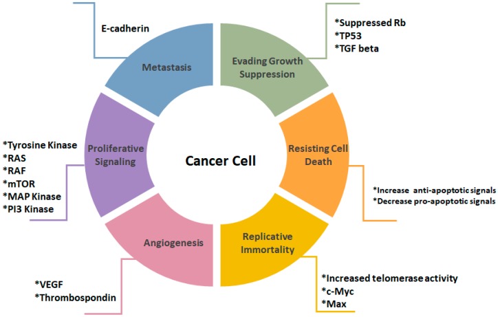
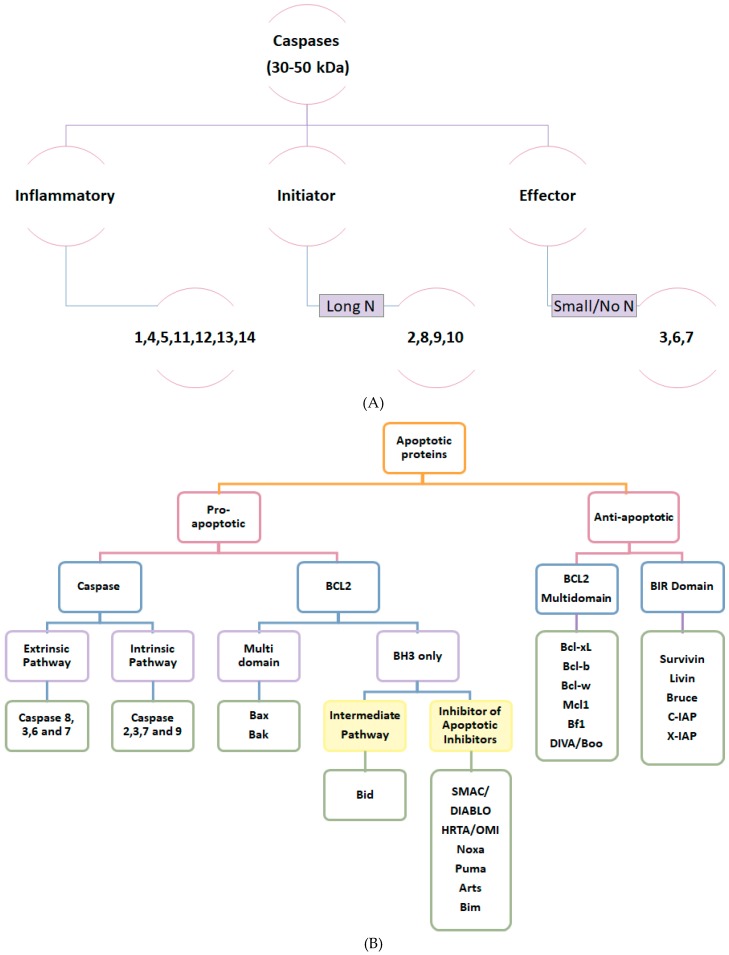
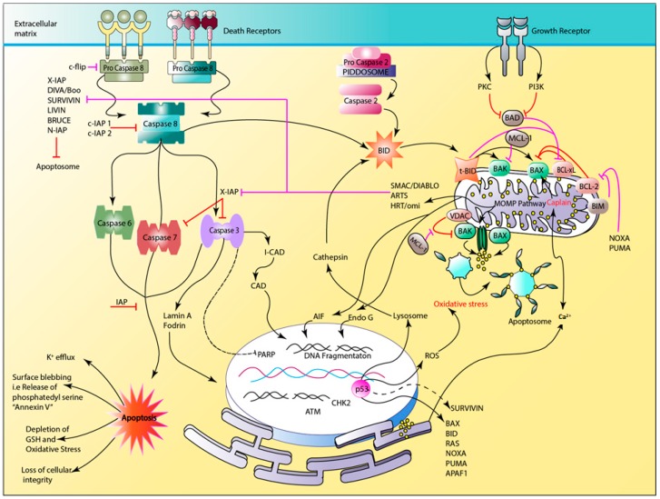
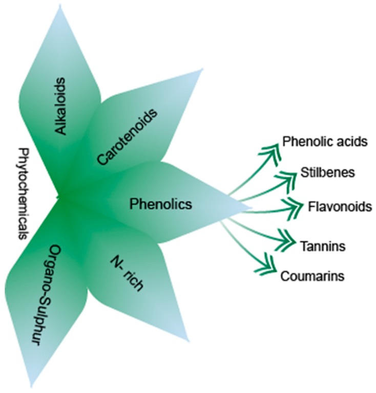
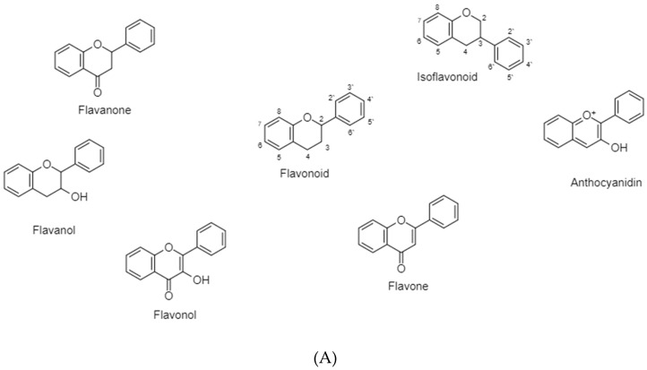
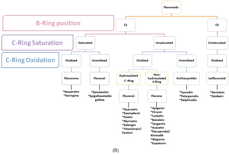
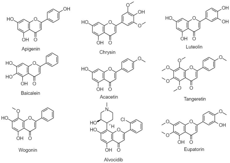
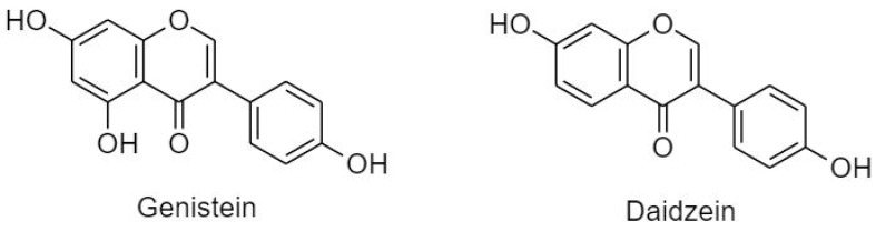
Cancer is the second leading cause of death globally. Although, there are many different approaches to cancer treatment, they are often painful due to adverse side effects and are sometimes ineffective due to increasing resistance to classical anti-cancer drugs or radiation therapy. Targeting delayed/inhibited apoptosis is a major approach in cancer treatment and a highly active area of research. Plant derived natural compounds are of major interest due to their high bioavailability, safety, minimal side effects and, most importantly, cost effectiveness. Flavonoids have gained importance as anti-cancer agents and have shown great potential as cytotoxic anti-cancer agents promoting apoptosis in cancer cells. In this review, a summary of flavonoids and their effectiveness in cancer treatment targeting apoptosis has been discussed.

Keywords: anti-cancer therapy; apoptosis; cancer; flavonoids; natural compounds; phytochemicals.


Conflict of interest statement

The authors declare no conflict of interest. The funders had no role in the design of the study; in the collection, analyses, or interpretation of data; in the writing of the manuscript, or in the decision to publish the results.

Figures

Figure 1
Figure 1
Illustration of cancer cell hallmarks. Evasion of growth suppression is mediated by inhibition of Rb, TP 53 and TGF beta. Resistance in cell death is signified by increase in anti-apoptotic and a decrease in pro-apoptotic factors. Replicative immortality is result of an increase in telomerase activity, c-Myc and Max. Angiogenesis is promoted due to the increase in VEGF and thrombospondin. The increased proliferative signaling is emphasized by increased tyrosine kinase activity, Ras, Raf, m-TOR, MAP kinase and PI3-kinase. Metastasis formation is marked by the loss of E-cadherin [4].
Figure 2
Figure 2
(A) Classification of caspases. Caspases are divided into three subtypes: inflammatory caspases (1,4,5,11,12,13,14), initiator caspases in intrinsic and extrinsic apoptotic pathways with long N terminal (2,8,9,10), and effector caspases known for their short/absent N-terminal (3,6,7) [6,8]. (B) Classification of apoptotic proteins according to their function in apoptosis. Pro-apoptotic proteins are classified into Caspases and BCL-2 families. Caspases are further subdivided according to their role in apoptotic pathway. Bcl-2 proteins are classified as multidomain proteins (Bax and Bak) and BH3 only domain that either serve in intermediate pathway (Bid and inhibitors of apoptotic inhibitors such as SMAC/Diablo, HRTA/Omi, Noxa, Puma, Arts and Bim). Anti-apoptotic proteins are classified as BCL-2 multi-domain proteins (Bcl-xl, Bcl-b, Bcl-w, Mcl-1, Bf1 and DIVA/Boo) and BIR domain family (survivin, livin, bruce, c-IAP and x-IAP).
Figure 3
Figure 3
Illustration of intrinsic, extrinsic, mitochondrial, and endoplasmic reticulum apoptotic pathways. The role of apoptotic proteins (black arrows), and their inhibition by anti-apoptotic proteins (Red) which are regulated by apoptotic inhibitors (Pink). An apoptotic cell is usually characterized by increased K+ efflux, surface blebbing, depletion of anti-oxidative markers such as GSH, chromatin condensation, and loss of cellular integrity.
Figure 4
Figure 4
Classification of phytochemicals. alkaloids, carotenoids, n-rich, organo-sulphur compounds, and phenolics, which are further classified as phenolic acids, stilbenes, flavonoids, tannins, and coumarins.
Figure 5
Figure 5
(A) Classification of flavonoids into six classes depending on structural differences: flavonol, flavanone, flavanol, flavone, anthocyanidin, and isoflavonoid. (B) Structural classification of plant flavonoids according to the position of B-ring on C2 or C3, B-ring saturation and oxidation.
Figure 5
Figure 5
(A) Classification of flavonoids into six classes depending on structural differences: flavonol, flavanone, flavanol, flavone, anthocyanidin, and isoflavonoid. (B) Structural classification of plant flavonoids according to the position of B-ring on C2 or C3, B-ring saturation and oxidation.
Figure 6
Figure 6
Flavanones. Hesperetin and naringenin.
Figure 7
Figure 7
Classification and structure of flavanols.
Figure 8
Figure 8
Classification and structure of flavonols.
Figure 9
Figure 9
Classification and structure of flavones.
Figure 10
Figure 10
Classification and structures of anthocyanidins.
Figure 11
Figure 11
Classification and structure of isoflavonoids.

References

    1. Igney F.H., Krammer P.H. Death and anti-death: Tumour resistance to apoptosis. Nat. Rev. 2002;2:277–288. doi: 10.1038/nrc776. - DOI - PubMed
    1. Safarzadeh E., Shotorbani S.S., Baradaran B. Herbal medicine as inducers of apoptosis in cancer treatment. Adv. Pharm. Bull. 2014;4:421–427. - PMC - PubMed
    1. Prakash O., Kumar A., Kumar P. Anticancer potential of plants and natural products: A review. Am. J. Pharmacol. Sci. 2013;1:104–115. doi: 10.12691/ajps-1-6-1. - DOI
    1. Hanahan D., Weinberg R.A. Review hallmarks of cancer: The next generation. Cell. 2011;144:646–674. doi: 10.1016/j.cell.2011.02.013. - DOI - PubMed
    1. Hanahan D., Weinberg R.A., Francisco S. The hallmarks of cancer review university of california at san francisco. Cell. 2000;100:57–70. doi: 10.1016/S0092-8674(00)81683-9. - DOI - PubMed

Grants and funding

LinkOut - more resources