. 2019 May:23:101083.
doi: 10.1016/j.redox.2018.101083. Epub 2018 Dec 18.

Advanced lipoxidation end products (ALEs) as RAGE binders: Mass spectrometric and computational studies to explain the reasons why

Affiliations

Advanced lipoxidation end products (ALEs) as RAGE binders: Mass spectrometric and computational studies to explain the reasons why

Marco Mol et al. Redox Biol. 2019 May.

Abstract

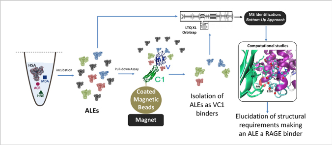
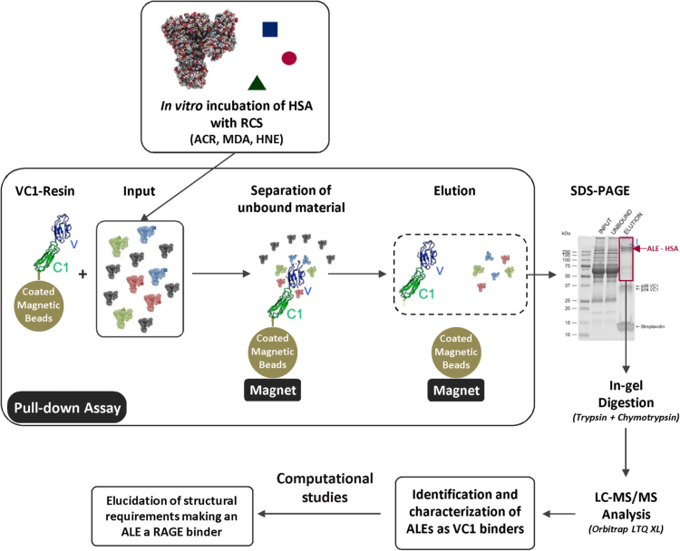
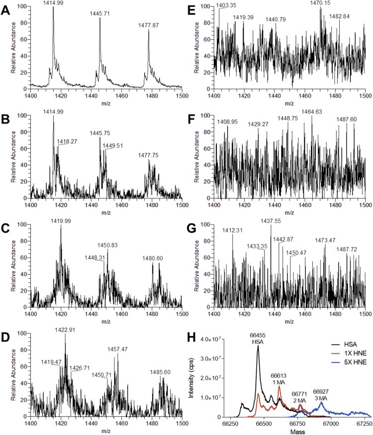
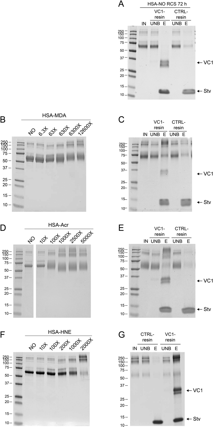
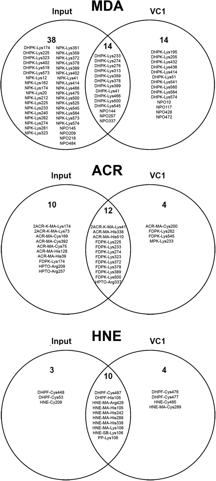
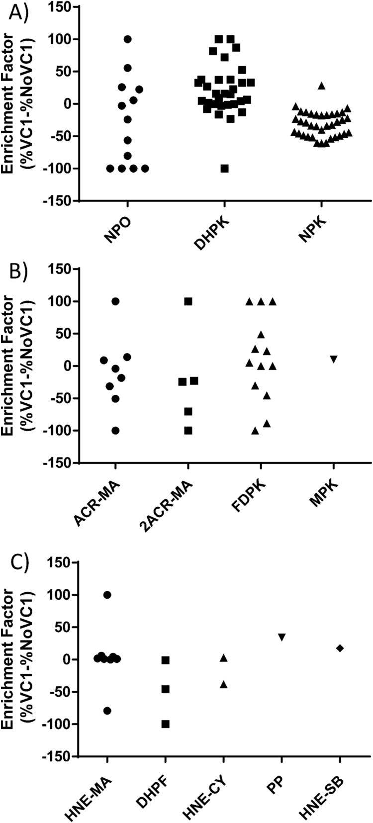
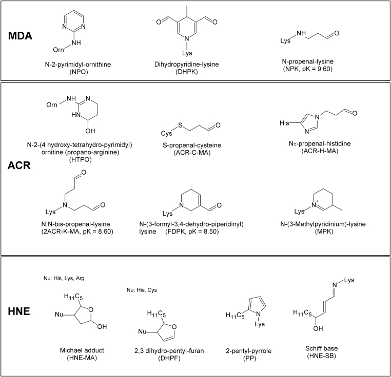
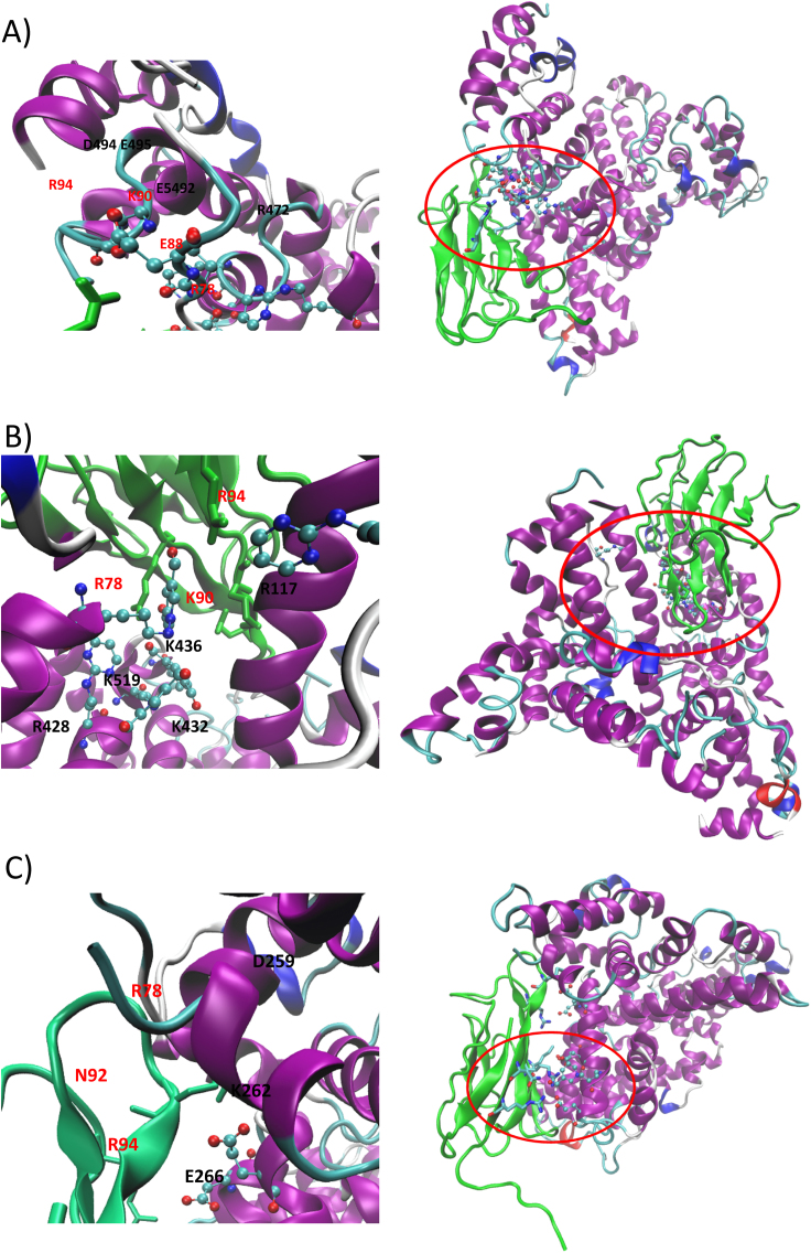
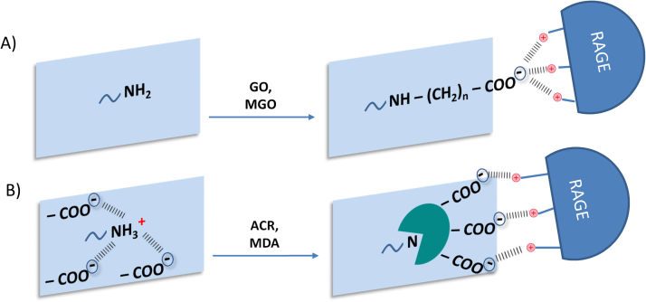
Advanced Lipoxidation End-products (ALEs) are modified proteins that can act as pathogenic factors in several chronic diseases. Several molecular mechanisms have so far been considered to explain the damaging action of ALEs and among these a pathway involving the receptor for advanced glycation end products (RAGE) should be considered. The aim of the present work is to understand if ALEs formed from lipid peroxidation derived reactive carbonyl species (RCS) are able to act as RAGE binders and also to gain a deeper insight into the molecular mechanisms involved in the protein-protein engagement. ALEs were produced in vitro, by incubating human serum albumin (HSA) with 4-hydroxy-trans- 2-nonenal (HNE), acrolein (ACR) and malondialdehyde (MDA). The identification of ALEs was performed by MS. ALEs were then subjected to the VC1 Pull-Down assay (VC1 is the ligand binding domain of RAGE) and the enrichment factor (the difference between the relative abundance in the enriched sample minus the amount in the untreated one) as an index of affinity, was determined. Computation studies were then carried out to explain the factors governing the affinity of the adducted moieties and the site of interaction on adducted HSA for VC1-binding. The in silico analyses revealed the key role played by those adducts which strongly reduce the basicity of the modified residues and thus occur at their neutral state at physiological conditions (e.g. the MDA adducts, dihydropyridine-Lysine (DHPK) and N-2-pyrimidyl-ornithine (NPO), and acrolein derivatives, N-(3-formyl-3,4-dehydro-piperidinyl) lysine, FDPK). These neutral adducts become unable to stabilize ion-pairs with the surrounding negative residues which thus can contact the RAGE positive residues. In conclusion, ALEs derived from lipid peroxidation-RCS are binders of RAGE and this affinity depends on the effect of the adduct moiety to reduce the basicity of the target amino acid and on the acid moieties surrounding the aminoacidic target.

Keywords: 4-hydroxy-trans− 2-nonenal (HNE); Acrolein (ACR) and malondialdehyde (MDA); Advanced lipoxidation end products (ALEs); Human serum albumin (HSA); Pull-down assay; RAGE; Reactive Carbonyl Species (RCS); VC1 domain.


Figures

fx1
Graphical abstract
Fig. 1
Fig. 1
Work flow of the study. ALEs-lipox were firstly prepared by incubating HSA with lipid peroxidation derived RCS (HNE, ACR and MDA) and then fully characterized by MS. The RAGE binding ability of each identified ALE was then determined by using the VC1 assay as previously reported . Computation studies were then carried out to explain the factors governing the affinity of the adducted moieties and the site of interaction on adducted HSA for VC1-binding.
Fig. 2
Fig. 2
Direct infusion ESI-MS analysis of native and HNE-modified HSA. Mass spectra of HSA recorded in a mass range between m/z 1400 and 1500. A) Native HSA shows sharp intense peaks referred to the charge ions 47 + , 46 + and 45 + ; the deconvoluted spectrum reports a MW 66,455 Da (H). When HSA is reacted with HNE at increasing molar ratios 1:1 (B), 1:5 (C), 1:10 (D), additional peaks relative to HNE adducts appear. At higher molar ratios 1:100 (E), 1:200 (F) and 1:1000 (G) the MS spectra lose resolution and become flat due to the presence of multiple adducts. H) Deconvoluted spectra showing the MS of HSA and protein adducts. MS spectra relative to HSA incubated with HNE at 1:100 M ratio and higher cannot be deconvoluted due to the extent of modification.
Fig. 3
Fig. 3
Modified albumin obtained by incubation of recombinant HSA in the presence of different molar ratio of RCS and VC1 pull-down assay. A) VC1 pull-down assay with untreated HSA. SDS PAGE analysis followed by Coomassie staining of the modified HSA obtained by 72 h incubation with the indicated molar ratio of RCS (panel B: MDA; panel D: ACR; panel F: HNE). The highest molar ratio of HSA-MDA (1:12,600), HSA-ACR (1:5000) or HSA-HNE (1:2000) were used as input (IN) in the pull-down assays with the VC1 and control (CTRL)-resins. The IN fractions, the unbound fractions (UNB) and eluates (E) were analyzed by SDS-PAGE followed by Coomassie staining. The gels show that untreated HSA does not bind VC1 (panel A), whereas high MW species of HSA-MDA (panel C), HSA-ACR (panel E) and HSA-HNE (panel G) are retained by the VC1-resin, but not by the CTRL-resin. Since the elution is performed in denaturing conditions, this step removes any associated molecule from the resin, including the two VC1 glycovariants (34 and 36 kDa) and streptavidin (Stv, 14 kDa), indicated by arrows.
Fig. 4
Fig. 4
Venn diagrams of the identified ALEs as reported inTable 1and2. ALEs are reported as the modified amino acid residues. The upper diagram refers to ALEs obtained by treating HSA with MDA, the middle with ACR and the lower with HNE. The input reports ALEs not retained by VC1 and present only in the input samples; VC1 are the ALEs identified only after VC1-enrichment; the intersections report the ALEs found both in the input and VC1-enriched samples.
Fig. 5
Fig. 5
Enrichment Factor (EF) graphical distribution. Enrichment Factor (EF) graphical distribution by means of vertical scatter plot overviewing the affinity of each identified ALEs towards VC1; all EF values spotted have been calculated as the differences between the relative abundance in the enriched sample minus the amount in the untreated one (Enrichment factor= %VC1-%NoVC1), and graphically grouped as: MDA-adducts (panel A), ACR-adducts (panel B) and HNE-adducts (panel C).
Fig. 6
Fig. 6
Chemical structures of ALEs-lipox formed by incubating HSA with MDA, ACR and HNE and identified by MS. For some ALEs the pK values are reported in brackets.
Fig. 7
Fig. 7
Putative RAGE-HSA complex as computed by protein-protein docking and induced by RP-Arg472 (6 A), DHPK-Lys436 (6B) and FDP-Lys262 (6 C). For each simulated adduct, the right panel shows the entire RAGE-HSA complex while the left panel focuses on key interactions stabilized by the adduct and its surrounding residues.
Fig. 8
Fig. 8
Mechanism explaining AGEs/ALEs and ALEs-lipox binding to VC1. A) VC1 engagement based on the acid residues of the protein ligand. Such a mechanism occurs when RCS, such as GO and MGO react with the basic residue forming a carboxylated adduct (CML and CEL) which directly contacts the RAGE residues. B) Panel B summarizes the mechanism described in the present paper for explaining the binding of ALEs-lipox to VC1 and called the “flowering effect”. It is based on a two-step process and involves exposed basic residues (mainly Lys and Arg) which in the non-adducted protein form a set of ionic bridges with the carboxylic groups of surrounding aspartate and glutamate residues. Lipid peroxidation derived RCS react with the basic residue and abolish or greatly reduce their basicity. Consequently, the adducted residues shift in a neutral form and such a change in the ionization state destabilizes the ionic bridges and renders the surrounding anionic residues more accessible and available to stabilize ion-pairs with the positive RAGE residues.

Similar articles

Cited by

References

    1. Gueraud F., Atalay M., Bresgen N., Cipak A., Eckl P.M., Huc L., Jouanin I., Siems W., Uchida K. Chemistry and biochemistry of lipid peroxidation products. Free Radic. Res. 2010;44(10):1098–1124. - PubMed
    1. Vistoli G., De Maddis D., Cipak A., Zarkovic N., Carini M., Aldini G. Advanced glycoxidation and lipoxidation end products (AGEs and ALEs): an overview of their mechanisms of formation. Free Radic. Res. 2013;47(Suppl 1):3–27. - PubMed
    1. Aldini G., Vistoli G., Stefek M., Chondrogianni N., Grune T., Sereikaite J., Sadowska-Bartosz I., Bartosz G. Molecular strategies to prevent, inhibit, and degrade advanced glycoxidation and advanced lipoxidation end products. Free Radic. Res. 2013;47(Suppl 1):93–137. - PubMed
    1. Mol M., Regazzoni L., Altomare A., Degani G., Carini M., Vistoli G., Aldini G. Enzymatic and non-enzymatic detoxification of 4-hydroxynonenal: methodological aspects and biological consequences. Free Radic. Biol. Med. 2017;111:328–344. - PubMed
    1. Curtis T. The role lipid aldehydes and ALEs in the pathogenesis of diabetic retinopathy. Free Radic. Biol. Med. 2014;75(Suppl 1):S8. - PubMed

Publication types