. 2019 Jan 15;116(3):874-879.
doi: 10.1073/pnas.1804042116. Epub 2018 Dec 31.

Patched1-ArhGAP36-PKA-Inversin axis determines the ciliary translocation of Smoothened for Sonic Hedgehog pathway activation

Affiliations

Patched1-ArhGAP36-PKA-Inversin axis determines the ciliary translocation of Smoothened for Sonic Hedgehog pathway activation

Boyan Zhang et al. Proc Natl Acad Sci U S A. .

Abstract

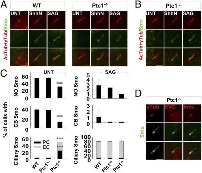
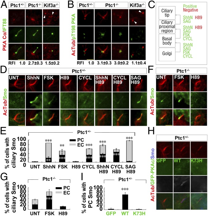
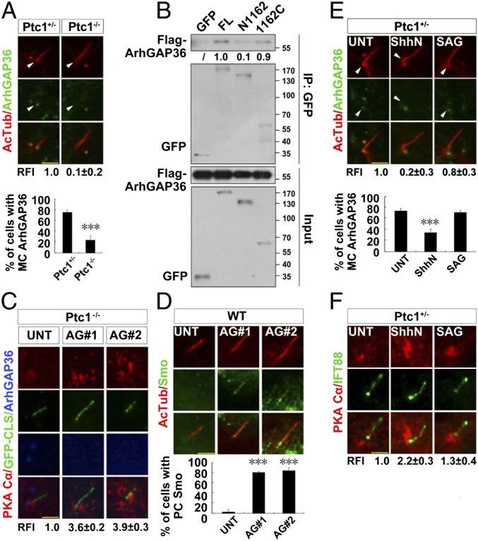
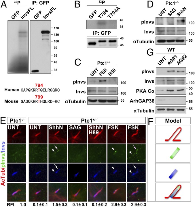
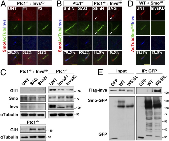
The Sonic Hedgehog (Shh) pathway conducts primarily in the primary cilium and plays important roles in cell proliferation, individual development, and tumorigenesis. Shh ligand binding with its ciliary membrane-localized transmembrane receptor Patched1 results in the removal of Patched1 from and the translocation of the transmembrane oncoprotein Smoothened into the cilium, leading to Shh signaling activation. However, how these processes are coupled remains unknown. Here, we show that the Patched1-ArhGAP36-PKA-Inversin axis determines the ciliary translocation of Smoothened. We find that Patched1 interacts with and stabilizes the PKA negative regulator ArhGAP36 to the centrosome. Activating the Shh pathway results in the removal of ArhGAP36 from the mother centriole and the centrosomal PKA accumulation. This PKA then phosphorylates Inversin and promotes its interaction with and the ciliary translocation of Smoothened. Knockdown of Inversin disrupts the ciliary translocation of Smoothened and Shh pathway activation. These findings reveal a regulatory molecular mechanism for the initial step of Shh pathway activation.

Keywords: Inversin; PKA; Patched1; Smoothened; Sonic Hedgehog pathway.


Conflict of interest statement

The authors declare no conflict of interest.

Figures

Fig. 1.
Fig. 1.
Smo mainly accumulates at the ciliary proximal region in Ptc1−/− cells. (AC) The ciliary proximal region localization of Smo in Ptc1−/− cells. G0 phase WT, Ptc1+/−, and Ptc1−/− MEF cells after the indicated treatments were immunostained for the indicated proteins. AcTub, acetylated α-tubulin; CB, ciliary base localization; EC, even ciliary localization; NO, no ciliary localization; PC, ciliary proximal region localization. In the statistics histograms for the results in A and B, the values are the mean ± SD; 50 cells per sample were randomly selected and counted in each of three independent experiments. ***P < 0.001. (D) A proportion of Smo accumulates at the Inversin compartment in Ptc1−/− cells. G0 phase Ptc1−/− cells were immunostained for the indicated proteins. Invs, Inversin; γTub, γ-tubulin. Note that only Inversin showed a partial colocalization with Smo at the ciliary proximal region. (Scale bars, 5 μm.)
Fig. 2.
Fig. 2.
The centrosomal accumulation of PKA results in the ciliary proximal localization of Smo in Ptc1−/− cells. (A and B) PKAc accumulates at the centrosome in Ptc1−/− cells. G0 phase Ptc1+/−, Ptc1−/−, and Kif3a−/− cells were immunostained for the indicated proteins. Arrowheads indicate the centrioles. The quantified relative fluorescent intensity (RFI) of centrosomal PKAc is shown Below each panel. The RFI results in A and B are shown as the mean ± SD; 50 cells were randomly selected for calculation. (C) A summary of the effects of the small molecules on the ciliary translocation of Smo. (D and E) PKA activity promotes Smo localization to the ciliary proximal region, downstream of Ptc1 inhibition by ShhN. G0 phase Ptc1+/− cells after the indicated treatments were immunostained for the indicated proteins. Note that PKA inhibition by H89 could abolish ShhN-induced, but had no effect on SAG- or cyclopamine-induced Smo ciliary translocation. (F and G) PKA activity accounts for Smo localization at the ciliary proximal region in Ptc1−/− cells. G0 phase Ptc1−/− cells after the indicated treatments were immunostained for the indicated proteins. (H and I) Increasing the amount of functional centrosomal PKAc promotes Smo localization at the ciliary proximal region. G0 phase Ptc1+/− cells stably expressing WT, K73H mutant GFP-PKAc, or GFP alone were immunostained for the indicated proteins. (Scale bars, 5 μm.) In the statistics histograms for results from E, G, and I, the values are the mean ± SD; 50 cells per sample were randomly selected and counted in each of three independent experiments. ***P < 0.001, **P < 0.01.
Fig. 3.
Fig. 3.
Removal of ciliary Ptc1 by Shh signaling abolishes centrosomal ArhGAP36 localization and increases the amount of centrosomal PKAc. (A) The mother centriolar (MC) localization of ArhGAP36 is impaired by Ptc1 knockout. G0 phase Ptc1+/− and Ptc1−/− cells were immunostained for the indicated proteins. Arrowheads indicate the MC localization of ArhGAP36. The RFIs and the ratio of MC-localized ArhGAP36 are shown Below each panel. (B) Ptc1 interacts with ArhGAP36 mainly via its C terminus. HEK 293T cells were cotransfected with the indicated plasmids, and arrested at the G0 phase, followed by an immunoprecipitation assay. The normalized results of immunoprecipitated Flag-ArhGAP36 by the indicated proteins are shown Below each panel; Flag-ArhGAP36 immunoprecipitated by full-length Ptc1-GFP was set as 1.0. (C) Knockdown of ArhGAP36 results in PKAc accumulation at the centrosome. The Ptc1−/− cells expressing a GFP-CLS sequence of Fibrocystin (53) were knocked down for ArhGAP36, arrested at the G0 phase, and immunostained for the indicated proteins. (D) Knockdown of ArhGAP36 results in Smo translocation to the ciliary proximal region. The WT cells were knocked down for ArhGAP36, arrested at the G0 phase, and immunostained for the indicated proteins. (E and F) Ptc1 inhibition by Shh signaling causes ArhGAP36 removal from the MC and PKAc accumulation at the centrosome. G0 phase Ptc1+/− cells after the indicated treatments were immunostained for the indicated proteins. Arrowheads indicate the MC localization of ArhGAP36. (Scale bars, 5 μm.) In the statistics histograms for results from A, D, and E, the values are the mean ± SD; 50 cells per sample were randomly selected and counted in each of three independent experiments. ***P < 0.001. The RFI results in A, C, E, and F are shown as the mean ± SD; 50 cells were randomly selected for calculation.
Fig. 4.
Fig. 4.
Shh signaling results in Inversin phosphorylation by PKA. (A and B) PKA phosphorylates Inversin at T794. HEK 293T cells were transfected with plasmids encoding the full length or fragments of human GFP-Inversin protein and arrested at the G0 phase. The GFP-tagged proteins were immunoprecipitated and subjected to in vitro kinase assay. T799 is the conserved site of the mouse Inversin protein analogous to the T794 in human protein. (C and D) Mouse Inversin is phosphorylated by PKA at T799. G0 phase Ptc1+/− cells after the indicated treatments were examined for the protein level of the indicated proteins. pInvs, phosphor-T799 Inversin. (E and F) Shh signaling leads to Inversin phosphorylation at T799 by PKA, with ciliary proximal region and tip localization. G0 phase Ptc1+/− and Ptc1−/− cells after the indicated treatments were immunostained for the indicated proteins. The localization pattern of the indicated proteins is shown in F. Arrowheads indicate the ciliary proximal region; arrows indicate the ciliary tip. (Scale bar, 5 μm.) The RFI results are shown as the mean ± SD; 50 cells were randomly selected for calculation. (G) Knockdown of ArhGAP36 results in Inversin phosphorylation at T799. WT cells were knocked down for ArhGAP36, arrested at the G0 phase, and examined for the indicated proteins.
Fig. 5.
Fig. 5.
Shh signaling-induced Inversin phosphorylation by PKA increases Inversin–Smo interaction. (A) Inversin interacts with Smo, and its T794 phosphorylation increases this interaction. HEK 293T cells were cotransfected with the indicated plasmids, arrested at the G0 phase, treated with or without FSK, and processed to immunoprecipitation assays. Note that losing the ability of T794 phosphorylation of Inversin by PKA impaired its binding with Smo. (B) Smo interacts with Inversin at the ciliary proximal region in Ptc1−/− cells. G0 phase Ptc1−/− cells were subjected to the PLA assay (see details in SI Appendix, Materials and Methods), singly labeled with Smo or Inversin, or colabeled with Smo and Inversin. (Scale bar, 5 μm.) (C and D) The phosphorylation-mimicking mutant of Inversin recruits Smo to the ciliary proximal region. WT cells stably expressing the WT or T794A/D mutant GFP-Inversin were arrested at the G0 phase and immunostained for the indicated proteins. The percentage of PC-patterned Smo in each cell line is shown in D. In each of three independent experiments, 50 cells per sample were randomly selected and counted. ***P < 0.001. (E) The T794D mutant Inversin results in a partial activation of Shh pathway. The parental cells (WT and Ptc1−/−) and the cells stably expressing the WT, T794A/D GFP-Inversin (labeled in green) were arrested at the G0 phase and examined for the indicated proteins. (F) Smo–Inversin interaction is increased by Shh pathway activation. HEK 293T cells were cotransfected with the indicated plasmids, arrested at the G0 phase, and processed to immunoprecipitation assays. The normalized results of total and immunoprecipitated upper band of Smo-Myc by the indicated proteins are shown Below each panel, and the amount of Smo-Myc under normal condition (UNT) was set as 1.0.
Fig. 6.
Fig. 6.
Inversin determines Smo ciliary translocation for Shh pathway activation. (A and B) Inversin is required for the ciliary translocation of Smo. Ptc1−/− or Ptc1+/− cells were knocked down for Inversin (InvsKD) and arrested at the G0 phase. After the indicated treatment, the cells were immunostained for the indicated proteins. Arrowheads indicate the ciliary base. The percentage of PC- and EC-localized Smo is shown. (C) Inversin is required for Shh signaling transduction. Ptc1+/−, Ptc1−/−, or Ptc1+/− cells knocked down for Inversin were arrested at the G0 phase; after the indicated treatment, the cells were lysed and the indicated proteins examined. (D) SmoM2 cannot translocate into the cilia of cells with Inversin knockdown. WT cells stably expressing SmoM2-GFP were knocked down for Inversin, arrested at the G0 phase, and immunostained for the indicated proteins. The percentage of EC-localized Smo is shown. In the statistics results from A, B, and D, the values are the mean ± SD; 50 cells per sample were randomly selected and counted in each of three independent experiments. (E) SmoM2 strongly interacts with Inversin in the absence of ShhN. HEK 293T cells were cotransfected with the indicated plasmids, arrested at the G0 phase, and processed to immunoprecipitation assays.

Similar articles

Cited by

References

    1. Briscoe J, Thérond PP. The mechanisms of Hedgehog signalling and its roles in development and disease. Nat Rev Mol Cell Biol. 2013;14:416–429. - PubMed
    1. Pak E, Segal RA. Hedgehog signal transduction: Key players, oncogenic drivers, and cancer therapy. Dev Cell. 2016;38:333–344. - PMC - PubMed
    1. Rohatgi R, Milenkovic L, Scott MP. Patched1 regulates Hedgehog signaling at the primary cilium. Science. 2007;317:372–376. - PubMed
    1. Pal K, et al. Smoothened determines β-arrestin-mediated removal of the G protein-coupled receptor Gpr161 from the primary cilium. J Cell Biol. 2016;212:861–875. - PMC - PubMed
    1. Bachmann VA, et al. Gpr161 anchoring of PKA consolidates GPCR and cAMP signaling. Proc Natl Acad Sci USA. 2016;113:7786–7791. - PMC - PubMed

Publication types