. 2019 Apr;60(4):869-879.
doi: 10.1194/jlr.M091322. Epub 2018 Dec 31.

An upstream enhancer regulates Gpihbp1 expression in a tissue-specific manner

Affiliations

An upstream enhancer regulates Gpihbp1 expression in a tissue-specific manner

Christopher M Allan et al. J Lipid Res. 2019 Apr.

Abstract

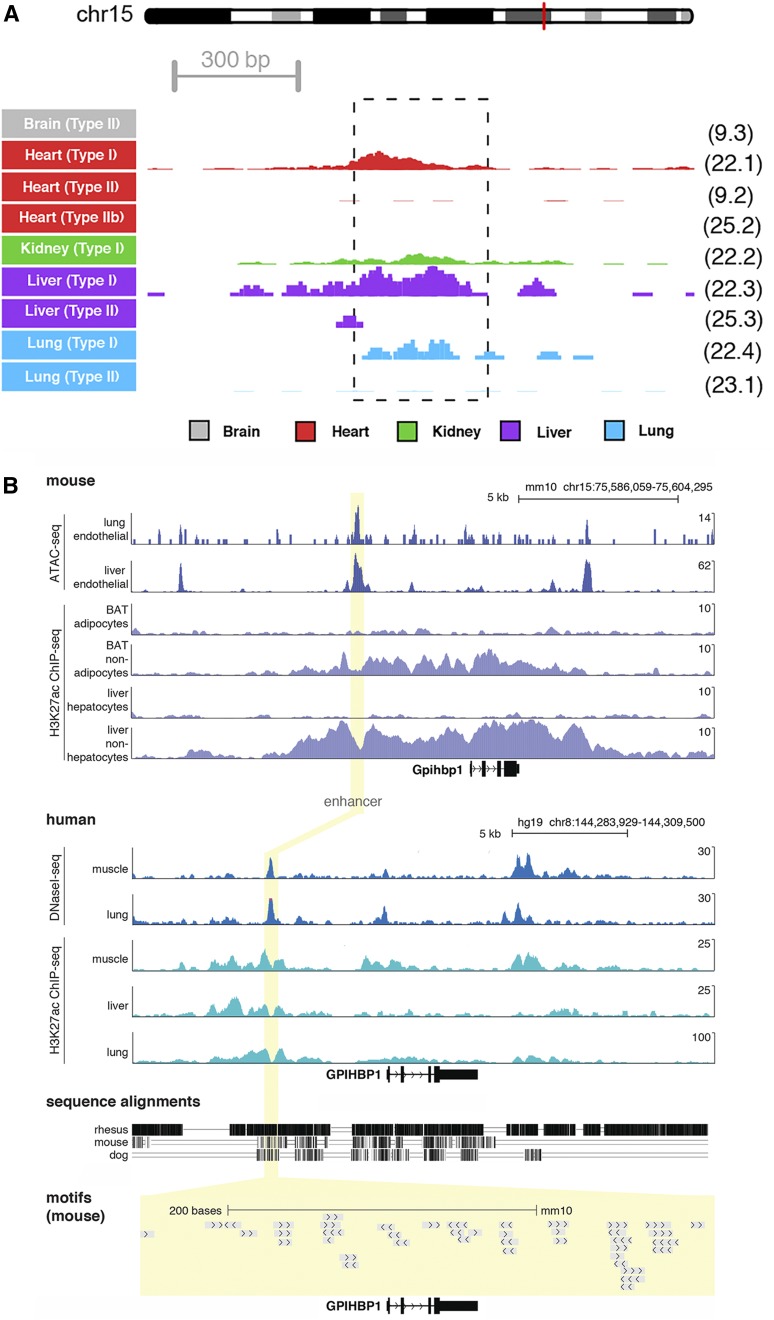
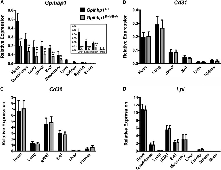
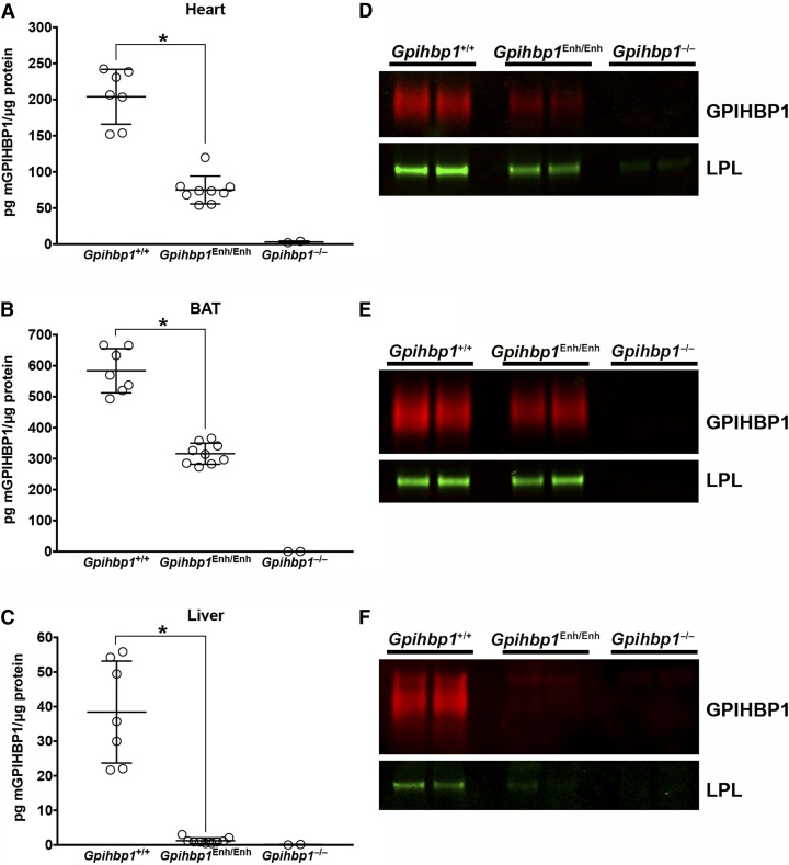
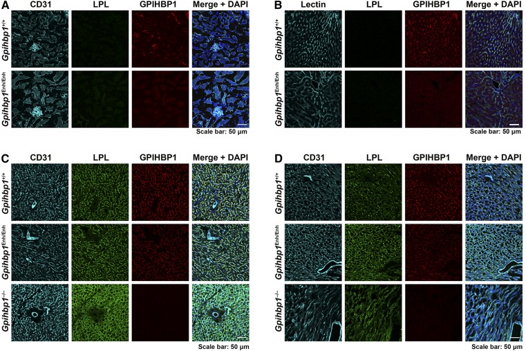
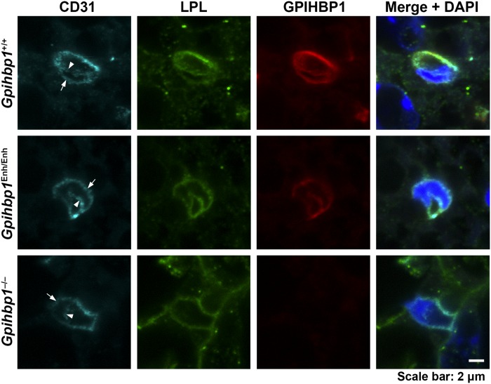
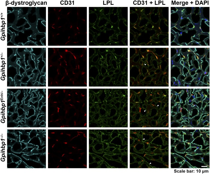
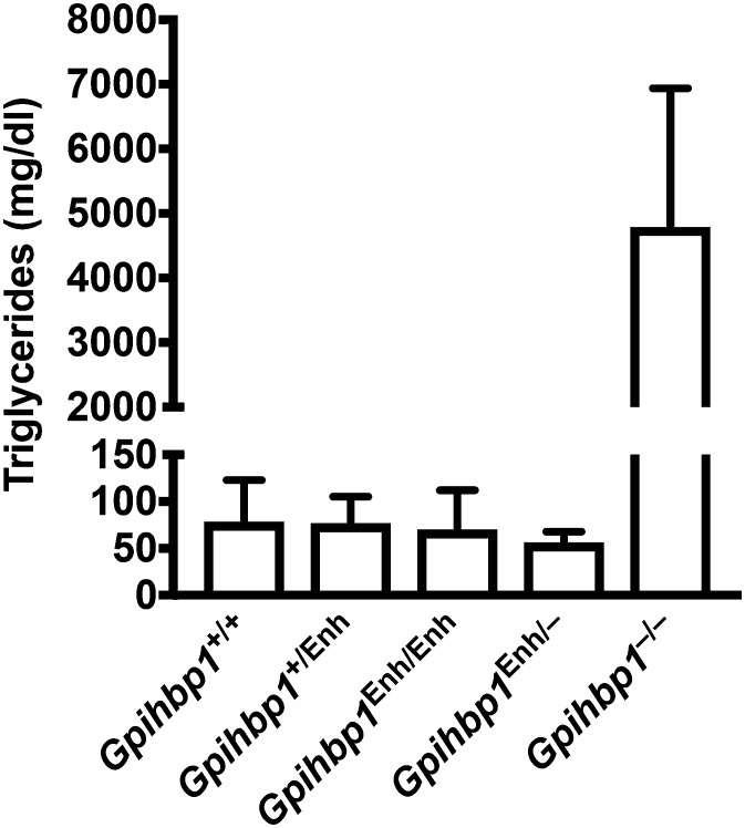
Glycosylphosphatidylinositol-anchored high density lipoprotein-binding protein 1 (GPIHBP1), the protein that shuttles LPL to the capillary lumen, is essential for plasma triglyceride metabolism. When GPIHBP1 is absent, LPL remains stranded within the interstitial spaces and plasma triglyceride hydrolysis is impaired, resulting in severe hypertriglyceridemia. While the functions of GPIHBP1 in intravascular lipolysis are reasonably well understood, no one has yet identified DNA sequences regulating GPIHBP1 expression. In the current studies, we identified an enhancer element located ∼3.6 kb upstream from exon 1 of mouse Gpihbp1. To examine the importance of the enhancer, we used CRISPR/Cas9 genome editing to create mice lacking the enhancer (Gpihbp1Enh/Enh). Removing the enhancer reduced Gpihbp1 expression by >90% in the liver and by ∼50% in heart and brown adipose tissue. The reduced expression of GPIHBP1 was insufficient to prevent LPL from reaching the capillary lumen, and it did not lead to hypertriglyceridemia-even when mice were fed a high-fat diet. Compound heterozygotes (Gpihbp1Enh/- mice) displayed further reductions in Gpihbp1 expression and exhibited partial mislocalization of LPL (increased amounts of LPL within the interstitial spaces of the heart), but the plasma triglyceride levels were not perturbed. The enhancer element that we identified represents the first insight into DNA sequences controlling Gpihbp1 expression.

Keywords: chylomicrons; endothelial cells; fatty acid metabolism; glycosylphosphatidylinositol-anchored high density lipoprotein–binding protein 1; lipids; lipolysis; triglycerides.


Conflict of interest statement

The authors have no financial interests to declare.

Figures

Fig. 1.
Fig. 1.
Epigenetic profiles for a putative Gpihbp1 enhancer. A: Single-cell chromatin accessibility profiles for mouse tissues identified nine types of endothelial cells with distinct accessibility profiles (13). Normalized sequence tag counts are shown at the Gpihbp1 enhancer in counts per million (CPM) reads; 5 CPM is the maximum on the y axis. The dashed box indicates the nucleotides deleted in the Gpihbp1Enh allele. The cell cluster identifier, labeled in the same manner as in the original publication (13), is shown to the right of each track. The red line on the depiction of Chr15 represents the location of the Gpihbp1 locus. B: Epigenomic profiles at the Gpihbp1 locus are shown for mouse (top) and the syntenic human region (bottom), detailing open chromatin profiles (ATAC-seq and DNaseI-seq) and H3K27ac ChIP-seq in different tissue/cell types. The yellow highlight depicts the region of the enhancer deletion. Sequence alignments and transcription factor binding motifs are shown below.
Fig. 2.
Fig. 2.
Gpihbp1 and Lpl transcript levels in 10-week-old Gpihbp1Enh/Enh and wild-type mice (Gpihbp1+/+). Gpihbp1 (A), Cd31 (B), Cd36 (C), and Lpl (D) transcript levels were measured by qRT-PCR (n = 10/group). Gpihbp1 expression was normalized to the expression of cyclophilin A. gWAT, gonadal white adipose tissue. Data show mean ± SD. P < 0.01; *P < 0.001; **P < 0.0001.
Fig. 3.
Fig. 3.
Reduced GPIHBP1 protein levels in Gpihbp1Enh/Enh mice. A–C: GPIHBP1 levels in tissue homogenates of heart (A), BAT (B), and liver (C) in 10-week-old mice were measured with a sandwich ELISA. Results are plotted as the mass of GPIHBP1 normalized to total protein (Gpihbp1+/+, n = 7; Gpihbp1Enh/Enh, n = 9; Gpihbp1−/−, n = 2). Data show mean ± SD; *P < 0.0001. D–F: GPIHBP1 and LPL levels in tissues of Gpihbp1Enh/Enh mice, as judged by Western blots. The GPIHBP1 (and any bound LPL) in 200 μg of heart (D) or BAT (E) tissue homogenates [or 1 mg of liver (F) homogenate] were immunoprecipitated with 25 μl of agarose beads coated with the GPIHBP1-specific antibody 11A12 (n = 2 mice/group). Relative amounts of GPIHBP1 and LPL in the immunoprecipitates were assessed by Western blotting with antibodies against GPIHBP1 (red) and LPL (green). GPIHBP1 signals, as judged by an infrared scanner, were quantified in Gpihbp1+/+ heart (1920000 and 1460000) and Gpihbp1Enh/Enh heart (406000 and 246000); Gpihbp1+/+ BAT (20600000 and 14900000) and Gpihbp1Enh/Enh BAT (7280000 and 9490000); and Gpihbp1+/+ liver (1110000 and 1320000) and Gpihbp1Enh/Enh liver (151000 and 163000). The LPL:GPIHBP1 ratio was calculated in Gpihbp1+/+ heart (0.427 and 0.633) and Gpihbp1Enh/Enh heart (1.138 and 1.341), Gpihbp1+/+ BAT (0.022 and 0.032) and Gpihbp1Enh/Enh BAT (0.047 and 0.045), and Gpihbp1+/+ liver (0.015 and 0.009) and Gpihbp1Enh/Enh liver (0.031 and 0.017).
Fig. 4.
Fig. 4.
Confocal immunofluorescence microscopy images of mouse tissues after staining with antibodies against CD31, GPIHBP1, and LPL. Sections of kidney (A), liver (B), BAT (C), and heart (D) were stained with antibodies against LPL (green) and GPIHBP1 (red). Kidney, BAT, and heart were stained with an antibody against CD31 (cyan); the liver was stained with tomato lectin (cyan). DNA was stained with DAPI (blue). Scale bar, 50 μm.
Fig. 5.
Fig. 5.
LPL reaches the capillary lumen in the BAT of Gpihbp1Enh/Enh mice despite reduced amounts of GPIHBP1 expression. Here, we examined the binding of CD31- (cyan), GPIHBP1- (red), and LPL-specific (green) antibodies to the BAT of Gpihbp1+/+, Gpihbp1Enh/Enh, and Gpihbp1−/− mice. To visualize the luminal and basolateral surfaces of capillary endothelial cells, we recorded confocal microscopy images of capillary cross-sections containing endothelial cell nuclei. In the BAT of Gpihbp1+/+ and Gpihbp1Enh/Enh mice, CD31, GPIHBP1, and LPL were visible on both the luminal (arrowheads) and basolateral surface of capillary endothelial cells (arrows). In Gpihbp1−/− mice, LPL was virtually undetectable along the luminal surface of capillary endothelial cells. DNA was stained with DAPI (blue). Scale bar, 2 μm.
Fig. 6.
Fig. 6.
LPL is partially mislocalized in the heart in Gpihbp1Enh/− mice, with increased amounts of LPL in the interstitial spaces near the surface of cardiomyocytes. Confocal microscopy studies were performed on sections of heart stained with antibodies for β-dystroglycan (cyan), CD31 (red), and LPL (green). β-Dystroglycan is located along the surface of cardiomyocytes. In comparing confocal images from Gpihbp1+/+ and Gpihbp1Enh/− mice, we observed more LPL outside of capillaries in Gpihbp1Enh/− mice (colocalizing with β-dystroglycan) (arrowheads in the CD31/LPL merged image point to several such regions). An even greater amount of interstitial LPL (colocalizing with β-dystroglycan) was observed in sections from Gpihbp1−/− mice (arrowheads). A small amount of LPL was mislocalized to the interstitial spaces in Gpihbp1+/− mice (arrowhead). DNA was stained with DAPI (blue). Scale bar, 10 μm.
Fig. 7.
Fig. 7.
Normal plasma triglycerides in Gpihbp1Enh/Enh and Gpihbp1Enh/− mice. Plasma triglyceride levels were measured in 10-week-old Gpihbp1+/+ (n = 16), Gpihbp1+/Enh (n = 20), Gpihbp1Enh/Enh (n = 17), Gpihbp1Enh/− (n = 8), and Gpihbp1−/− (n = 6) mice. Data show mean ± SD.

References

    1. Goldberg I. J. 1996. Lipoprotein lipase and lipolysis: Central roles in lipoprotein metabolism and atherogenesis. J. Lipid Res. 37: 693–707. - PubMed
    1. Fong L. G., Young S. G., Beigneux A. P., Bensadoun A., Oberer M., Jiang H., and Ploug M.. 2016. GPIHBP1 and plasma triglyceride metabolism. Trends Endocrinol. Metab. 27: 455–469. - PMC - PubMed
    1. Allan C. M., Larsson M., Jung R. S., Ploug M., Bensadoun A., Beigneux A. P., Fong L. G., and Young S. G.. 2017. Mobility of “HSPG-bound” LPL explains how LPL is able to reach GPIHBP1 on capillaries. J. Lipid Res. 58: 216–225. - PMC - PubMed
    1. Kristensen K. K., Midtgaard S. R., Mysling S., Kovrov O., Hansen L. B., Skar-Gislinge N., Beigneux A. P., Kragelund B. B., Olivecrona G., Young S. G., et al. . 2018. A disordered acidic domain in GPIHBP1 harboring a sulfated tyrosine regulates lipoprotein lipase. Proc. Natl. Acad. Sci. USA. 115: E6020–E6029. - PMC - PubMed
    1. Davies B. S., Beigneux A. P., Barnes R. H. 2nd, Tu Y., Gin P., Weinstein M. M., Nobumori C., Nyren R., Goldberg I., Olivecrona G., et al. . 2010. GPIHBP1 is responsible for the entry of lipoprotein lipase into capillaries. Cell Metab. 12: 42–52. - PMC - PubMed

Publication types