. 2019 Mar;47(3):194-202.
doi: 10.1124/dmd.118.084772. Epub 2018 Dec 31.

Commensal Gut Bacteria Convert the Immunosuppressant Tacrolimus to Less Potent Metabolites

Affiliations

Commensal Gut Bacteria Convert the Immunosuppressant Tacrolimus to Less Potent Metabolites

Yukuang Guo et al. Drug Metab Dispos. 2019 Mar.

Abstract

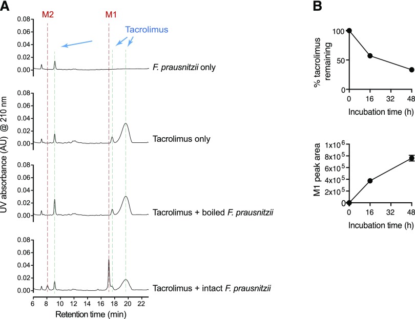
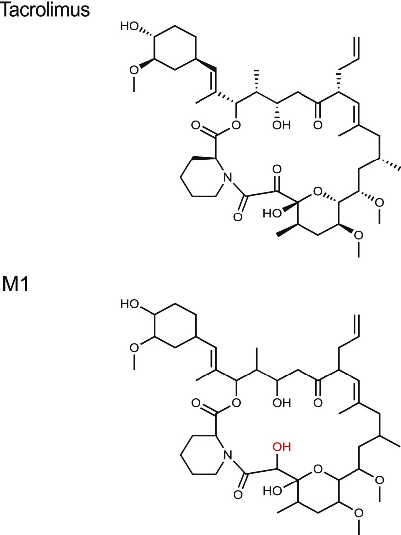
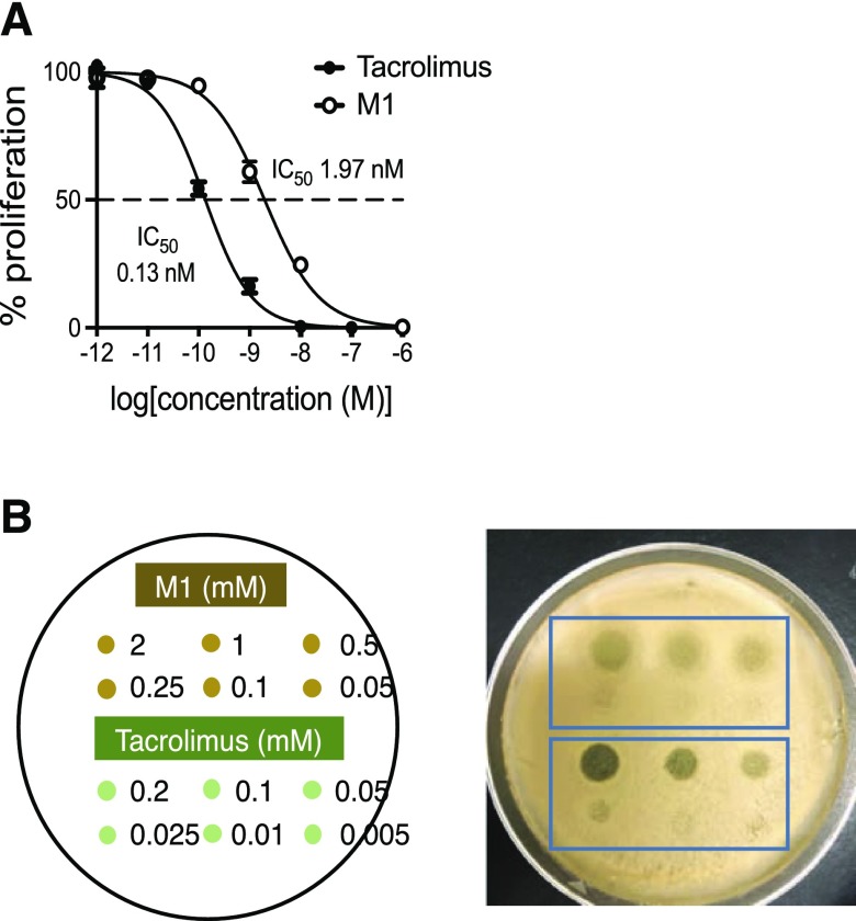
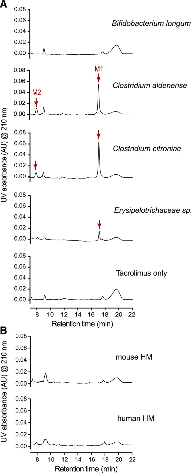
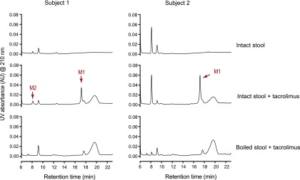
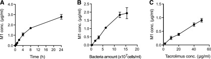
Tacrolimus exhibits low and variable drug exposure after oral dosing, but the contributing factors remain unclear. Based on our recent report showing a positive correlation between fecal abundance of Faecalibacterium prausnitzii and oral tacrolimus dose in kidney transplant patients, we tested whether F. prausnitzii and other gut abundant bacteria are capable of metabolizing tacrolimus. Incubation of F. prausnitzii with tacrolimus led to production of two compounds (the major one named M1), which was not observed upon tacrolimus incubation with hepatic microsomes. Isolation, purification, and structure elucidation using mass spectrometry and nuclear magnetic resonance spectroscopy indicated that M1 is a C-9 keto-reduction product of tacrolimus. Pharmacological activity testing using human peripheral blood mononuclear cells demonstrated that M1 is 15-fold less potent than tacrolimus as an immunosuppressant. Screening of 22 gut bacteria species revealed that most Clostridiales bacteria are extensive tacrolimus metabolizers. Tacrolimus conversion to M1 was verified in fresh stool samples from two healthy adults. M1 was also detected in the stool samples from kidney transplant recipients who had been taking tacrolimus orally. Together, this study presents gut bacteria metabolism as a previously unrecognized elimination route of tacrolimus, potentially contributing to the low and variable tacrolimus exposure after oral dosing.


Figures

Fig. 1.
Fig. 1.
F. prausnitzii metabolizes tacrolimus. (A) F. prausnitzii (OD600 2.6) cultured in YCFA media was incubated with tacrolimus (100 μg/ml) anaerobically at 37°C for 48 hours. The mixture was analyzed by using HPLC/UV. (B) Time profiles of tacrolimus disappearance and M1 appearance upon anaerobic incubation of tacrolimus (100 μg/ml) with F. prausnitzii.
Fig. 2.
Fig. 2.
Chemical structures of tacrolimus and F. prausnitzii–derived metabolite M1. M1 structure was identified using mass spectrometry and nuclear magnetic resonance spectroscopy.
Fig. 3.
Fig. 3.
M1 is less potent than tacrolimus as an immunosuppressant and antifungal agent. (A) Immunosuppressant activities of tacrolimus and M1 were examined in PBMCs by measuring cell proliferation after treatment with a T-lymphocyte mitogen in the presence of tacrolimus or M1. (B) Antifungal activities of tacrolimus and M1 were examined using Malassezia sympodialis. The yeast was inoculated on a modified Dixon agar plate. After 1 hour incubation, an aliquot of tacrolimus or M1 at different concentrations was placed on the plate, as shown in the left panel, and incubated at 37°C for 2 days.
Fig. 4.
Fig. 4.
Multiple commensal gut bacteria convert tacrolimus to M1. (A) Representative chromatograms of bacteria incubated with tacrolimus. M1 nonproducer (Bifidobacterium longum) or producer (Clostridium aldenense, Clostridium citroniae, and Erysipelotrichaceae sp.) cultured overnight in YCFA media was incubated with tacrolimus (100 μg/ml) anaerobically at 37°C for 48 hours. The mixture was analyzed by using HPLC/UV at 210 nm. (B) Mouse or human hepatic microsomes [(HMs); 3 mg microsomal protein/ml] were incubated with tacrolimus (100 μg/ml) at 37°C for 2 hours aerobically. The mixture was analyzed by using HPLC/UV.
Fig. 5.
Fig. 5.
Human gut microbiota convert tacrolimus to M1. Tacrolimus (100 μg/ml) was incubated anaerobically with human stool samples from two different subjects (100 mg wet weight/ml) for 48 hours at 37°C. A separate set of samples was boiled for 10 minutes before incubation with tacrolimus. The incubation mixtures were analyzed by HPLC/UV.
Fig. 6.
Fig. 6.
M1 formation by small intestinal bacteria may be extensive. (A) Tacrolimus (10 μg/ml) was incubated anaerobically with F. prausnitzii (6.3 × 10⁷ cells/ml) for varying amounts of time. (B) Tacrolimus (10 μg/ml) was incubated with varying amount of F. parusnitzii in PBS at 37°C for 2 hours. (C) Tacrolimus at varying concentrations was incubated with F. prausnitzii (6.3 × 10⁷ cells/ml) for 1 hour. M1 concentrations in the reaction mixtures were analyzed by liquid chromatography–MS/MS.

References

    1. Altschul SF, Gish W, Miller W, Myers EW, Lipman DJ. (1990) Basic local alignment search tool. J Mol Biol 215:403–410. - PubMed
    1. Arumugam M, Raes J, Pelletier E, Le Paslier D, Yamada T, Mende DR, Fernandes GR, Tap J, Bruls T, Batto JM, et al. MetaHIT Consortium (2011) Enterotypes of the human gut microbiome. Nature 473:174–180. - PMC - PubMed
    1. Dubbelboer IR, Pohanka A, Said R, Rosenborg S, Beck O. (2012) Quantification of tacrolimus and three demethylated metabolites in human whole blood using LC-ESI-MS/MS. Ther Drug Monit 34:134–142. - PubMed
    1. Edgar RC. (2013) UPARSE: highly accurate OTU sequences from microbial amplicon reads. Nat Methods 10:996–998. - PubMed
    1. Floren LC, Bekersky I, Benet LZ, Mekki Q, Dressler D, Lee JW, Roberts JP, Hebert MF. (1997) Tacrolimus oral bioavailability doubles with coadministration of ketoconazole. Clin Pharmacol Ther 62:41–49. - PubMed

Publication types

MeSH terms

LinkOut - more resources